SlideShare a Scribd company logo
Dr D Nagaraju, Asst.Prof. of Botany, Govt. Degree College, Warangal, TS
Lichens (Algae+Fungi)
(Structure and Reproduction; Ecological and
Economic Importance)
Lichens are a small group of curious plants, formed from a combination of a fungal
partner (mycobiont) and an algal partner (phycobiont). The fungal filaments surround and
grow into the algal cells, and provide the majority of the lichen's physical bulk and shape.
Lichen is derived from the Greek word ‘Leprous’ and refers to medicine used for treatment of
skin diseases because of their appearance as peeling skin. Theophrastus (371-284 B.C) coined
the term ‘lichen’ for superficial growth on the bark of olive tree. Lichen species collectively
called as ‘stone flower’ in English; in Sanskrit-Sailaja, Saileya, Shilapushpa; in Hindi-Charila,
Pathar-ka-phool, Shaik; in Telugu-Rathipuvvu. The study of Lichens is called Lichenology.
Lichenization is the process by which fungal hyphae and algal cells literally grow together to
form a mutualistic association.
Erik Acharius (1810) a Swedish botanist, referred to as Father of Lichenology, coined
several terms for the structures peculiar to lichens and described many new genera and numerous
new species on the basis of external morphology in his monumental works. His four major works
of research are Lichenographisuecic prodromus (1798), Methodus Lichenum (1803),
Lichenographia Universalis (1810), and Synopsis Methodica Lichenum(1814) formed the
foundations of modern lichenology.
Dual Nature of Lichens:
A Swedish Scientist Schwendner (1867-68) first studied in detail nature of lichens (dual
nature) and explained that they formed as a result of close association between a fungus and an
alga. The lichen thallus thus represents as Idyllic-marriage- as a consortium between the
fungus and alga. The two components behave like husband and wife benefitting one another.
Mycobiont:
The fungal component of lichen is called the mycobiont. The mycobiont may be an
Ascomycete or Basidiomycete. The associated lichens are called either ascolichens or
basidiolichens, respectively. Living as a symbiont in a lichen appears to be a successful
way for a fungus to derive essential nutrients, since about 20% of all fungal species have
acquired this mode of life. Most of the lichenized fungi are Ascomycotina, 12 orders include
mostly lichenized members (Arthoniomycetes , Eurotiomycetes , Leucanomycetes ,
Lichenomycetes ). Some are Basidiomycotina – Aphyllophorales, few Agaricales and Some
are Deuteromycota. 20% of all fungi & 40% of Ascomycotina are lichenised ( Krik et al.,2008).
So far there is no evidence about the fungi belonging to Mastigomycotina and Zygomycotina
forming lichens.
Dr D Nagaraju, Asst.Prof. of Botany, Govt. Degree College, Warangal, TS
Photobionts:
The photosynthetic partner in lichen is called a photobiont. The photobiont in lichens
come from a wide variety of simple prokaryotic and eukaryotic organisms. The majority of
the lichens contain eukaryotic photobionts that are green algae (Chlorophyta) or yellowgreen
algae (Xanthophyta). The prokaryotes are blue green "algae" (cyanobacteria). Algal
photobionts are called phycobionts. Cyanobacterium photobionts are called
cyanobionts. About 90% of all known lichens have phycobionts, and about 10 have
cyanobionts. Approximately 100 species of photosynthetic partners from 40 genera and five
distinct classes (prokaryotic: Cyanophyceae; eukaryotic: Trebouxiophyceae, Phaeophyceae,
Chlorophyceae) have been found to associate with the lichen-forming fungi.
Common algal photobionts are from the genus Trebouxia, Trentepohlia, Pseudotrebouxia,
or Myrmecia (algae). Trebouxia is the most common genus of green algae in lichens,
occurring in about 40% of all lichens. The second most commonly represented
green alga genus is Trentepohlia. Overall, about 100 species of eukaryotes are known to
occur as photobionts in lichens. All the algae are probably able to exist independently in
nature as well as in the lichen.
A "cyanolichen" is lichen with a cyanobacterium as its main photosynthetic component
(photobiont). The most commonly occurring cyanobacteria genus is Nostoc. Other
common cyanobacterium photobionts are from Scytonema. Many cyanolichens are
small and black, and have limestone as the substrate. Another cyanolichen group, the jelly
lichens (e.g. from the genera Collema or Leptogium) are large and foliose (e.g., species of
Peltigera, Lobaria and Degelia.
Toby Spribille and John McCutcheon (2016) discovered third partner (trinity) in lichen
symbiosis, for the past150 years, scientists assumed that the lichens consisted of such a
symbiosis of fungi and algae. However, attempts by lichenologists to artificially meld fungi and
algae into lichens never yielded the same results one would see in nature. Something seemed to
be missing. They found that many lichens also have feature specific basidiomycete (yeasts)
in their cortex, means the symbiosis involves not one but two fungi, and one algae,
discovered this missing link, (in Bryoria) by marked each partner with its own fluorescent
molecule (red, green, blue) that the trinity was clearly revealed. This discovery is changing
the paradigm of lichen biology.
Evolution and palaeontology:
The fossil record for lichens is poor. The extreme habitats that lichens dominate, such as tundra,
mountains, and deserts, are not ordinarily conducive to producing fossils.
Endosymbiotic Glomeromycota comparable with living Geosiphon may extend back into the
Proterozoic in the form of 1500 million year old Horodyskia and 2200 million year old
Diskagma. Discovery of these fossils suggest that fungi developed symbiotic partnerships with
photoautotrophs long before the evolution of vascular plants.
The oldest fossil lichens in which both symbiotic partners have been recovered date to the Early
Devonian Rhynie chert, about 400 million years old. The slightly older fossil Spongiophyton has
also been interpreted as a lichen on morphological and isotopic grounds, although the isotopic
basis is decidedly shaky. It has been demonstrated that Silurian Devonian fossils Nematothallus
Dr D Nagaraju, Asst.Prof. of Botany, Govt. Degree College, Warangal, TS
and Prototaxites were lichenized. Thus lichenized Ascomycota and Basidiomycota were a
component of early Silurian Devonian
terrestrial ecosystems. Lutzoni et al. (2001) indicate that lichenization probably evolved earlier
and was followed by multiple independent losses. Some non lichen forming
fungi may have secondarily lost the ability to form a lichen association. As a result, lichenization
has been viewed as a highly successful nutritional strategy.
Growth rate: Lichens often have a regular but very slow growth rate of less than a millimetre
per year. Different lichen species have been measured to grow as slowly as 0.5 mm, and as fast
as 0.5 meter per year. In crustose lichens, the area along the margin is where the most active
growth is taking place. Most crustose lichens grow only 1–2 mm in diameter per year. Climatic
factors most favourable for the growth of lichens are direct light, a moderate or cold temperature,
constant moisture and unpolluted atmosphere.
Nutrition: The major partner of lichens, the mycobiont is heterotropic in nutrition. The
minor partner of lichens, the phycobiont is an autotrphic organism which can carryout
photosynthesis. Hence, algal partner helps the fungal partner in nutrition. In some lichens the
fungal hyphae sends haustoria into algal cells to absorb the nutrients. The fungi get vitamins
essential for growth like thiamine, protein and others from algal cells. The carbohydrates found
in algal cells formed through photosynthesis are transferred to fungal hyphae, the carbohydrates
are varies in algae with species Nostoc form glucose, Trebouxia produce ribitol. The fungal
hyphae store the carbohydrates in the form of mannitol. When photobiont is a Cyanophycean
member it fixes atmospheric nitrogen in the form of ammonia, and supplies it to the fungal
partner. The fungal hyphae absorb water and mineral from the surroundings through the upper
cortex and form substrate through rhizenes and supply them to the algal partner.
Occurrence: Lichens are pioneer species, among the first living things to grow on bare rock or
areas denuded of life by a disaster. Lichens are amongst the slowest-growing organisms,
but their tolerance of environmental extremes enables them to colonise habitats where few other
macroscopic organisms can grow. They grow where neither the fungal partner nor the
photosynthetic partner could survive alone, because they benefit from their unique symbiotic
association. A major ecophysiological advantage of lichens is that they are poikilohydric
(poikilo-variable, hydric- relating to water), meaning that though they have little control over
the status of their hydration, they can tolerate irregular and extended periods of severe
desiccation. Like some mosses, liverworts, ferns, and a few "resurrection plants", upon
desiccation, lichens enter a metabolic suspension or stasis (known as cryptobiosis) in which the
cells of the lichen symbionts are dehydrated to a degree that halts most biochemical activity. In
this cryptobiotic state, lichens can survive wider extremes of temperature, radiation and drought
in the harsh environments they often inhabit.
Lichens do not have roots and do not need to tap continuous reservoirs of water like most higher
plants, thus they can grow in locations impossible for most plants, such as bare rock, sterile soil
or sand, and various artificial structures such as walls, roofs and monuments. Many lichens also
grow as epiphytes (epi- on the surface, phyte- plant) on plants, particularly on the trunks and
branches of trees. These are not parasites on the plants they grow on, but only use them as a
substrate to grow on, growing on leaves may have the appearance of being parasites on the
Dr D Nagaraju, Asst.Prof. of Botany, Govt. Degree College, Warangal, TS
leaves, but they are not. However, some lichens are known to parasitise other lichens.
Diploschistes muscorum starts its development in the tissue of a host Cladonia species.
The fungal species that form unique symbiotic relationships with algae are called lichenized
fungi. They typically belong to the fungal classes Ascomycetes or Basidiomycetes. Another
term used by lichenologists is "lichenicolous." This refers to specialized fungi that live on the
thallus or the ascocarps of lichens. They are different from the lichenized fungi that form the
main body of lichens. Lichenicolous fungi belong to three fungal classes, the Ascomycetes,
Deuteromycetes and Basiodiomycetes. A black lichenicolous fungus Lichenochora verrucicola
is parasitic on the the grayish-brown crustose lichen Aspicilia cuprea.
Lichens form easily distinguishable coloured patches on tree barks, rocks and soil. They are
Universally distributed organisms. Microchemical Methods used for the Identification of Lichens
by Alan Orange et al. (2010). Found Components of lichen may react with certain test chemicals
to give colour reactions which assist in the identification of a species, Commonly used test
chemicals are: K potassium hydroxide, C – bleach, I – iodine, Pd paraphenyl-enediamine, a
weak acid putting a drop on a lichen turns an area bright yellow to orange, this helps identify it
as belonging to either the genus Cladonia or Lecanora.
Lichens are most successful symbiotic organisms in nature, dominating 10% or more of the
earth’s terrestrial area (Kumar et al. 2010) and known to be the earliest colonizers of terrestrial
habitats with a worldwide distribution. These are distributed in almost all the phytogeographical
regions of the world in tropics, subtropics, temperate, from plain to mountain regions and arctic
to tropical regions. They are most abundant in tropical rain forests and evergreen forests in
temperate regions, in cold regions, even also found in desert areas on exposed rocks. Lichens
generally grow on stems, branches and bark of forest trees, on wood, ground and surface of rocks
etc. it form easily distinguishable coloured patches Depending on the substrates, lichens are
described as:
1. Saxicolous: The lichens grow on Rocks eg: Peltigera, Dermatocarpon, Verrucaria etc.
2. Corticolous: The lichens grow on Barks and tree trunks eg: Parmelia, Usnea etc.
3. Terricolous: The lichens grow on Soil, ground as a substrate eg: Cladonia floerkeana,
Lecidea etc.
4. Lignicolous: The lichens grows on wood from which the bark has been stripped
eg: Cyphellum, Calicium, Chanetheca etc.
5. Epilithic Lichens: Crustose lichens that grow on the rocks.
6. Endolithic Lichens: Crustose lichens grow immersed inside rock, growing between
the crystals with only their fruiting bodies exposed to the air
7. Endophloidic Lichens or Endophloidal Lichens: Lichens grow immersed inside
plant Tissues.
8. Epiphyllous or Follicolous Lichens: Lichens that use leaves as substrates, whether
the leaf is still on the tree or on the ground. eg. Byssoloma sp. Sporopodium
vezdeanum, Strigula smaragdula.
9. Umbillicate Lichens: Foliose lichens that are attached to the substrate at only one
Dr D Nagaraju, Asst.Prof. of Botany, Govt. Degree College, Warangal, TS
point. eg. Lasallia papulosa, Lobaria linita.
10. Vagrant Lichens These are not attached to a substrate at all, and lives its life being
blown around by the wind. eg. Xanthoparmelia semiviridis and Xanthomaculina
convolute.
11. Vitricolous lichens: Lichens grow on glass. The term is derived from the Latin
vitrum (glass) and cola (indicating inhabitant), so the word vitricolous literally means
'glass inhabiting'. eg. Xanthparmelia mougeotina. Diploicia canescens, Lepraria flava
(Chrysothrix candelaris) and Pertusaria leucosora.
12. Fresh water Lichens: Lichens grow on hard siliceous rocks in fresh water
eg. Hymenelia lacustris.
13. Marine lichens: Lichens grow on silicious rocky shore of sea eg. Caloplaca marina,
Verrucaria mucosa.
Saxicolous Terricolous Corticolous Lignicolous
Follicolous Fresh water lichen Marine lichen Vitricolous
Fig. 3. 1. Types of Lichens based on Substrates.
P.A. Micheli described above 300 species of lichens in his Nova Plantarum Genera. Lichens of
India are being studied since 1753 AD by Linnaeus. India has a rich diversity of lichens
represented by more than 2,040 species (Awasthi, 2000), which is about 10% of the total 20,000
species known from the world. Singh and Sinha (1997) divided India in to eight lichenographic
regions. Among them according to recent observation undoubtedly Western Ghats records the
highest number lichen taxa (1096 spp.) out of these 257 species endemic to Western Ghats.
Among different states Tamilnadu have the highest number of lichens with 736 species followed
by Karnataka, Kerala, Maharastra with 455, 391, and 184 species respectively, and Eastern &
Dr D Nagaraju, Asst.Prof. of Botany, Govt. Degree College, Warangal, TS
Western Himalayas (850 spp.). Belanger (1838) initiated the study of lichens in the Western
Ghats and described a total of 40 taxa from Pondicherry and Coramandal Coast. Quraishi (1928)
and Chopra were studied Himalayan Lichens. Chopra (1934) in his monograph on the “Lichens
of Darjeeling and the Sikkim Himalayas” described 80 species belonging to 38 genera of
Ascolichens. Dr. D. D. Awasthi “Father of Indian Lichenology” in the late forties of the last
century established in India and a number of lichenological investigations related with
monographic, revisionary and floristic studies were initiated in the country, he written “A Hand
Book of Lichens” (2000), Compendium of the Macrolichens from India, Nepal and Srilanka
(2007) and Singh, K.P., Sinha, G.P. and Bujarbarua, P. (2004). Endemic lichens of India.
The first record of lichen community studies from India was provided by Dudgeon (1923) who
studied succession of epiphytic lichens on Quercus leucotrichophora tree in Western Himalayas.
The collected specimens were investigated morphologically, anatomically and chemically at
Lichenology laboratory of the National Botanical Research Institute, Lucknow. In Andhra
Pradesh and Telangana Prof. C. Manoharachary and his associates at Osmania University,
carried out an extensive survey on lichens and reported that there are 17 genera and 65 species in
different parts of the states. Among the 65 species recorded, 58 species occur on stem and bark,
while sexicolous and terricolous lichens are very few. Morphologically 38 are foliose lichens, 12
fruiticose lichens and 15 are crustose lichens. The predominant and major genera include
Parmelia, Cladonia, Leconora, Physcia, Derinaria, Heterodermium, Hyperphyscia, Graphis,
Pyxia etc. all these are Ascolichens.
Nature of Association (Physiology):
The association of autotrophic phycobiont with heterotrophic mycobiont is described
variously by different scientists. Three different hypothesis have been put forth to explain the
nature of the association in this plant complex.
The parasitism: Flank (1913) and others opinion that the fungus lives during all or part of its
life in parasitic relation with the algal host and also sustains a relation with an organic or
inorganic substratum. Geitler (1927) considered the relationship as one of parasitism, the fungus
being parasitic on the alga, as evidenced in some cases by the presence of fungus haustoria in the
algal cells. The alga as considered a mere victim of the fungus, the fungi in lichens are mildly
parasitic upon the algal cells, it permits most of the algal cells to live. The following two facts
support their view:
➢ In some lichens the fungal hyphae give out haustoria or appressoria. These penetrate the
algal cells to obtain nutrition for the fungus mycelium. The haustoria produced by some
fungi penetrate and eventually kill some of the algal cells but more often a hyphal branch
tip becomes closely adpresed to the algal cell without entering it. In some cases, the algal
cells which have died in the lichen thallus, are digested and used by the fungal
component.
➢ When the two components of the lichen thallus are artificially separated the alga is able
to live as an independent individual. It grows and multiplies like an independent
organism. But the fungus is unable to do so. It invariably dies.
Dr D Nagaraju, Asst.Prof. of Botany, Govt. Degree College, Warangal, TS
This experimental evidence is cited as a positive proof of the fact that the fungus lives as a
parasite on alga in the lichen thallus. The supporters of this hypothesis do not see any real
benefit which the alga derives from the fungal partner. They argue that the water and minerals in
solution. The alga could get in the free living state as well by itself as in the association with the
fungus.
Helotism: The relationship between the fungus and the algal partner in the lichen thallus is an
example of symbiosis but the fungus in this partnership has the upper hand. The algal partner
lives as a prisoner or as a subordinate partner (slave). The association between the two partners is
thus described as master-slavery relationship. The supporters (Schwendner) of this view suggest
the term helotism to this kind of association.
Symbiosis: The two partners in the lichen thallus derive mutual benefit from their partnership.
There are advantages on both sides. They hold that the fungus with its rhizoids absorbs water and
minerals in solution from the substratum. It also absorbs moisture from the fog and moist air, the
water and minerals thus absorbed are passed on to the alga. The fungal hyphae are also
somewhat gelatinous. They absorb water readily and hold tenaciously. In this way, they afford
protection to the algal partner from fatal drying when the air humidity is very low. The alga is
thus able to live in exposed places where it would have been impossible for it to grow in the free
state. In addition the fungal hyphae, which form the body of lichen, provide shelter to the alga.
They thus protect it from intense light, drought, and other adverse weather conditions. The duty
of alga is to synthesize the necessary carbohydrates with the help of its green chloroplasts and in
the case of cyanophycophilous lichens, nitrogen as well. This it does both for itself and also for
the fungus. The alga, therefore, provide food for the fungus. In the course of providing food to
the fungal partner, a few of the algal cells may be exhausted. They may perish. The bulk of them,
however, live. They grow and multiply within the thallus. According to this view, the nature of
association between the two components of the lichen thallus appears to be a mutualistic one.
The following evidences support this view:
➢ The high resolution radio autography experiment of Jacob and Ahmdjear (1971) in
Cladonia lends support to this hypothesis. In this experiment C14-labeled sodium
bicarbonate was provided as a source of carbon dioxide for photosynthesis to a segment
of the lichen thallus. It appeared in the organic compounds first in the algal component
and 15 minutes later in the hyphae of fungal component. This evidence supports the
movement of materials from the alga to the fungus in the lichen thallus.
➢ Ultra structural studies have revealed three kinds of physical relationship between the
fungus and the alga. In the first type, there is very close wall to wall contact between the
hypha and the algal cell. The wall becomes thin over the area of contact. In the second
type, the hypha invaginates deeply into the algal cells but do not rupture the cell wall. In
the third type, the hypha penetrates the cell wall and cause invagination of the algal
plasmalemma. It thus appears that the penetration of algal cells by the fungus is not
necessary in lichens.
Dr D Nagaraju, Asst.Prof. of Botany, Govt. Degree College, Warangal, TS
➢ The fungal component provides protection to the alga which is very susceptible to
dessication. Fungi store mannitol, which lowers the osmotic potential so that water
accumulation occurs in fungi even in environments of lower water vailability. The fungus
is intimately associated with the alga, so some of the water becomes available to the
photobiont which thus continues its metabolic activities. When the thallus is drying out, it
is the fungus medulla which provides water to the rest of the thallus.
➢ Lichens release many chemical substances like lecanoric acid, physodic acid etc. which
protect both the fungi and the algae from sunlight, insects and make them unpalatable to
many herbivores. These substances cannot be produced either by the mycobiont or the
phycobiont individually. These are produced only symbiotically. This property of lichens
helps them in extending their distribution to the areas where environmental conditions are
highly unfavourable and none of the component could survive individually.
The famous German mycologist Anton von deBary (1887) described the association as
symbiosis. Reinke (1896) described the association as mutualism or consortium in the plant
world. Elenkin (1902) described it as endosaprophytism. Some lichens may have more than
one algal partner. Based on this the association is described as polysymbiosis or parasymbiosis
by well known Indian lichenologist Awasthi.
Thallus structure and types:
Morphology: The vegetative part of the lichen is the thallus, and this can develop into a
morphologically diverse range of structures: multiply branched tufts, flat, leaf-like structures,
filamentous structures, aggregations of tiny plates, thin crusts, layers of powdery granules, or
hardly any visible thallus at all. Smith et al. (2009) Recognized as belonging to mainly different
groups viz. Crustose lichens (Placodiod crustose), Foliose lichens, Fruiticose lichens,
Squammulose lichens, Leprose lichens, Filamentous lichens, Gelatinous lichens, Byssoid
lichens , Lichens with no visible thallus.
Crustose lichens: The thallus forms a crust over the substrate and is thin firmly attached to it.
There is an upper cortex, at least in early development, but no lower cortex and the medulla is in
direct contact with the substrate and commonly grows into it to some extent. Consequently, the
lichen normally cannot be collected intact without collecting a portion of the substrate along with
it. Close examination of the surface, often with a hand-lens, is essential for identification. eg.
Graphis scripta, Caloplaca ochracea, Diploschistes scruposus, Verrucaria, Rhizocarpon.
Placodioid: Crustose, but with lobes towards the margin. The thallus is generally crustose, but
the margin extends into distinct, radiating lobes. eg. Caloplaca thallincola, Lecanora muralis,
Diploicia canescens.
Foliose lichens: The thallus generally forms flat, leaf-like lobes, with differentiated layers of
tissue, the upper and lower cortices, forming the upper and lower surfaces. The lobes are
commonly, but not always, appressed to the substrate surface, but can be lifted away. The lower
cortex is often differently coloured, frequently brown or black and usually bears rhizines. In
Peltigera the lower surface is ecorticate. eg. Parmotrema perlatum, Parmelia.
Dr D Nagaraju, Asst.Prof. of Botany, Govt. Degree College, Warangal, TS
Fruiticose lichens: The thallus is extended up into a tufted or pendant branched structure, the
branches being covered by a single cortex. In fruticose lichens with flattened branches, e.g.
Ramalina spp., the cortex extends round both surfaces of the branch. Consequently, they differ
from foliose lichens with branched, aerial lobes such as Evernia, Usnea, Cladonia, Ramalina etc.
Squammulose lichens: the thallus is composed of usually small, flat, usually massed, often
overlapping scales –‘squamules’. They differ anatomically from the smaller foliose lichens in
that the squamules do not have a lower cortex, or at most it is weakly differentiated, though the
underside may be differently coloured from the rest of the medulla and sometimes (as in
Catapyrenium and Placidium), rhizoidal hyphae may be produced. eg. Cladonia digitata,
Cladonia foliacea.
Leprose lichens: The thallus surface is composed of powdery mass granules containing algal
cells and fungal hyphae with no overlying cortex, even during early stages. There can be a thin,
underlying medulla but even this may be missing, the lichen then being a film of granules with
little, if any, further fungal matrix. eg. Dirina massiliensis, Lecanora expallens and Porpidia
tuberculosa.
Filamentous lichens: In filamentous lichens, the fungal hyphae form sheaths around filaments
of the alga (Trentepohlia or trichomeforming cyanobacteria). The lichen retains the morphology
of the algal component, though will generally be a little more robust and generally darker in
colour. In cyanolichens, e.g. Ephebe lanata, the lichen may actually differ little in appearance
from the free-living cyanobacterium (Stigonema in the case of Ephebe). Fruiting bodies are
unknown in some species (Cystocoleus ebeneus, Racodium rupestre), generally rare in others.
Lichens with no visible thallus: Thallus is inside a rock surface or another lichen, or quickly
disappears, this crustose nature is not fully developed or not apparent. Pyrenocarpous lichens
(those with perithecia as fruiting bodies) commonly occur as black dots embedded in the
substrate, with no visible thallus on the surface. eg. Acrocordia conoidea, Collemopsidium
foveolatum.
Gelatinous Lichens: Thallus is jelly like filaments with stringy or hair like matter with no
internal structures for its parts. eg. Collema nigrescens.
Byssoid Lichen: (Flax-like or silk-like) Lichen thalluses with a wispy growth form, like teased
wool, structure less. eg. Coenogonium implexum, Jarmania tristis.
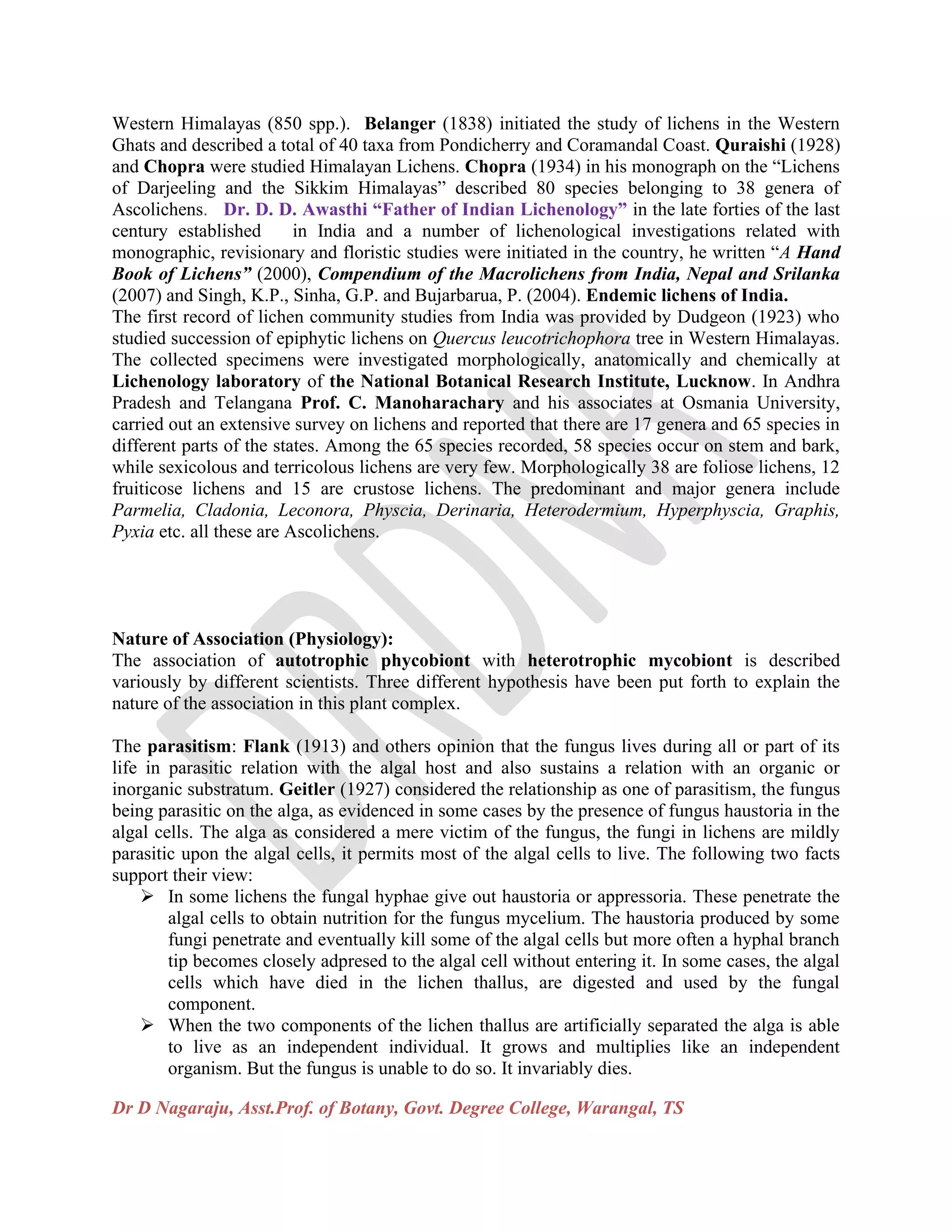
Lecanora muralis Parmotrema perlatum Roccella phycopsis
(Crustose Lichen) (Foliose Lichen) (Fruiticose Lichen)
Caloplaca chrysodeta Collema nigrescens
(Leprose Lichen) (Gelatinous Lichen)
Dr D Nagaraju, Asst.Prof. of Botany, Govt. Degree College, Warangal, TS
Fig. 3.2-Types of thallus in lichens based on thallus structure.
Internal Structure of lichen thallus:
On the basis of internal structure of thallus, the lichens are divided in to two groups namely
homoiomerous and heteromerous.
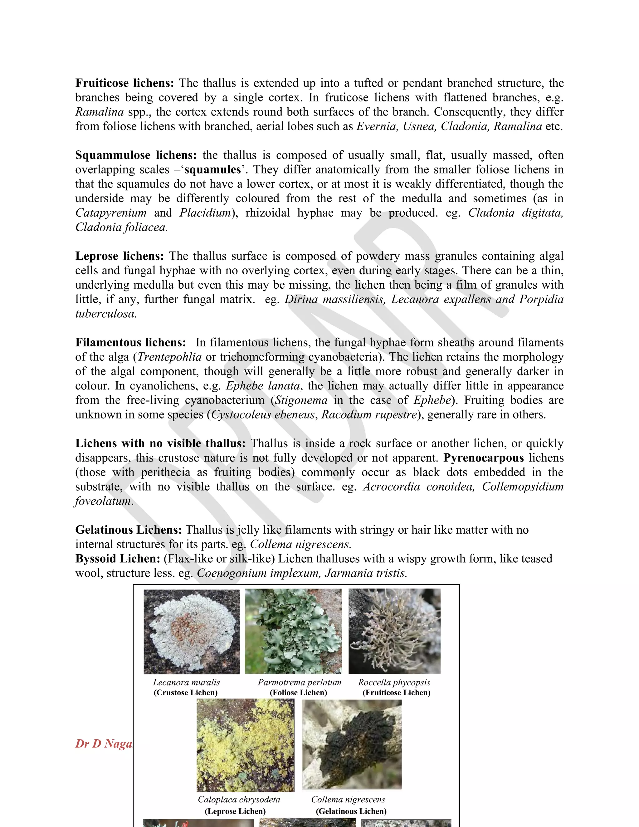
Fig. 3. 3 -Lichen thallus internal structures A. Homoiomerous thallus B. Heteromerous thallus
Homoiomerous thallus: In most of the crustose lichens, the thallus shows a simple structure
with little differentiation. It consists of a loosely interwoven mass of fungal hyphae with algal
cells equally and uniformly distributed throughout the thallus. The algal component is a
blue-green one with the gelatinous cells arranged in unbranched trichomes. The lichen thallus
Dr D Nagaraju, Asst.Prof. of Botany, Govt. Degree College, Warangal, TS
with the algal component scattered uniformly between the fungal hyphae throughout the thallus
is called homoiomerous. eg. Collema, Leptogium. In gelatinous, byssoid, Filamentous and
leprose lichens lack a cortex (are called ecorticate), and generally have only undifferentiated
tissue, similar to only having a symbiont layer.
Heteromerous thallus: In most of the foliose and fruiticose lichens, thallus is differentiated into
several layers of tissues are called heteromerous. The algal component in the heteromerous
thallus is restricted to a specific zone or layer. A vertical section through the foliose thallus
reveals the four distinct zones that are upper cortex, algal zone, medulla and lower cortex. eg.
Parmelia, Xanthoria.
Upper cortex: it forms the upper surface which is generally thick and protective. The fugal
hyphae in this region grow more or less vertically and are compactly interwoven to roduce a
tissue like layer (Plectenchyma or Pseudoparanchyma) called the upper cortex. The fungal cells
in the upper cortex are either closely packed without intercellular spaces between them or with
intercellular spaces filled with gelatinous material generally the upper cortex has epidermis-like
configuration on the surface.
Algal zone: it is the blue-green or the green zone which lies immediately beneath the upper
cortex. It consists of a tangled network of loosely interwoven fungal hyphae with the algal cells
of a green alga (in Xanthoria) or of a blue-green alga (in Peltigera canina) intermixed with the
fungal hyphae. Common among the unicellular green algae present in this layer are Chlorella,
Pluerococcus, and Cystococcus, Gloeocapsa is a common example of a unicellular blue-green
alga present in this region. The filamentous blue green algae found in the algal zone are Nostoc,
Rivularia. The algal region is the photosynthetic region of the lichen thallus. Formerly it was
called as the gondial layer a misnomer. The algal cells multiply by cell division or aplanospore
formation. The enveloping fungal hyphae. In some species send haustoria into the algal cells.
The haustoria absorb nutrition for the fungus.
Medulla: it forms the central core of the thallus. It is less compact and consists of loosely
interwoven hyphae with large spaces between them in certain regions. The fungal hyphae in this
region are scattered and usually run longitudinally. They become very thick in the region of the
vein and thin at the margin. Here and there they give out anastomosing strands.
Lower cortex: it forms the lower surface of the thallus and is composed of densely compacted
hyphae. They may run perpendicular to the surface of the thallus or parallel to it. Bundles of
hyphae (rhizinae) often arise from the surface of the lower cortex and penetrate the substratum to
function as anchoring organs. In some fruiticose lichen species the lower cortex is absent. Its
place is taken up by a thin sheet of hyphae constituting the hypothallus. It persists chiefly at the
margins of the thallus. The rhizines in these species arise directly from the thicker part of the
medullary region.
Special structures associated with lichen thallus:
In addition to the general structure, lichens bear certain peculiar vegetative structures. Those
structures are:
Dr D Nagaraju, Asst.Prof. of Botany, Govt. Degree College, Warangal, TS
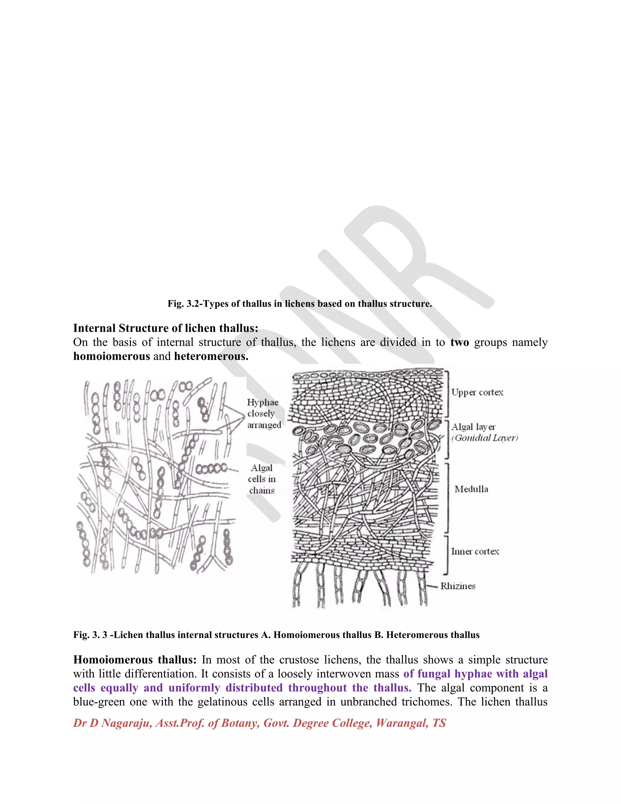
Fig. 3.4-Special structures A. Breathing pores B. Cyphellae, (C, D, E)-Vegetative reproductive structures C. Isidium
D. Cephalodia E. Soridia.
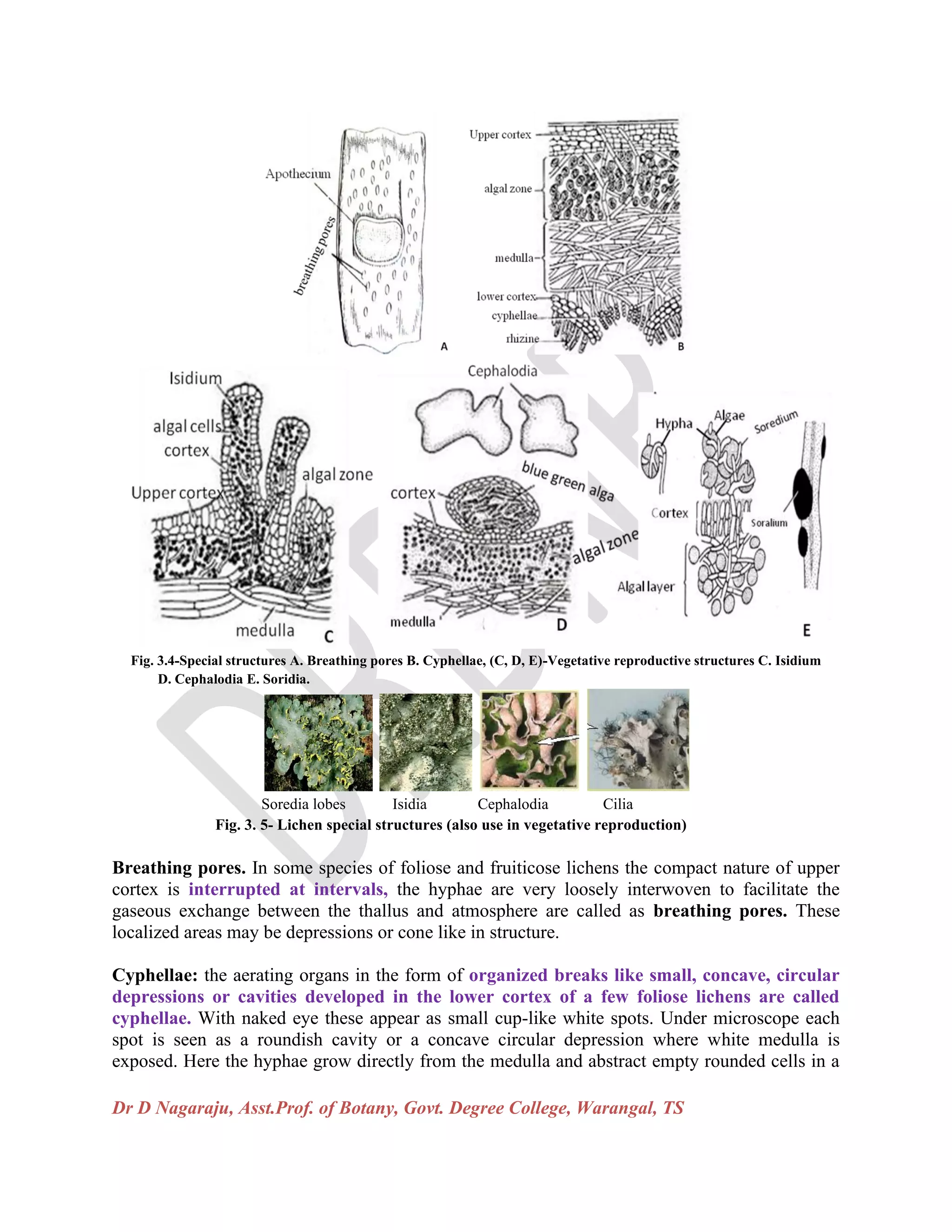
Soredia lobes Isidia Cephalodia Cilia
Fig. 3. 5- Lichen special structures (also use in vegetative reproduction)
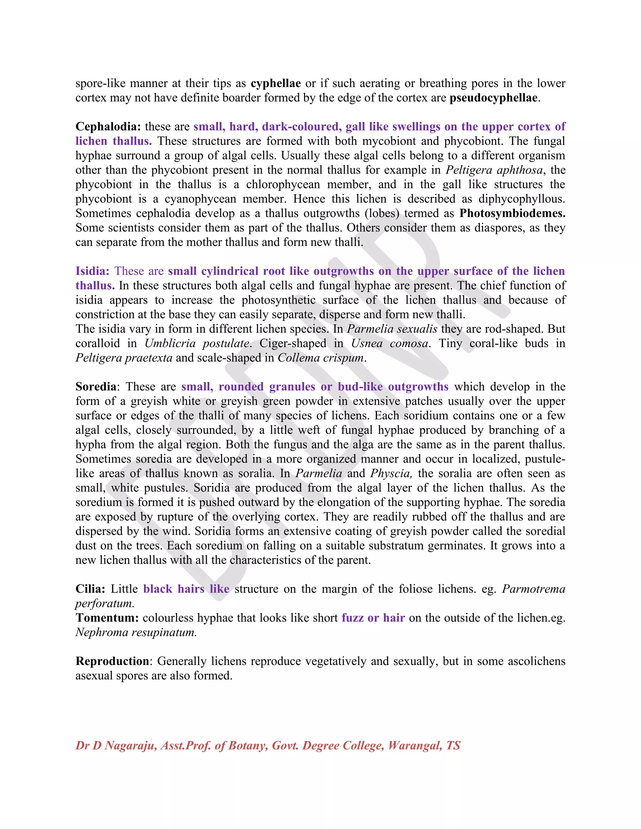
Breathing pores. In some species of foliose and fruiticose lichens the compact nature of upper
cortex is interrupted at intervals, the hyphae are very loosely interwoven to facilitate the
gaseous exchange between the thallus and atmosphere are called as breathing pores. These
localized areas may be depressions or cone like in structure.
Cyphellae: the aerating organs in the form of organized breaks like small, concave, circular
depressions or cavities developed in the lower cortex of a few foliose lichens are called
cyphellae. With naked eye these appear as small cup-like white spots. Under microscope each
spot is seen as a roundish cavity or a concave circular depression where white medulla is
exposed. Here the hyphae grow directly from the medulla and abstract empty rounded cells in a
Dr D Nagaraju, Asst.Prof. of Botany, Govt. Degree College, Warangal, TS
spore-like manner at their tips as cyphellae or if such aerating or breathing pores in the lower
cortex may not have definite boarder formed by the edge of the cortex are pseudocyphellae.
Cephalodia: these are small, hard, dark-coloured, gall like swellings on the upper cortex of
lichen thallus. These structures are formed with both mycobiont and phycobiont. The fungal
hyphae surround a group of algal cells. Usually these algal cells belong to a different organism
other than the phycobiont present in the normal thallus for example in Peltigera aphthosa, the
phycobiont in the thallus is a chlorophycean member, and in the gall like structures the
phycobiont is a cyanophycean member. Hence this lichen is described as diphycophyllous.
Sometimes cephalodia develop as a thallus outgrowths (lobes) termed as Photosymbiodemes.
Some scientists consider them as part of the thallus. Others consider them as diaspores, as they
can separate from the mother thallus and form new thalli.
Isidia: These are small cylindrical root like outgrowths on the upper surface of the lichen
thallus. In these structures both algal cells and fungal hyphae are present. The chief function of
isidia appears to increase the photosynthetic surface of the lichen thallus and because of
constriction at the base they can easily separate, disperse and form new thalli.
The isidia vary in form in different lichen species. In Parmelia sexualis they are rod-shaped. But
coralloid in Umblicria postulate. Ciger-shaped in Usnea comosa. Tiny coral-like buds in
Peltigera praetexta and scale-shaped in Collema crispum.
Soredia: These are small, rounded granules or bud-like outgrowths which develop in the
form of a greyish white or greyish green powder in extensive patches usually over the upper
surface or edges of the thalli of many species of lichens. Each soridium contains one or a few
algal cells, closely surrounded, by a little weft of fungal hyphae produced by branching of a
hypha from the algal region. Both the fungus and the alga are the same as in the parent thallus.
Sometimes soredia are developed in a more organized manner and occur in localized, pustule-
like areas of thallus known as soralia. In Parmelia and Physcia, the soralia are often seen as
small, white pustules. Soridia are produced from the algal layer of the lichen thallus. As the
soredium is formed it is pushed outward by the elongation of the supporting hyphae. The soredia
are exposed by rupture of the overlying cortex. They are readily rubbed off the thallus and are
dispersed by the wind. Soridia forms an extensive coating of greyish powder called the soredial
dust on the trees. Each soredium on falling on a suitable substratum germinates. It grows into a
new lichen thallus with all the characteristics of the parent.
Cilia: Little black hairs like structure on the margin of the foliose lichens. eg. Parmotrema
perforatum.
Tomentum: colourless hyphae that looks like short fuzz or hair on the outside of the lichen.eg.
Nephroma resupinatum.
Reproduction: Generally lichens reproduce vegetatively and sexually, but in some ascolichens
asexual spores are also formed.
Dr D Nagaraju, Asst.Prof. of Botany, Govt. Degree College, Warangal, TS
Vegetative reproduction: It occurs mainly through 4 methods viz. Fragmentation of the thallus,
formation of propagules and diaspores like sordia, isidia and cephalodia (given in Special
structures associated with lichen thallus).
Fragmentation: When thallus fragments because of physical forces or other reasons (ageing,
accidental severing), each fragment can form a new lichen thallus. If older cells in the posterior
or basal parts of the thallus die and separate the branches or lobes, each of these continues to
grow independently by apical growth. If larger portion of the established thalli (contain both
symbionts) are accidentally broken off the parent plant, develop into a new lichen individual. In
some fruiticose lichens, these detached portions are carried by the wind to other trees, there they
develop into new lichen plants. Fragmentation is an efficient means of propagation in lichens. It
leads to increase in the number of individuals of the species as well as constant of new territories.
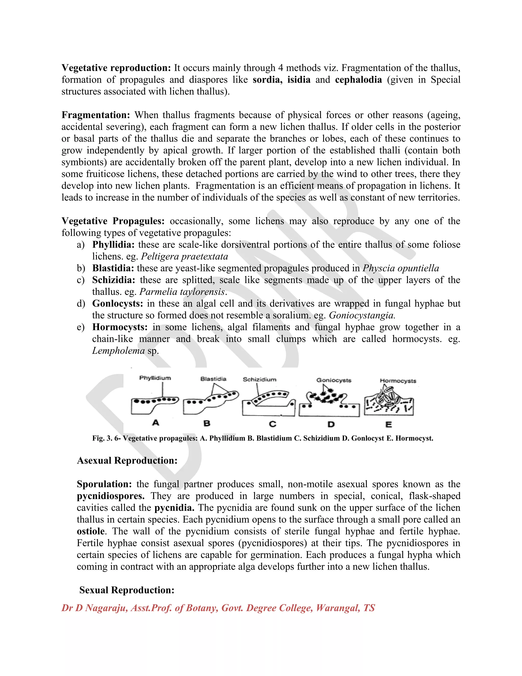
Vegetative Propagules: occasionally, some lichens may also reproduce by any one of the
following types of vegetative propagules:
a) Phyllidia: these are scale-like dorsiventral portions of the entire thallus of some foliose
lichens. eg. Peltigera praetextata
b) Blastidia: these are yeast-like segmented propagules produced in Physcia opuntiella
c) Schizidia: these are splitted, scale like segments made up of the upper layers of the
thallus. eg. Parmelia taylorensis.
d) Gonlocysts: in these an algal cell and its derivatives are wrapped in fungal hyphae but
the structure so formed does not resemble a soralium. eg. Goniocystangia.
e) Hormocysts: in some lichens, algal filaments and fungal hyphae grow together in a
chain-like manner and break into small clumps which are called hormocysts. eg.
Lempholema sp.
Fig. 3. 6- Vegetative propagules: A. Phyllidium B. Blastidium C. Schizidium D. Gonlocyst E. Hormocyst.
Asexual Reproduction:
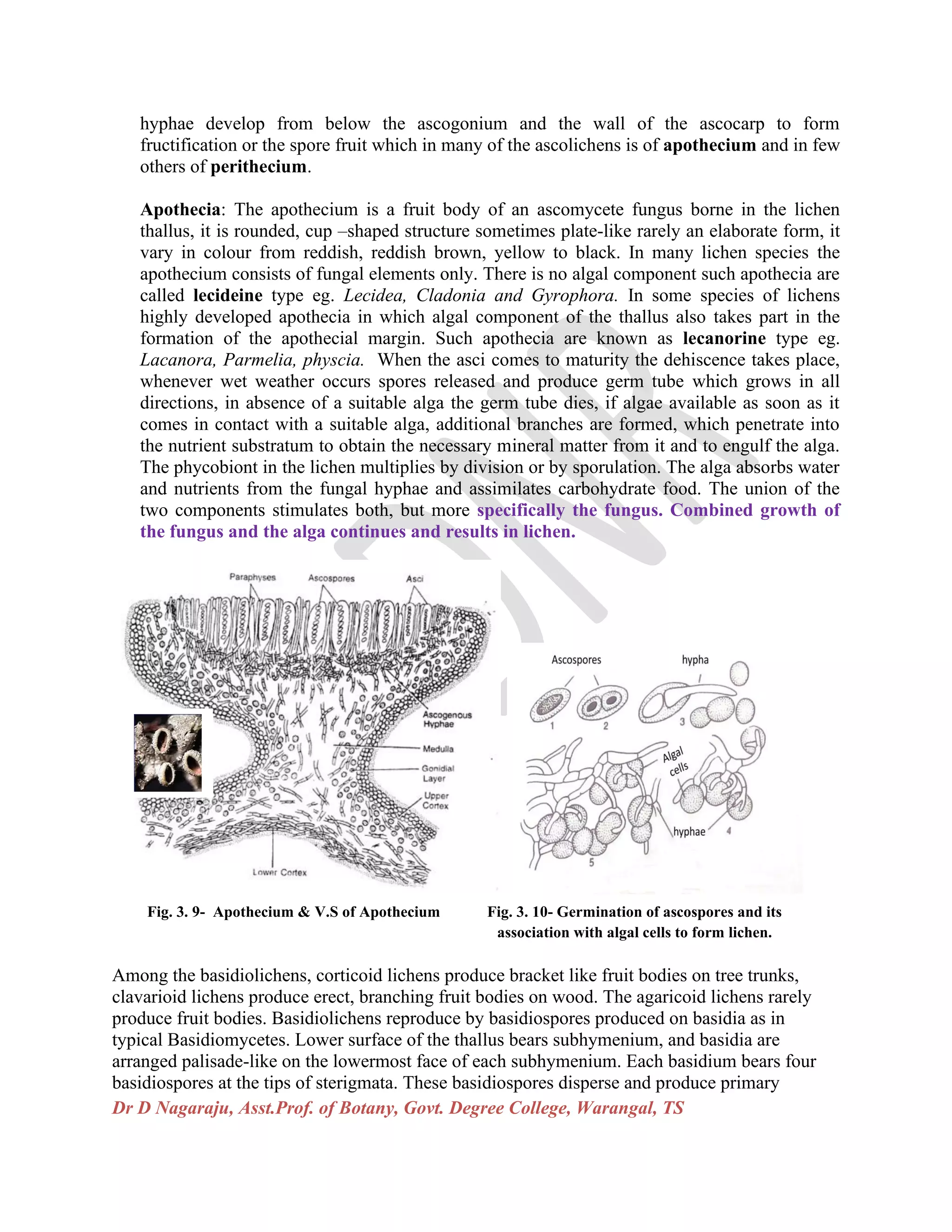
Sporulation: the fungal partner produces small, non-motile asexual spores known as the
pycnidiospores. They are produced in large numbers in special, conical, flask-shaped
cavities called the pycnidia. The pycnidia are found sunk on the upper surface of the lichen
thallus in certain species. Each pycnidium opens to the surface through a small pore called an
ostiole. The wall of the pycnidium consists of sterile fungal hyphae and fertile hyphae.
Fertile hyphae consist asexual spores (pycnidiospores) at their tips. The pycnidiospores in
certain species of lichens are capable for germination. Each produces a fungal hypha which
coming in contract with an appropriate alga develops further into a new lichen thallus.
Sexual Reproduction:
Dr D Nagaraju, Asst.Prof. of Botany, Govt. Degree College, Warangal, TS
In lichens sexual reproduction is exhibited exclusively by the mycobiont. The photobiont has
no contribution to the sexual process. The male reproductive organ is called the
spermogonium and the female organ is carpogonium. The mode of sexual reproduction in
Ascolichens is similar to that of Ascomycotina, while in Basidiolichens it is similar to that
of Basidiomycotina, and sexual reproduction is absent in Dueterolichens.
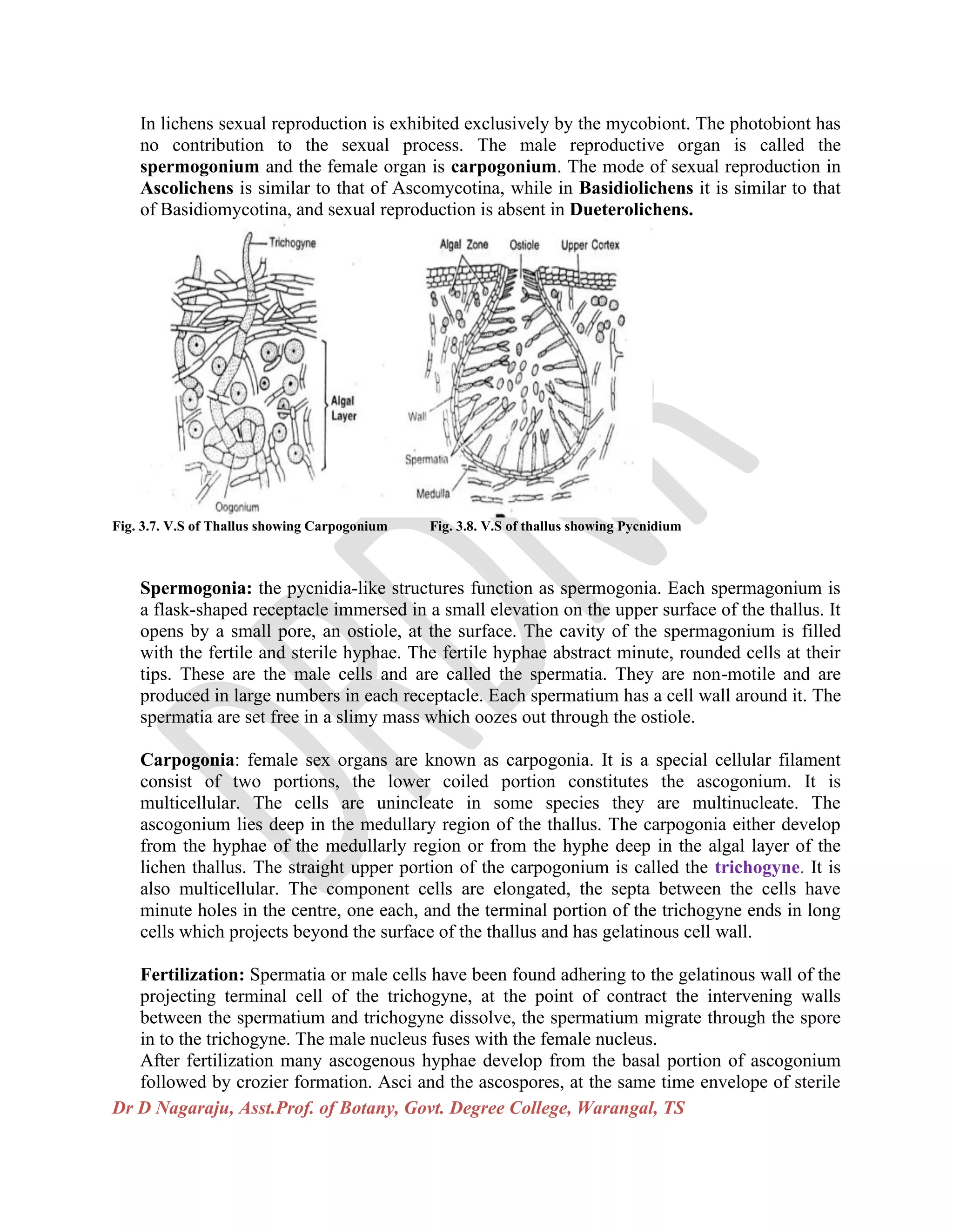
Fig. 3.7. V.S of Thallus showing Carpogonium Fig. 3.8. V.S of thallus showing Pycnidium
Spermogonia: the pycnidia-like structures function as spermogonia. Each spermagonium is
a flask-shaped receptacle immersed in a small elevation on the upper surface of the thallus. It
opens by a small pore, an ostiole, at the surface. The cavity of the spermagonium is filled
with the fertile and sterile hyphae. The fertile hyphae abstract minute, rounded cells at their
tips. These are the male cells and are called the spermatia. They are non-motile and are
produced in large numbers in each receptacle. Each spermatium has a cell wall around it. The
spermatia are set free in a slimy mass which oozes out through the ostiole.
Carpogonia: female sex organs are known as carpogonia. It is a special cellular filament
consist of two portions, the lower coiled portion constitutes the ascogonium. It is
multicellular. The cells are unincleate in some species they are multinucleate. The
ascogonium lies deep in the medullary region of the thallus. The carpogonia either develop
from the hyphae of the medullarly region or from the hyphe deep in the algal layer of the
lichen thallus. The straight upper portion of the carpogonium is called the trichogyne. It is
also multicellular. The component cells are elongated, the septa between the cells have
minute holes in the centre, one each, and the terminal portion of the trichogyne ends in long
cells which projects beyond the surface of the thallus and has gelatinous cell wall.
Fertilization: Spermatia or male cells have been found adhering to the gelatinous wall of the
projecting terminal cell of the trichogyne, at the point of contract the intervening walls
between the spermatium and trichogyne dissolve, the spermatium migrate through the spore
in to the trichogyne. The male nucleus fuses with the female nucleus.
After fertilization many ascogenous hyphae develop from the basal portion of ascogonium
followed by crozier formation. Asci and the ascospores, at the same time envelope of sterile
Dr D Nagaraju, Asst.Prof. of Botany, Govt. Degree College, Warangal, TS
hyphae develop from below the ascogonium and the wall of the ascocarp to form
fructification or the spore fruit which in many of the ascolichens is of apothecium and in few
others of perithecium.
Apothecia: The apothecium is a fruit body of an ascomycete fungus borne in the lichen
thallus, it is rounded, cup –shaped structure sometimes plate-like rarely an elaborate form, it
vary in colour from reddish, reddish brown, yellow to black. In many lichen species the
apothecium consists of fungal elements only. There is no algal component such apothecia are
called lecideine type eg. Lecidea, Cladonia and Gyrophora. In some species of lichens
highly developed apothecia in which algal component of the thallus also takes part in the
formation of the apothecial margin. Such apothecia are known as lecanorine type eg.
Lacanora, Parmelia, physcia. When the asci comes to maturity the dehiscence takes place,
whenever wet weather occurs spores released and produce germ tube which grows in all
directions, in absence of a suitable alga the germ tube dies, if algae available as soon as it
comes in contact with a suitable alga, additional branches are formed, which penetrate into
the nutrient substratum to obtain the necessary mineral matter from it and to engulf the alga.
The phycobiont in the lichen multiplies by division or by sporulation. The alga absorbs water
and nutrients from the fungal hyphae and assimilates carbohydrate food. The union of the
two components stimulates both, but more specifically the fungus. Combined growth of
the fungus and the alga continues and results in lichen.
Fig. 3. 9- Apothecium & V.S of Apothecium Fig. 3. 10- Germination of ascospores and its
association with algal cells to form lichen.
Among the basidiolichens, corticoid lichens produce bracket like fruit bodies on tree trunks,
clavarioid lichens produce erect, branching fruit bodies on wood. The agaricoid lichens rarely
produce fruit bodies. Basidiolichens reproduce by basidiospores produced on basidia as in
typical Basidiomycetes. Lower surface of the thallus bears subhymenium, and basidia are
arranged palisade-like on the lowermost face of each subhymenium. Each basidium bears four
basidiospores at the tips of sterigmata. These basidiospores disperse and produce primary
Dr D Nagaraju, Asst.Prof. of Botany, Govt. Degree College, Warangal, TS
mycelium, secondary mycelium and tertiary mycelium as in normal fungi. When secondary or
tertiary mycelium comes in contact with a suitable algal partner, it forms lichen.
Pseudolichens: There are forms which have great resemblance with the true lichens. Most of
them grow on the bark of trees. But their mode of life is different from the true lichens. Some of
the pseudolichens are saprophytes whereas others are adapted to parasitic mode of life. These do
not posses algae.
Some important Genera of Lichens
1.Cladonia rangiferina 2. Parmelia saxatilis 3.Usnea subfloridana 4. Cetraria islandica 5. Peltigera canina
Fig. 3.11- Some Important Lichens (1,2,3,4 & 5)
Special Interest Features of Lichens:
➢ The lichens are dual organisms, each consisting of two different individuals, an alga
and a fungus, having a composite thalloid structure.
➢ The fungal component are mainly Ascomycotina and Basidiomycotina, on the basis
of fungal component lichens devided into Ascolichens and Basidiolichens. Algal
components are mainly Chlorophyceae and Cyanophyceae.
➢ On the basis of the thallus, lichens have beed classified in to Crustose, Foliose,
Fruiticose, Squamulose, Filamentous etc. types.
➢ The lichens having special structures viz. Breathing pores, Cyphellae, Cilia,
Cephalodia, Isidia, Soridia.
➢ The lichens reproduce vegetatively by Fragmentation, vegetative propagules and
diaspores like Cephalodia, Isidia, Soridia.
➢ Only fungal component of lichens is involved in sexual reproduction, female sex
organ is carpogonium and male sex organ is spermagonium.
➢ The fruit bodies of lichens are cup-shaped apothecium or flask-shaped perithesium.
➢ Ascospores under suitable conditions germinates to produce a fungal hyphae only
when it comes in contact with a proper alga, develops into a new lichen.
➢ The thallus like plant body is made up of a fungus and an alga living in closest
association. Fungus provides the body of the organism whereas alga synthesizes
carbohydrate food for itself and the fungus. The nature of association between the two
partners is the best example of symbiosis in the plant community.
Dr D Nagaraju, Asst.Prof. of Botany, Govt. Degree College, Warangal, TS
➢ The lichens are the slowest growing of all the plants. The average growth rate of
lichen thallus ranges from 1mm to 10mm in radius a year. It can grow on barren
substratum under conditions of drought and starvation and in situations where no
other vegetation can exist.
➢ The lichens have an extraordinary ability to withstand complete drying and for a very
long period. The completely dried up thalli, on wetting absorb moisture and resume
growth.
➢ The thallus of certain species of lichens contains characteristic kinds of lichen acids
found in no other plants. The lichen acids are important from the taxonomic point of
view.
➢ The lichens play a very important role in soil formation.
Difference between Lichens and Fungi:
Lichens Fungi
➢ Lichens flourish the most in
the temperate and colder
regions of the globe.
➢ It grows freely exposed to air
and light and dislike smooky
atmosphere of towns.
➢ It can grow on barren
substratum under drought and
starvation conditions.
➢ It grows generlly as epiphytes
or as terrestrial autophytes.
➢ The thallus is generally
conspicuously coloured due to
the presence of various
organic acids.
➢ The thallus is generally tough,
leathery in texture and in many
lichens it is soft as a rubber
sponge.
➢ Fungi thrive best in the tropical
and subtropical regions of the
world which are warm.
➢ It prefers moist, shady, dark
places and vitiated atmosphere of
towns.
➢ It requires dead or living organic
matter on which they feed and
demand much moisture.
➢ It grows either as parasites or as
saprophytes.
➢ The fungal body is generally
colourless.
➢ The thallus is usually a
filamentous mycelium, delicate in
texture, immersed in substratum.
In many it is slimy and gelatinous.
Dr D Nagaraju, Asst.Prof. of Botany, Govt. Degree College, Warangal, TS
** *

More Related Content

What's hot

Lichen powerpoint presentation
Lichen powerpoint  presentationLichen powerpoint  presentation
Lichen powerpoint presentation
rashediqbal7
 
Pentoxylon
PentoxylonPentoxylon
Vaucheria
VaucheriaVaucheria
Vaucheria
Tahir Shahzad
 
Xanthophyta by sohail riaz hashmi
Xanthophyta by sohail riaz hashmiXanthophyta by sohail riaz hashmi
Xanthophyta by sohail riaz hashmi
Quaid-e-Azam University, Islamabad
 
Economic importance of bryophytes
Economic importance of bryophytesEconomic importance of bryophytes
Economic importance of bryophytes
gohil sanjay bhagvanji
 
Xanthophyceae
XanthophyceaeXanthophyceae
Xanthophyceae
NikkiM12
 
Cyanophyta
CyanophytaCyanophyta
Cyanophyta
Sangeeta Das
 
Takhtajan System of Classification.pdf
Takhtajan System of Classification.pdfTakhtajan System of Classification.pdf
Takhtajan System of Classification.pdf
IqraAli875037
 
CHARA.pptx
CHARA.pptxCHARA.pptx
Soral evolution in filicopsida
Soral evolution in filicopsida Soral evolution in filicopsida
Soral evolution in filicopsida
subhashis das
 
Evolution of sporophyte in bryotphytes
Evolution of sporophyte in bryotphytesEvolution of sporophyte in bryotphytes
Evolution of sporophyte in bryotphytes
Sangeeta Das
 
Classification of brayophyta
Classification of brayophytaClassification of brayophyta
Classification of brayophyta
vaishalidandge3
 
General Characters of Gymnosperms.pptx
General Characters of Gymnosperms.pptxGeneral Characters of Gymnosperms.pptx
General Characters of Gymnosperms.pptx
Aliya Fathima Ilyas
 
Selaginella ppt
Selaginella pptSelaginella ppt
Selaginella ppt
Naresh Kumar
 
Gymnosperms - General characteristics and Economic importance.pptx
Gymnosperms - General characteristics and Economic importance.pptxGymnosperms - General characteristics and Economic importance.pptx
Gymnosperms - General characteristics and Economic importance.pptx
MEGHASHREE A M
 
ALGAE - THALLUS ORGANIZATION.pptx
ALGAE - THALLUS ORGANIZATION.pptxALGAE - THALLUS ORGANIZATION.pptx
ALGAE - THALLUS ORGANIZATION.pptx
G S G College Umarkhed Dist Yavatmal
 
Angiosperm anatomy.ppt
Angiosperm anatomy.pptAngiosperm anatomy.ppt
Angiosperm anatomy.ppt
Dr. Harwinder Kaur
 
Pteridophyte ppt
Pteridophyte pptPteridophyte ppt
Pteridophyte ppt
sumitachoudhary
 
Sphenophyllum
SphenophyllumSphenophyllum
Sphenophyllum
J K COLLEGE,PURULIA
 
General characteristics of pteridophytes
General characteristics of pteridophytesGeneral characteristics of pteridophytes
General characteristics of pteridophytes
SankritaShankarGaonk
 

What's hot (20)

Lichen powerpoint presentation
Lichen powerpoint  presentationLichen powerpoint  presentation
Lichen powerpoint presentation
 
Pentoxylon
PentoxylonPentoxylon
Pentoxylon
 
Vaucheria
VaucheriaVaucheria
Vaucheria
 
Xanthophyta by sohail riaz hashmi
Xanthophyta by sohail riaz hashmiXanthophyta by sohail riaz hashmi
Xanthophyta by sohail riaz hashmi
 
Economic importance of bryophytes
Economic importance of bryophytesEconomic importance of bryophytes
Economic importance of bryophytes
 
Xanthophyceae
XanthophyceaeXanthophyceae
Xanthophyceae
 
Cyanophyta
CyanophytaCyanophyta
Cyanophyta
 
Takhtajan System of Classification.pdf
Takhtajan System of Classification.pdfTakhtajan System of Classification.pdf
Takhtajan System of Classification.pdf
 
CHARA.pptx
CHARA.pptxCHARA.pptx
CHARA.pptx
 
Soral evolution in filicopsida
Soral evolution in filicopsida Soral evolution in filicopsida
Soral evolution in filicopsida
 
Evolution of sporophyte in bryotphytes
Evolution of sporophyte in bryotphytesEvolution of sporophyte in bryotphytes
Evolution of sporophyte in bryotphytes
 
Classification of brayophyta
Classification of brayophytaClassification of brayophyta
Classification of brayophyta
 
General Characters of Gymnosperms.pptx
General Characters of Gymnosperms.pptxGeneral Characters of Gymnosperms.pptx
General Characters of Gymnosperms.pptx
 
Selaginella ppt
Selaginella pptSelaginella ppt
Selaginella ppt
 
Gymnosperms - General characteristics and Economic importance.pptx
Gymnosperms - General characteristics and Economic importance.pptxGymnosperms - General characteristics and Economic importance.pptx
Gymnosperms - General characteristics and Economic importance.pptx
 
ALGAE - THALLUS ORGANIZATION.pptx
ALGAE - THALLUS ORGANIZATION.pptxALGAE - THALLUS ORGANIZATION.pptx
ALGAE - THALLUS ORGANIZATION.pptx
 
Angiosperm anatomy.ppt
Angiosperm anatomy.pptAngiosperm anatomy.ppt
Angiosperm anatomy.ppt
 
Pteridophyte ppt
Pteridophyte pptPteridophyte ppt
Pteridophyte ppt
 
Sphenophyllum
SphenophyllumSphenophyllum
Sphenophyllum
 
General characteristics of pteridophytes
General characteristics of pteridophytesGeneral characteristics of pteridophytes
General characteristics of pteridophytes
 

Similar to Lichen General Characters by Dr D.Nagaraju

Lichens
LichensLichens
methanogens,chytrids_and_microbes_and_insects.pdf
methanogens,chytrids_and_microbes_and_insects.pdfmethanogens,chytrids_and_microbes_and_insects.pdf
methanogens,chytrids_and_microbes_and_insects.pdf
MidhatSarfraz
 
Soil Flora
Soil FloraSoil Flora
Soil Flora
Hemant Mahara
 
Biological classification
Biological classificationBiological classification
Biological classification
Samarji
 
Chap 5 fungi and other organism (mutualistic)
Chap 5 fungi and other organism (mutualistic)Chap 5 fungi and other organism (mutualistic)
Chap 5 fungi and other organism (mutualistic)
Alia Najiha
 
R.H.wittaker 5 kingom of classification
R.H.wittaker 5 kingom of classificationR.H.wittaker 5 kingom of classification
R.H.wittaker 5 kingom of classification
Ayushi Mukherjee
 
Microbes, diversity, discovery & development
Microbes, diversity, discovery & developmentMicrobes, diversity, discovery & development
Microbes, diversity, discovery & development
OsamaZafar16
 
Lichens (Microbiology and Pathology)
Lichens  (Microbiology and Pathology) Lichens  (Microbiology and Pathology)
Lichens (Microbiology and Pathology)
nishakataria10
 
Intro to fungi
Intro to fungiIntro to fungi
Intro to fungi
joygtablante
 
Micro organisms
Micro  organisms Micro  organisms
Micro organisms
Harshita68
 
Introduction and significance of Symbiotic algae
Introduction and significance of  Symbiotic algaeIntroduction and significance of  Symbiotic algae
Introduction and significance of Symbiotic algae
kushbuR
 
Biology Form 4 Chapter 8 :Dynamic Ecosystem Part 2
Biology Form 4 Chapter 8 :Dynamic Ecosystem  Part 2Biology Form 4 Chapter 8 :Dynamic Ecosystem  Part 2
Biology Form 4 Chapter 8 :Dynamic Ecosystem Part 2
Nirmala Josephine
 
Fungus
FungusFungus
Treacheophytes
TreacheophytesTreacheophytes
Treacheophytes
SabaIshfaq2
 
Overview of fungi
Overview of fungiOverview of fungi
Overview of fungi
Roksana Aftab Ruhi
 
Introductiontoalgalcharacteristicsanddiversity
IntroductiontoalgalcharacteristicsanddiversityIntroductiontoalgalcharacteristicsanddiversity
Introductiontoalgalcharacteristicsanddiversity
Muhammad Fahad Saleh
 
additional topics for midterms.pptx
additional topics for midterms.pptxadditional topics for midterms.pptx
additional topics for midterms.pptx
MissFaith2
 
Microbiology -Bacterial Growth & nutrition
Microbiology -Bacterial Growth & nutritionMicrobiology -Bacterial Growth & nutrition
Microbiology -Bacterial Growth & nutrition
Nistarini College, Purulia (W.B) India
 
lichens-a-general-account.ppt
lichens-a-general-account.pptlichens-a-general-account.ppt
lichens-a-general-account.ppt
jaya1992
 
Grade 11
Grade 11Grade 11
Grade 11
Peace Kgotso
 

Similar to Lichen General Characters by Dr D.Nagaraju (20)

Lichens
LichensLichens
Lichens
 
methanogens,chytrids_and_microbes_and_insects.pdf
methanogens,chytrids_and_microbes_and_insects.pdfmethanogens,chytrids_and_microbes_and_insects.pdf
methanogens,chytrids_and_microbes_and_insects.pdf
 
Soil Flora
Soil FloraSoil Flora
Soil Flora
 
Biological classification
Biological classificationBiological classification
Biological classification
 
Chap 5 fungi and other organism (mutualistic)
Chap 5 fungi and other organism (mutualistic)Chap 5 fungi and other organism (mutualistic)
Chap 5 fungi and other organism (mutualistic)
 
R.H.wittaker 5 kingom of classification
R.H.wittaker 5 kingom of classificationR.H.wittaker 5 kingom of classification
R.H.wittaker 5 kingom of classification
 
Microbes, diversity, discovery & development
Microbes, diversity, discovery & developmentMicrobes, diversity, discovery & development
Microbes, diversity, discovery & development
 
Lichens (Microbiology and Pathology)
Lichens  (Microbiology and Pathology) Lichens  (Microbiology and Pathology)
Lichens (Microbiology and Pathology)
 
Intro to fungi
Intro to fungiIntro to fungi
Intro to fungi
 
Micro organisms
Micro  organisms Micro  organisms
Micro organisms
 
Introduction and significance of Symbiotic algae
Introduction and significance of  Symbiotic algaeIntroduction and significance of  Symbiotic algae
Introduction and significance of Symbiotic algae
 
Biology Form 4 Chapter 8 :Dynamic Ecosystem Part 2
Biology Form 4 Chapter 8 :Dynamic Ecosystem  Part 2Biology Form 4 Chapter 8 :Dynamic Ecosystem  Part 2
Biology Form 4 Chapter 8 :Dynamic Ecosystem Part 2
 
Fungus
FungusFungus
Fungus
 
Treacheophytes
TreacheophytesTreacheophytes
Treacheophytes
 
Overview of fungi
Overview of fungiOverview of fungi
Overview of fungi
 
Introductiontoalgalcharacteristicsanddiversity
IntroductiontoalgalcharacteristicsanddiversityIntroductiontoalgalcharacteristicsanddiversity
Introductiontoalgalcharacteristicsanddiversity
 
additional topics for midterms.pptx
additional topics for midterms.pptxadditional topics for midterms.pptx
additional topics for midterms.pptx
 
Microbiology -Bacterial Growth & nutrition
Microbiology -Bacterial Growth & nutritionMicrobiology -Bacterial Growth & nutrition
Microbiology -Bacterial Growth & nutrition
 
lichens-a-general-account.ppt
lichens-a-general-account.pptlichens-a-general-account.ppt
lichens-a-general-account.ppt
 
Grade 11
Grade 11Grade 11
Grade 11
 

More from Nagaraj Deshaboina

Dr D Nagaraju, Botany
Dr D Nagaraju, BotanyDr D Nagaraju, Botany
Dr D Nagaraju, Botany
Nagaraj Deshaboina
 
Fungi structure Reproduction Classification, Life cycles Bot coaching mater...
Fungi structure  Reproduction Classification, Life cycles  Bot coaching mater...Fungi structure  Reproduction Classification, Life cycles  Bot coaching mater...
Fungi structure Reproduction Classification, Life cycles Bot coaching mater...
Nagaraj Deshaboina
 
Herbarium DrDNR
Herbarium  DrDNRHerbarium  DrDNR
Herbarium DrDNR
Nagaraj Deshaboina
 
UGC LOCF-Botany- DRDNR
UGC LOCF-Botany- DRDNRUGC LOCF-Botany- DRDNR
UGC LOCF-Botany- DRDNR
Nagaraj Deshaboina
 
MSc-Botany CPGCET 19 Question Paper for All Telangana Universities
MSc-Botany  CPGCET 19 Question Paper for All Telangana UniversitiesMSc-Botany  CPGCET 19 Question Paper for All Telangana Universities
MSc-Botany CPGCET 19 Question Paper for All Telangana Universities
Nagaraj Deshaboina
 
Covid 19 CORONA Virus Awareness Assessment By Dr D. Nagaraju
Covid 19 CORONA Virus Awareness Assessment By Dr D. NagarajuCovid 19 CORONA Virus Awareness Assessment By Dr D. Nagaraju
Covid 19 CORONA Virus Awareness Assessment By Dr D. Nagaraju
Nagaraj Deshaboina
 
Importance of Lichens (Ecological and Economic Importance) by DrD Nagaarju
Importance of Lichens (Ecological and Economic Importance) by DrD NagaarjuImportance of Lichens (Ecological and Economic Importance) by DrD Nagaarju
Importance of Lichens (Ecological and Economic Importance) by DrD Nagaarju
Nagaraj Deshaboina
 
BSc_Botany Syllabus CBCS System
BSc_Botany Syllabus CBCS System BSc_Botany Syllabus CBCS System
BSc_Botany Syllabus CBCS System
Nagaraj Deshaboina
 

More from Nagaraj Deshaboina (8)

Dr D Nagaraju, Botany
Dr D Nagaraju, BotanyDr D Nagaraju, Botany
Dr D Nagaraju, Botany
 
Fungi structure Reproduction Classification, Life cycles Bot coaching mater...
Fungi structure  Reproduction Classification, Life cycles  Bot coaching mater...Fungi structure  Reproduction Classification, Life cycles  Bot coaching mater...
Fungi structure Reproduction Classification, Life cycles Bot coaching mater...
 
Herbarium DrDNR
Herbarium  DrDNRHerbarium  DrDNR
Herbarium DrDNR
 
UGC LOCF-Botany- DRDNR
UGC LOCF-Botany- DRDNRUGC LOCF-Botany- DRDNR
UGC LOCF-Botany- DRDNR
 
MSc-Botany CPGCET 19 Question Paper for All Telangana Universities
MSc-Botany  CPGCET 19 Question Paper for All Telangana UniversitiesMSc-Botany  CPGCET 19 Question Paper for All Telangana Universities
MSc-Botany CPGCET 19 Question Paper for All Telangana Universities
 
Covid 19 CORONA Virus Awareness Assessment By Dr D. Nagaraju
Covid 19 CORONA Virus Awareness Assessment By Dr D. NagarajuCovid 19 CORONA Virus Awareness Assessment By Dr D. Nagaraju
Covid 19 CORONA Virus Awareness Assessment By Dr D. Nagaraju
 
Importance of Lichens (Ecological and Economic Importance) by DrD Nagaarju
Importance of Lichens (Ecological and Economic Importance) by DrD NagaarjuImportance of Lichens (Ecological and Economic Importance) by DrD Nagaarju
Importance of Lichens (Ecological and Economic Importance) by DrD Nagaarju
 
BSc_Botany Syllabus CBCS System
BSc_Botany Syllabus CBCS System BSc_Botany Syllabus CBCS System
BSc_Botany Syllabus CBCS System
 

Recently uploaded

bordetella pertussis.................................ppt
bordetella pertussis.................................pptbordetella pertussis.................................ppt
bordetella pertussis.................................ppt
kejapriya1
 
ANAMOLOUS SECONDARY GROWTH IN DICOT ROOTS.pptx
ANAMOLOUS SECONDARY GROWTH IN DICOT ROOTS.pptxANAMOLOUS SECONDARY GROWTH IN DICOT ROOTS.pptx
ANAMOLOUS SECONDARY GROWTH IN DICOT ROOTS.pptx
RASHMI M G
 
Shallowest Oil Discovery of Turkiye.pptx
Shallowest Oil Discovery of Turkiye.pptxShallowest Oil Discovery of Turkiye.pptx
Shallowest Oil Discovery of Turkiye.pptx
Gokturk Mehmet Dilci
 
Oedema_types_causes_pathophysiology.pptx
Oedema_types_causes_pathophysiology.pptxOedema_types_causes_pathophysiology.pptx
Oedema_types_causes_pathophysiology.pptx
muralinath2
 
Phenomics assisted breeding in crop improvement
Phenomics assisted breeding in crop improvementPhenomics assisted breeding in crop improvement
Phenomics assisted breeding in crop improvement
IshaGoswami9
 
如何办理(uvic毕业证书)维多利亚大学毕业证本科学位证书原版一模一样
如何办理(uvic毕业证书)维多利亚大学毕业证本科学位证书原版一模一样如何办理(uvic毕业证书)维多利亚大学毕业证本科学位证书原版一模一样
如何办理(uvic毕业证书)维多利亚大学毕业证本科学位证书原版一模一样
yqqaatn0
 
Deep Software Variability and Frictionless Reproducibility
Deep Software Variability and Frictionless ReproducibilityDeep Software Variability and Frictionless Reproducibility
Deep Software Variability and Frictionless Reproducibility
University of Rennes, INSA Rennes, Inria/IRISA, CNRS
 
Sharlene Leurig - Enabling Onsite Water Use with Net Zero Water
Sharlene Leurig - Enabling Onsite Water Use with Net Zero WaterSharlene Leurig - Enabling Onsite Water Use with Net Zero Water
Sharlene Leurig - Enabling Onsite Water Use with Net Zero Water
Texas Alliance of Groundwater Districts
 
aziz sancar nobel prize winner: from mardin to nobel
aziz sancar nobel prize winner: from mardin to nobelaziz sancar nobel prize winner: from mardin to nobel
aziz sancar nobel prize winner: from mardin to nobel
İsa Badur
 
原版制作(carleton毕业证书)卡尔顿大学毕业证硕士文凭原版一模一样
原版制作(carleton毕业证书)卡尔顿大学毕业证硕士文凭原版一模一样原版制作(carleton毕业证书)卡尔顿大学毕业证硕士文凭原版一模一样
原版制作(carleton毕业证书)卡尔顿大学毕业证硕士文凭原版一模一样
yqqaatn0
 
Chapter 12 - climate change and the energy crisis
Chapter 12 - climate change and the energy crisisChapter 12 - climate change and the energy crisis
Chapter 12 - climate change and the energy crisis
tonzsalvador2222
 
Comparing Evolved Extractive Text Summary Scores of Bidirectional Encoder Rep...
Comparing Evolved Extractive Text Summary Scores of Bidirectional Encoder Rep...Comparing Evolved Extractive Text Summary Scores of Bidirectional Encoder Rep...
Comparing Evolved Extractive Text Summary Scores of Bidirectional Encoder Rep...
University of Maribor
 
Micronuclei test.M.sc.zoology.fisheries.
Micronuclei test.M.sc.zoology.fisheries.Micronuclei test.M.sc.zoology.fisheries.
Micronuclei test.M.sc.zoology.fisheries.
Aditi Bajpai
 
Deep Behavioral Phenotyping in Systems Neuroscience for Functional Atlasing a...
Deep Behavioral Phenotyping in Systems Neuroscience for Functional Atlasing a...Deep Behavioral Phenotyping in Systems Neuroscience for Functional Atlasing a...
Deep Behavioral Phenotyping in Systems Neuroscience for Functional Atlasing a...
Ana Luísa Pinho
 
NuGOweek 2024 Ghent programme overview flyer
NuGOweek 2024 Ghent programme overview flyerNuGOweek 2024 Ghent programme overview flyer
NuGOweek 2024 Ghent programme overview flyer
pablovgd
 
THEMATIC APPERCEPTION TEST(TAT) cognitive abilities, creativity, and critic...
THEMATIC  APPERCEPTION  TEST(TAT) cognitive abilities, creativity, and critic...THEMATIC  APPERCEPTION  TEST(TAT) cognitive abilities, creativity, and critic...
THEMATIC APPERCEPTION TEST(TAT) cognitive abilities, creativity, and critic...
Abdul Wali Khan University Mardan,kP,Pakistan
 
Applied Science: Thermodynamics, Laws & Methodology.pdf
Applied Science: Thermodynamics, Laws & Methodology.pdfApplied Science: Thermodynamics, Laws & Methodology.pdf
Applied Science: Thermodynamics, Laws & Methodology.pdf
University of Hertfordshire
 
Randomised Optimisation Algorithms in DAPHNE
Randomised Optimisation Algorithms in DAPHNERandomised Optimisation Algorithms in DAPHNE
Randomised Optimisation Algorithms in DAPHNE
University of Maribor
 
ESR spectroscopy in liquid food and beverages.pptx
ESR spectroscopy in liquid food and beverages.pptxESR spectroscopy in liquid food and beverages.pptx
ESR spectroscopy in liquid food and beverages.pptx
PRIYANKA PATEL
 
Nucleic Acid-its structural and functional complexity.
Nucleic Acid-its structural and functional complexity.Nucleic Acid-its structural and functional complexity.
Nucleic Acid-its structural and functional complexity.
Nistarini College, Purulia (W.B) India
 

Recently uploaded (20)

bordetella pertussis.................................ppt
bordetella pertussis.................................pptbordetella pertussis.................................ppt
bordetella pertussis.................................ppt
 
ANAMOLOUS SECONDARY GROWTH IN DICOT ROOTS.pptx
ANAMOLOUS SECONDARY GROWTH IN DICOT ROOTS.pptxANAMOLOUS SECONDARY GROWTH IN DICOT ROOTS.pptx
ANAMOLOUS SECONDARY GROWTH IN DICOT ROOTS.pptx
 
Shallowest Oil Discovery of Turkiye.pptx
Shallowest Oil Discovery of Turkiye.pptxShallowest Oil Discovery of Turkiye.pptx
Shallowest Oil Discovery of Turkiye.pptx
 
Oedema_types_causes_pathophysiology.pptx
Oedema_types_causes_pathophysiology.pptxOedema_types_causes_pathophysiology.pptx
Oedema_types_causes_pathophysiology.pptx
 
Phenomics assisted breeding in crop improvement
Phenomics assisted breeding in crop improvementPhenomics assisted breeding in crop improvement
Phenomics assisted breeding in crop improvement
 
如何办理(uvic毕业证书)维多利亚大学毕业证本科学位证书原版一模一样
如何办理(uvic毕业证书)维多利亚大学毕业证本科学位证书原版一模一样如何办理(uvic毕业证书)维多利亚大学毕业证本科学位证书原版一模一样
如何办理(uvic毕业证书)维多利亚大学毕业证本科学位证书原版一模一样
 
Deep Software Variability and Frictionless Reproducibility
Deep Software Variability and Frictionless ReproducibilityDeep Software Variability and Frictionless Reproducibility
Deep Software Variability and Frictionless Reproducibility
 
Sharlene Leurig - Enabling Onsite Water Use with Net Zero Water
Sharlene Leurig - Enabling Onsite Water Use with Net Zero WaterSharlene Leurig - Enabling Onsite Water Use with Net Zero Water
Sharlene Leurig - Enabling Onsite Water Use with Net Zero Water
 
aziz sancar nobel prize winner: from mardin to nobel
aziz sancar nobel prize winner: from mardin to nobelaziz sancar nobel prize winner: from mardin to nobel
aziz sancar nobel prize winner: from mardin to nobel
 
原版制作(carleton毕业证书)卡尔顿大学毕业证硕士文凭原版一模一样
原版制作(carleton毕业证书)卡尔顿大学毕业证硕士文凭原版一模一样原版制作(carleton毕业证书)卡尔顿大学毕业证硕士文凭原版一模一样
原版制作(carleton毕业证书)卡尔顿大学毕业证硕士文凭原版一模一样
 
Chapter 12 - climate change and the energy crisis
Chapter 12 - climate change and the energy crisisChapter 12 - climate change and the energy crisis
Chapter 12 - climate change and the energy crisis
 
Comparing Evolved Extractive Text Summary Scores of Bidirectional Encoder Rep...
Comparing Evolved Extractive Text Summary Scores of Bidirectional Encoder Rep...Comparing Evolved Extractive Text Summary Scores of Bidirectional Encoder Rep...
Comparing Evolved Extractive Text Summary Scores of Bidirectional Encoder Rep...
 
Micronuclei test.M.sc.zoology.fisheries.
Micronuclei test.M.sc.zoology.fisheries.Micronuclei test.M.sc.zoology.fisheries.
Micronuclei test.M.sc.zoology.fisheries.
 
Deep Behavioral Phenotyping in Systems Neuroscience for Functional Atlasing a...
Deep Behavioral Phenotyping in Systems Neuroscience for Functional Atlasing a...Deep Behavioral Phenotyping in Systems Neuroscience for Functional Atlasing a...
Deep Behavioral Phenotyping in Systems Neuroscience for Functional Atlasing a...
 
NuGOweek 2024 Ghent programme overview flyer
NuGOweek 2024 Ghent programme overview flyerNuGOweek 2024 Ghent programme overview flyer
NuGOweek 2024 Ghent programme overview flyer
 
THEMATIC APPERCEPTION TEST(TAT) cognitive abilities, creativity, and critic...
THEMATIC  APPERCEPTION  TEST(TAT) cognitive abilities, creativity, and critic...THEMATIC  APPERCEPTION  TEST(TAT) cognitive abilities, creativity, and critic...
THEMATIC APPERCEPTION TEST(TAT) cognitive abilities, creativity, and critic...
 
Applied Science: Thermodynamics, Laws & Methodology.pdf
Applied Science: Thermodynamics, Laws & Methodology.pdfApplied Science: Thermodynamics, Laws & Methodology.pdf
Applied Science: Thermodynamics, Laws & Methodology.pdf
 
Randomised Optimisation Algorithms in DAPHNE
Randomised Optimisation Algorithms in DAPHNERandomised Optimisation Algorithms in DAPHNE
Randomised Optimisation Algorithms in DAPHNE
 
ESR spectroscopy in liquid food and beverages.pptx
ESR spectroscopy in liquid food and beverages.pptxESR spectroscopy in liquid food and beverages.pptx
ESR spectroscopy in liquid food and beverages.pptx
 
Nucleic Acid-its structural and functional complexity.
Nucleic Acid-its structural and functional complexity.Nucleic Acid-its structural and functional complexity.
Nucleic Acid-its structural and functional complexity.
 

Lichen General Characters by Dr D.Nagaraju

  • 1. Dr D Nagaraju, Asst.Prof. of Botany, Govt. Degree College, Warangal, TS Lichens (Algae+Fungi) (Structure and Reproduction; Ecological and Economic Importance) Lichens are a small group of curious plants, formed from a combination of a fungal partner (mycobiont) and an algal partner (phycobiont). The fungal filaments surround and grow into the algal cells, and provide the majority of the lichen's physical bulk and shape. Lichen is derived from the Greek word ‘Leprous’ and refers to medicine used for treatment of skin diseases because of their appearance as peeling skin. Theophrastus (371-284 B.C) coined the term ‘lichen’ for superficial growth on the bark of olive tree. Lichen species collectively called as ‘stone flower’ in English; in Sanskrit-Sailaja, Saileya, Shilapushpa; in Hindi-Charila, Pathar-ka-phool, Shaik; in Telugu-Rathipuvvu. The study of Lichens is called Lichenology. Lichenization is the process by which fungal hyphae and algal cells literally grow together to form a mutualistic association. Erik Acharius (1810) a Swedish botanist, referred to as Father of Lichenology, coined several terms for the structures peculiar to lichens and described many new genera and numerous new species on the basis of external morphology in his monumental works. His four major works of research are Lichenographisuecic prodromus (1798), Methodus Lichenum (1803), Lichenographia Universalis (1810), and Synopsis Methodica Lichenum(1814) formed the foundations of modern lichenology. Dual Nature of Lichens: A Swedish Scientist Schwendner (1867-68) first studied in detail nature of lichens (dual nature) and explained that they formed as a result of close association between a fungus and an alga. The lichen thallus thus represents as Idyllic-marriage- as a consortium between the fungus and alga. The two components behave like husband and wife benefitting one another. Mycobiont: The fungal component of lichen is called the mycobiont. The mycobiont may be an Ascomycete or Basidiomycete. The associated lichens are called either ascolichens or basidiolichens, respectively. Living as a symbiont in a lichen appears to be a successful way for a fungus to derive essential nutrients, since about 20% of all fungal species have acquired this mode of life. Most of the lichenized fungi are Ascomycotina, 12 orders include mostly lichenized members (Arthoniomycetes , Eurotiomycetes , Leucanomycetes , Lichenomycetes ). Some are Basidiomycotina – Aphyllophorales, few Agaricales and Some are Deuteromycota. 20% of all fungi & 40% of Ascomycotina are lichenised ( Krik et al.,2008). So far there is no evidence about the fungi belonging to Mastigomycotina and Zygomycotina forming lichens.
  • 2. Dr D Nagaraju, Asst.Prof. of Botany, Govt. Degree College, Warangal, TS Photobionts: The photosynthetic partner in lichen is called a photobiont. The photobiont in lichens come from a wide variety of simple prokaryotic and eukaryotic organisms. The majority of the lichens contain eukaryotic photobionts that are green algae (Chlorophyta) or yellowgreen algae (Xanthophyta). The prokaryotes are blue green "algae" (cyanobacteria). Algal photobionts are called phycobionts. Cyanobacterium photobionts are called cyanobionts. About 90% of all known lichens have phycobionts, and about 10 have cyanobionts. Approximately 100 species of photosynthetic partners from 40 genera and five distinct classes (prokaryotic: Cyanophyceae; eukaryotic: Trebouxiophyceae, Phaeophyceae, Chlorophyceae) have been found to associate with the lichen-forming fungi. Common algal photobionts are from the genus Trebouxia, Trentepohlia, Pseudotrebouxia, or Myrmecia (algae). Trebouxia is the most common genus of green algae in lichens, occurring in about 40% of all lichens. The second most commonly represented green alga genus is Trentepohlia. Overall, about 100 species of eukaryotes are known to occur as photobionts in lichens. All the algae are probably able to exist independently in nature as well as in the lichen. A "cyanolichen" is lichen with a cyanobacterium as its main photosynthetic component (photobiont). The most commonly occurring cyanobacteria genus is Nostoc. Other common cyanobacterium photobionts are from Scytonema. Many cyanolichens are small and black, and have limestone as the substrate. Another cyanolichen group, the jelly lichens (e.g. from the genera Collema or Leptogium) are large and foliose (e.g., species of Peltigera, Lobaria and Degelia. Toby Spribille and John McCutcheon (2016) discovered third partner (trinity) in lichen symbiosis, for the past150 years, scientists assumed that the lichens consisted of such a symbiosis of fungi and algae. However, attempts by lichenologists to artificially meld fungi and algae into lichens never yielded the same results one would see in nature. Something seemed to be missing. They found that many lichens also have feature specific basidiomycete (yeasts) in their cortex, means the symbiosis involves not one but two fungi, and one algae, discovered this missing link, (in Bryoria) by marked each partner with its own fluorescent molecule (red, green, blue) that the trinity was clearly revealed. This discovery is changing the paradigm of lichen biology. Evolution and palaeontology: The fossil record for lichens is poor. The extreme habitats that lichens dominate, such as tundra, mountains, and deserts, are not ordinarily conducive to producing fossils. Endosymbiotic Glomeromycota comparable with living Geosiphon may extend back into the Proterozoic in the form of 1500 million year old Horodyskia and 2200 million year old Diskagma. Discovery of these fossils suggest that fungi developed symbiotic partnerships with photoautotrophs long before the evolution of vascular plants. The oldest fossil lichens in which both symbiotic partners have been recovered date to the Early Devonian Rhynie chert, about 400 million years old. The slightly older fossil Spongiophyton has also been interpreted as a lichen on morphological and isotopic grounds, although the isotopic basis is decidedly shaky. It has been demonstrated that Silurian Devonian fossils Nematothallus
  • 3. Dr D Nagaraju, Asst.Prof. of Botany, Govt. Degree College, Warangal, TS and Prototaxites were lichenized. Thus lichenized Ascomycota and Basidiomycota were a component of early Silurian Devonian terrestrial ecosystems. Lutzoni et al. (2001) indicate that lichenization probably evolved earlier and was followed by multiple independent losses. Some non lichen forming fungi may have secondarily lost the ability to form a lichen association. As a result, lichenization has been viewed as a highly successful nutritional strategy. Growth rate: Lichens often have a regular but very slow growth rate of less than a millimetre per year. Different lichen species have been measured to grow as slowly as 0.5 mm, and as fast as 0.5 meter per year. In crustose lichens, the area along the margin is where the most active growth is taking place. Most crustose lichens grow only 1–2 mm in diameter per year. Climatic factors most favourable for the growth of lichens are direct light, a moderate or cold temperature, constant moisture and unpolluted atmosphere. Nutrition: The major partner of lichens, the mycobiont is heterotropic in nutrition. The minor partner of lichens, the phycobiont is an autotrphic organism which can carryout photosynthesis. Hence, algal partner helps the fungal partner in nutrition. In some lichens the fungal hyphae sends haustoria into algal cells to absorb the nutrients. The fungi get vitamins essential for growth like thiamine, protein and others from algal cells. The carbohydrates found in algal cells formed through photosynthesis are transferred to fungal hyphae, the carbohydrates are varies in algae with species Nostoc form glucose, Trebouxia produce ribitol. The fungal hyphae store the carbohydrates in the form of mannitol. When photobiont is a Cyanophycean member it fixes atmospheric nitrogen in the form of ammonia, and supplies it to the fungal partner. The fungal hyphae absorb water and mineral from the surroundings through the upper cortex and form substrate through rhizenes and supply them to the algal partner. Occurrence: Lichens are pioneer species, among the first living things to grow on bare rock or areas denuded of life by a disaster. Lichens are amongst the slowest-growing organisms, but their tolerance of environmental extremes enables them to colonise habitats where few other macroscopic organisms can grow. They grow where neither the fungal partner nor the photosynthetic partner could survive alone, because they benefit from their unique symbiotic association. A major ecophysiological advantage of lichens is that they are poikilohydric (poikilo-variable, hydric- relating to water), meaning that though they have little control over the status of their hydration, they can tolerate irregular and extended periods of severe desiccation. Like some mosses, liverworts, ferns, and a few "resurrection plants", upon desiccation, lichens enter a metabolic suspension or stasis (known as cryptobiosis) in which the cells of the lichen symbionts are dehydrated to a degree that halts most biochemical activity. In this cryptobiotic state, lichens can survive wider extremes of temperature, radiation and drought in the harsh environments they often inhabit. Lichens do not have roots and do not need to tap continuous reservoirs of water like most higher plants, thus they can grow in locations impossible for most plants, such as bare rock, sterile soil or sand, and various artificial structures such as walls, roofs and monuments. Many lichens also grow as epiphytes (epi- on the surface, phyte- plant) on plants, particularly on the trunks and branches of trees. These are not parasites on the plants they grow on, but only use them as a substrate to grow on, growing on leaves may have the appearance of being parasites on the
  • 4. Dr D Nagaraju, Asst.Prof. of Botany, Govt. Degree College, Warangal, TS leaves, but they are not. However, some lichens are known to parasitise other lichens. Diploschistes muscorum starts its development in the tissue of a host Cladonia species. The fungal species that form unique symbiotic relationships with algae are called lichenized fungi. They typically belong to the fungal classes Ascomycetes or Basidiomycetes. Another term used by lichenologists is "lichenicolous." This refers to specialized fungi that live on the thallus or the ascocarps of lichens. They are different from the lichenized fungi that form the main body of lichens. Lichenicolous fungi belong to three fungal classes, the Ascomycetes, Deuteromycetes and Basiodiomycetes. A black lichenicolous fungus Lichenochora verrucicola is parasitic on the the grayish-brown crustose lichen Aspicilia cuprea. Lichens form easily distinguishable coloured patches on tree barks, rocks and soil. They are Universally distributed organisms. Microchemical Methods used for the Identification of Lichens by Alan Orange et al. (2010). Found Components of lichen may react with certain test chemicals to give colour reactions which assist in the identification of a species, Commonly used test chemicals are: K potassium hydroxide, C – bleach, I – iodine, Pd paraphenyl-enediamine, a weak acid putting a drop on a lichen turns an area bright yellow to orange, this helps identify it as belonging to either the genus Cladonia or Lecanora. Lichens are most successful symbiotic organisms in nature, dominating 10% or more of the earth’s terrestrial area (Kumar et al. 2010) and known to be the earliest colonizers of terrestrial habitats with a worldwide distribution. These are distributed in almost all the phytogeographical regions of the world in tropics, subtropics, temperate, from plain to mountain regions and arctic to tropical regions. They are most abundant in tropical rain forests and evergreen forests in temperate regions, in cold regions, even also found in desert areas on exposed rocks. Lichens generally grow on stems, branches and bark of forest trees, on wood, ground and surface of rocks etc. it form easily distinguishable coloured patches Depending on the substrates, lichens are described as: 1. Saxicolous: The lichens grow on Rocks eg: Peltigera, Dermatocarpon, Verrucaria etc. 2. Corticolous: The lichens grow on Barks and tree trunks eg: Parmelia, Usnea etc. 3. Terricolous: The lichens grow on Soil, ground as a substrate eg: Cladonia floerkeana, Lecidea etc. 4. Lignicolous: The lichens grows on wood from which the bark has been stripped eg: Cyphellum, Calicium, Chanetheca etc. 5. Epilithic Lichens: Crustose lichens that grow on the rocks. 6. Endolithic Lichens: Crustose lichens grow immersed inside rock, growing between the crystals with only their fruiting bodies exposed to the air 7. Endophloidic Lichens or Endophloidal Lichens: Lichens grow immersed inside plant Tissues. 8. Epiphyllous or Follicolous Lichens: Lichens that use leaves as substrates, whether the leaf is still on the tree or on the ground. eg. Byssoloma sp. Sporopodium vezdeanum, Strigula smaragdula. 9. Umbillicate Lichens: Foliose lichens that are attached to the substrate at only one
  • 5. Dr D Nagaraju, Asst.Prof. of Botany, Govt. Degree College, Warangal, TS point. eg. Lasallia papulosa, Lobaria linita. 10. Vagrant Lichens These are not attached to a substrate at all, and lives its life being blown around by the wind. eg. Xanthoparmelia semiviridis and Xanthomaculina convolute. 11. Vitricolous lichens: Lichens grow on glass. The term is derived from the Latin vitrum (glass) and cola (indicating inhabitant), so the word vitricolous literally means 'glass inhabiting'. eg. Xanthparmelia mougeotina. Diploicia canescens, Lepraria flava (Chrysothrix candelaris) and Pertusaria leucosora. 12. Fresh water Lichens: Lichens grow on hard siliceous rocks in fresh water eg. Hymenelia lacustris. 13. Marine lichens: Lichens grow on silicious rocky shore of sea eg. Caloplaca marina, Verrucaria mucosa. Saxicolous Terricolous Corticolous Lignicolous Follicolous Fresh water lichen Marine lichen Vitricolous Fig. 3. 1. Types of Lichens based on Substrates. P.A. Micheli described above 300 species of lichens in his Nova Plantarum Genera. Lichens of India are being studied since 1753 AD by Linnaeus. India has a rich diversity of lichens represented by more than 2,040 species (Awasthi, 2000), which is about 10% of the total 20,000 species known from the world. Singh and Sinha (1997) divided India in to eight lichenographic regions. Among them according to recent observation undoubtedly Western Ghats records the highest number lichen taxa (1096 spp.) out of these 257 species endemic to Western Ghats. Among different states Tamilnadu have the highest number of lichens with 736 species followed by Karnataka, Kerala, Maharastra with 455, 391, and 184 species respectively, and Eastern &
  • 6. Dr D Nagaraju, Asst.Prof. of Botany, Govt. Degree College, Warangal, TS Western Himalayas (850 spp.). Belanger (1838) initiated the study of lichens in the Western Ghats and described a total of 40 taxa from Pondicherry and Coramandal Coast. Quraishi (1928) and Chopra were studied Himalayan Lichens. Chopra (1934) in his monograph on the “Lichens of Darjeeling and the Sikkim Himalayas” described 80 species belonging to 38 genera of Ascolichens. Dr. D. D. Awasthi “Father of Indian Lichenology” in the late forties of the last century established in India and a number of lichenological investigations related with monographic, revisionary and floristic studies were initiated in the country, he written “A Hand Book of Lichens” (2000), Compendium of the Macrolichens from India, Nepal and Srilanka (2007) and Singh, K.P., Sinha, G.P. and Bujarbarua, P. (2004). Endemic lichens of India. The first record of lichen community studies from India was provided by Dudgeon (1923) who studied succession of epiphytic lichens on Quercus leucotrichophora tree in Western Himalayas. The collected specimens were investigated morphologically, anatomically and chemically at Lichenology laboratory of the National Botanical Research Institute, Lucknow. In Andhra Pradesh and Telangana Prof. C. Manoharachary and his associates at Osmania University, carried out an extensive survey on lichens and reported that there are 17 genera and 65 species in different parts of the states. Among the 65 species recorded, 58 species occur on stem and bark, while sexicolous and terricolous lichens are very few. Morphologically 38 are foliose lichens, 12 fruiticose lichens and 15 are crustose lichens. The predominant and major genera include Parmelia, Cladonia, Leconora, Physcia, Derinaria, Heterodermium, Hyperphyscia, Graphis, Pyxia etc. all these are Ascolichens. Nature of Association (Physiology): The association of autotrophic phycobiont with heterotrophic mycobiont is described variously by different scientists. Three different hypothesis have been put forth to explain the nature of the association in this plant complex. The parasitism: Flank (1913) and others opinion that the fungus lives during all or part of its life in parasitic relation with the algal host and also sustains a relation with an organic or inorganic substratum. Geitler (1927) considered the relationship as one of parasitism, the fungus being parasitic on the alga, as evidenced in some cases by the presence of fungus haustoria in the algal cells. The alga as considered a mere victim of the fungus, the fungi in lichens are mildly parasitic upon the algal cells, it permits most of the algal cells to live. The following two facts support their view: ➢ In some lichens the fungal hyphae give out haustoria or appressoria. These penetrate the algal cells to obtain nutrition for the fungus mycelium. The haustoria produced by some fungi penetrate and eventually kill some of the algal cells but more often a hyphal branch tip becomes closely adpresed to the algal cell without entering it. In some cases, the algal cells which have died in the lichen thallus, are digested and used by the fungal component. ➢ When the two components of the lichen thallus are artificially separated the alga is able to live as an independent individual. It grows and multiplies like an independent organism. But the fungus is unable to do so. It invariably dies.
  • 7. Dr D Nagaraju, Asst.Prof. of Botany, Govt. Degree College, Warangal, TS This experimental evidence is cited as a positive proof of the fact that the fungus lives as a parasite on alga in the lichen thallus. The supporters of this hypothesis do not see any real benefit which the alga derives from the fungal partner. They argue that the water and minerals in solution. The alga could get in the free living state as well by itself as in the association with the fungus. Helotism: The relationship between the fungus and the algal partner in the lichen thallus is an example of symbiosis but the fungus in this partnership has the upper hand. The algal partner lives as a prisoner or as a subordinate partner (slave). The association between the two partners is thus described as master-slavery relationship. The supporters (Schwendner) of this view suggest the term helotism to this kind of association. Symbiosis: The two partners in the lichen thallus derive mutual benefit from their partnership. There are advantages on both sides. They hold that the fungus with its rhizoids absorbs water and minerals in solution from the substratum. It also absorbs moisture from the fog and moist air, the water and minerals thus absorbed are passed on to the alga. The fungal hyphae are also somewhat gelatinous. They absorb water readily and hold tenaciously. In this way, they afford protection to the algal partner from fatal drying when the air humidity is very low. The alga is thus able to live in exposed places where it would have been impossible for it to grow in the free state. In addition the fungal hyphae, which form the body of lichen, provide shelter to the alga. They thus protect it from intense light, drought, and other adverse weather conditions. The duty of alga is to synthesize the necessary carbohydrates with the help of its green chloroplasts and in the case of cyanophycophilous lichens, nitrogen as well. This it does both for itself and also for the fungus. The alga, therefore, provide food for the fungus. In the course of providing food to the fungal partner, a few of the algal cells may be exhausted. They may perish. The bulk of them, however, live. They grow and multiply within the thallus. According to this view, the nature of association between the two components of the lichen thallus appears to be a mutualistic one. The following evidences support this view: ➢ The high resolution radio autography experiment of Jacob and Ahmdjear (1971) in Cladonia lends support to this hypothesis. In this experiment C14-labeled sodium bicarbonate was provided as a source of carbon dioxide for photosynthesis to a segment of the lichen thallus. It appeared in the organic compounds first in the algal component and 15 minutes later in the hyphae of fungal component. This evidence supports the movement of materials from the alga to the fungus in the lichen thallus. ➢ Ultra structural studies have revealed three kinds of physical relationship between the fungus and the alga. In the first type, there is very close wall to wall contact between the hypha and the algal cell. The wall becomes thin over the area of contact. In the second type, the hypha invaginates deeply into the algal cells but do not rupture the cell wall. In the third type, the hypha penetrates the cell wall and cause invagination of the algal plasmalemma. It thus appears that the penetration of algal cells by the fungus is not necessary in lichens.
  • 8. Dr D Nagaraju, Asst.Prof. of Botany, Govt. Degree College, Warangal, TS ➢ The fungal component provides protection to the alga which is very susceptible to dessication. Fungi store mannitol, which lowers the osmotic potential so that water accumulation occurs in fungi even in environments of lower water vailability. The fungus is intimately associated with the alga, so some of the water becomes available to the photobiont which thus continues its metabolic activities. When the thallus is drying out, it is the fungus medulla which provides water to the rest of the thallus. ➢ Lichens release many chemical substances like lecanoric acid, physodic acid etc. which protect both the fungi and the algae from sunlight, insects and make them unpalatable to many herbivores. These substances cannot be produced either by the mycobiont or the phycobiont individually. These are produced only symbiotically. This property of lichens helps them in extending their distribution to the areas where environmental conditions are highly unfavourable and none of the component could survive individually. The famous German mycologist Anton von deBary (1887) described the association as symbiosis. Reinke (1896) described the association as mutualism or consortium in the plant world. Elenkin (1902) described it as endosaprophytism. Some lichens may have more than one algal partner. Based on this the association is described as polysymbiosis or parasymbiosis by well known Indian lichenologist Awasthi. Thallus structure and types: Morphology: The vegetative part of the lichen is the thallus, and this can develop into a morphologically diverse range of structures: multiply branched tufts, flat, leaf-like structures, filamentous structures, aggregations of tiny plates, thin crusts, layers of powdery granules, or hardly any visible thallus at all. Smith et al. (2009) Recognized as belonging to mainly different groups viz. Crustose lichens (Placodiod crustose), Foliose lichens, Fruiticose lichens, Squammulose lichens, Leprose lichens, Filamentous lichens, Gelatinous lichens, Byssoid lichens , Lichens with no visible thallus. Crustose lichens: The thallus forms a crust over the substrate and is thin firmly attached to it. There is an upper cortex, at least in early development, but no lower cortex and the medulla is in direct contact with the substrate and commonly grows into it to some extent. Consequently, the lichen normally cannot be collected intact without collecting a portion of the substrate along with it. Close examination of the surface, often with a hand-lens, is essential for identification. eg. Graphis scripta, Caloplaca ochracea, Diploschistes scruposus, Verrucaria, Rhizocarpon. Placodioid: Crustose, but with lobes towards the margin. The thallus is generally crustose, but the margin extends into distinct, radiating lobes. eg. Caloplaca thallincola, Lecanora muralis, Diploicia canescens. Foliose lichens: The thallus generally forms flat, leaf-like lobes, with differentiated layers of tissue, the upper and lower cortices, forming the upper and lower surfaces. The lobes are commonly, but not always, appressed to the substrate surface, but can be lifted away. The lower cortex is often differently coloured, frequently brown or black and usually bears rhizines. In Peltigera the lower surface is ecorticate. eg. Parmotrema perlatum, Parmelia.
  • 9. Dr D Nagaraju, Asst.Prof. of Botany, Govt. Degree College, Warangal, TS Fruiticose lichens: The thallus is extended up into a tufted or pendant branched structure, the branches being covered by a single cortex. In fruticose lichens with flattened branches, e.g. Ramalina spp., the cortex extends round both surfaces of the branch. Consequently, they differ from foliose lichens with branched, aerial lobes such as Evernia, Usnea, Cladonia, Ramalina etc. Squammulose lichens: the thallus is composed of usually small, flat, usually massed, often overlapping scales –‘squamules’. They differ anatomically from the smaller foliose lichens in that the squamules do not have a lower cortex, or at most it is weakly differentiated, though the underside may be differently coloured from the rest of the medulla and sometimes (as in Catapyrenium and Placidium), rhizoidal hyphae may be produced. eg. Cladonia digitata, Cladonia foliacea. Leprose lichens: The thallus surface is composed of powdery mass granules containing algal cells and fungal hyphae with no overlying cortex, even during early stages. There can be a thin, underlying medulla but even this may be missing, the lichen then being a film of granules with little, if any, further fungal matrix. eg. Dirina massiliensis, Lecanora expallens and Porpidia tuberculosa. Filamentous lichens: In filamentous lichens, the fungal hyphae form sheaths around filaments of the alga (Trentepohlia or trichomeforming cyanobacteria). The lichen retains the morphology of the algal component, though will generally be a little more robust and generally darker in colour. In cyanolichens, e.g. Ephebe lanata, the lichen may actually differ little in appearance from the free-living cyanobacterium (Stigonema in the case of Ephebe). Fruiting bodies are unknown in some species (Cystocoleus ebeneus, Racodium rupestre), generally rare in others. Lichens with no visible thallus: Thallus is inside a rock surface or another lichen, or quickly disappears, this crustose nature is not fully developed or not apparent. Pyrenocarpous lichens (those with perithecia as fruiting bodies) commonly occur as black dots embedded in the substrate, with no visible thallus on the surface. eg. Acrocordia conoidea, Collemopsidium foveolatum. Gelatinous Lichens: Thallus is jelly like filaments with stringy or hair like matter with no internal structures for its parts. eg. Collema nigrescens. Byssoid Lichen: (Flax-like or silk-like) Lichen thalluses with a wispy growth form, like teased wool, structure less. eg. Coenogonium implexum, Jarmania tristis. Lecanora muralis Parmotrema perlatum Roccella phycopsis (Crustose Lichen) (Foliose Lichen) (Fruiticose Lichen) Caloplaca chrysodeta Collema nigrescens (Leprose Lichen) (Gelatinous Lichen)
  • 10. Dr D Nagaraju, Asst.Prof. of Botany, Govt. Degree College, Warangal, TS Fig. 3.2-Types of thallus in lichens based on thallus structure. Internal Structure of lichen thallus: On the basis of internal structure of thallus, the lichens are divided in to two groups namely homoiomerous and heteromerous. Fig. 3. 3 -Lichen thallus internal structures A. Homoiomerous thallus B. Heteromerous thallus Homoiomerous thallus: In most of the crustose lichens, the thallus shows a simple structure with little differentiation. It consists of a loosely interwoven mass of fungal hyphae with algal cells equally and uniformly distributed throughout the thallus. The algal component is a blue-green one with the gelatinous cells arranged in unbranched trichomes. The lichen thallus
  • 11. Dr D Nagaraju, Asst.Prof. of Botany, Govt. Degree College, Warangal, TS with the algal component scattered uniformly between the fungal hyphae throughout the thallus is called homoiomerous. eg. Collema, Leptogium. In gelatinous, byssoid, Filamentous and leprose lichens lack a cortex (are called ecorticate), and generally have only undifferentiated tissue, similar to only having a symbiont layer. Heteromerous thallus: In most of the foliose and fruiticose lichens, thallus is differentiated into several layers of tissues are called heteromerous. The algal component in the heteromerous thallus is restricted to a specific zone or layer. A vertical section through the foliose thallus reveals the four distinct zones that are upper cortex, algal zone, medulla and lower cortex. eg. Parmelia, Xanthoria. Upper cortex: it forms the upper surface which is generally thick and protective. The fugal hyphae in this region grow more or less vertically and are compactly interwoven to roduce a tissue like layer (Plectenchyma or Pseudoparanchyma) called the upper cortex. The fungal cells in the upper cortex are either closely packed without intercellular spaces between them or with intercellular spaces filled with gelatinous material generally the upper cortex has epidermis-like configuration on the surface. Algal zone: it is the blue-green or the green zone which lies immediately beneath the upper cortex. It consists of a tangled network of loosely interwoven fungal hyphae with the algal cells of a green alga (in Xanthoria) or of a blue-green alga (in Peltigera canina) intermixed with the fungal hyphae. Common among the unicellular green algae present in this layer are Chlorella, Pluerococcus, and Cystococcus, Gloeocapsa is a common example of a unicellular blue-green alga present in this region. The filamentous blue green algae found in the algal zone are Nostoc, Rivularia. The algal region is the photosynthetic region of the lichen thallus. Formerly it was called as the gondial layer a misnomer. The algal cells multiply by cell division or aplanospore formation. The enveloping fungal hyphae. In some species send haustoria into the algal cells. The haustoria absorb nutrition for the fungus. Medulla: it forms the central core of the thallus. It is less compact and consists of loosely interwoven hyphae with large spaces between them in certain regions. The fungal hyphae in this region are scattered and usually run longitudinally. They become very thick in the region of the vein and thin at the margin. Here and there they give out anastomosing strands. Lower cortex: it forms the lower surface of the thallus and is composed of densely compacted hyphae. They may run perpendicular to the surface of the thallus or parallel to it. Bundles of hyphae (rhizinae) often arise from the surface of the lower cortex and penetrate the substratum to function as anchoring organs. In some fruiticose lichen species the lower cortex is absent. Its place is taken up by a thin sheet of hyphae constituting the hypothallus. It persists chiefly at the margins of the thallus. The rhizines in these species arise directly from the thicker part of the medullary region. Special structures associated with lichen thallus: In addition to the general structure, lichens bear certain peculiar vegetative structures. Those structures are:
  • 12. Dr D Nagaraju, Asst.Prof. of Botany, Govt. Degree College, Warangal, TS Fig. 3.4-Special structures A. Breathing pores B. Cyphellae, (C, D, E)-Vegetative reproductive structures C. Isidium D. Cephalodia E. Soridia. Soredia lobes Isidia Cephalodia Cilia Fig. 3. 5- Lichen special structures (also use in vegetative reproduction) Breathing pores. In some species of foliose and fruiticose lichens the compact nature of upper cortex is interrupted at intervals, the hyphae are very loosely interwoven to facilitate the gaseous exchange between the thallus and atmosphere are called as breathing pores. These localized areas may be depressions or cone like in structure. Cyphellae: the aerating organs in the form of organized breaks like small, concave, circular depressions or cavities developed in the lower cortex of a few foliose lichens are called cyphellae. With naked eye these appear as small cup-like white spots. Under microscope each spot is seen as a roundish cavity or a concave circular depression where white medulla is exposed. Here the hyphae grow directly from the medulla and abstract empty rounded cells in a
  • 13. Dr D Nagaraju, Asst.Prof. of Botany, Govt. Degree College, Warangal, TS spore-like manner at their tips as cyphellae or if such aerating or breathing pores in the lower cortex may not have definite boarder formed by the edge of the cortex are pseudocyphellae. Cephalodia: these are small, hard, dark-coloured, gall like swellings on the upper cortex of lichen thallus. These structures are formed with both mycobiont and phycobiont. The fungal hyphae surround a group of algal cells. Usually these algal cells belong to a different organism other than the phycobiont present in the normal thallus for example in Peltigera aphthosa, the phycobiont in the thallus is a chlorophycean member, and in the gall like structures the phycobiont is a cyanophycean member. Hence this lichen is described as diphycophyllous. Sometimes cephalodia develop as a thallus outgrowths (lobes) termed as Photosymbiodemes. Some scientists consider them as part of the thallus. Others consider them as diaspores, as they can separate from the mother thallus and form new thalli. Isidia: These are small cylindrical root like outgrowths on the upper surface of the lichen thallus. In these structures both algal cells and fungal hyphae are present. The chief function of isidia appears to increase the photosynthetic surface of the lichen thallus and because of constriction at the base they can easily separate, disperse and form new thalli. The isidia vary in form in different lichen species. In Parmelia sexualis they are rod-shaped. But coralloid in Umblicria postulate. Ciger-shaped in Usnea comosa. Tiny coral-like buds in Peltigera praetexta and scale-shaped in Collema crispum. Soredia: These are small, rounded granules or bud-like outgrowths which develop in the form of a greyish white or greyish green powder in extensive patches usually over the upper surface or edges of the thalli of many species of lichens. Each soridium contains one or a few algal cells, closely surrounded, by a little weft of fungal hyphae produced by branching of a hypha from the algal region. Both the fungus and the alga are the same as in the parent thallus. Sometimes soredia are developed in a more organized manner and occur in localized, pustule- like areas of thallus known as soralia. In Parmelia and Physcia, the soralia are often seen as small, white pustules. Soridia are produced from the algal layer of the lichen thallus. As the soredium is formed it is pushed outward by the elongation of the supporting hyphae. The soredia are exposed by rupture of the overlying cortex. They are readily rubbed off the thallus and are dispersed by the wind. Soridia forms an extensive coating of greyish powder called the soredial dust on the trees. Each soredium on falling on a suitable substratum germinates. It grows into a new lichen thallus with all the characteristics of the parent. Cilia: Little black hairs like structure on the margin of the foliose lichens. eg. Parmotrema perforatum. Tomentum: colourless hyphae that looks like short fuzz or hair on the outside of the lichen.eg. Nephroma resupinatum. Reproduction: Generally lichens reproduce vegetatively and sexually, but in some ascolichens asexual spores are also formed.
  • 14. Dr D Nagaraju, Asst.Prof. of Botany, Govt. Degree College, Warangal, TS Vegetative reproduction: It occurs mainly through 4 methods viz. Fragmentation of the thallus, formation of propagules and diaspores like sordia, isidia and cephalodia (given in Special structures associated with lichen thallus). Fragmentation: When thallus fragments because of physical forces or other reasons (ageing, accidental severing), each fragment can form a new lichen thallus. If older cells in the posterior or basal parts of the thallus die and separate the branches or lobes, each of these continues to grow independently by apical growth. If larger portion of the established thalli (contain both symbionts) are accidentally broken off the parent plant, develop into a new lichen individual. In some fruiticose lichens, these detached portions are carried by the wind to other trees, there they develop into new lichen plants. Fragmentation is an efficient means of propagation in lichens. It leads to increase in the number of individuals of the species as well as constant of new territories. Vegetative Propagules: occasionally, some lichens may also reproduce by any one of the following types of vegetative propagules: a) Phyllidia: these are scale-like dorsiventral portions of the entire thallus of some foliose lichens. eg. Peltigera praetextata b) Blastidia: these are yeast-like segmented propagules produced in Physcia opuntiella c) Schizidia: these are splitted, scale like segments made up of the upper layers of the thallus. eg. Parmelia taylorensis. d) Gonlocysts: in these an algal cell and its derivatives are wrapped in fungal hyphae but the structure so formed does not resemble a soralium. eg. Goniocystangia. e) Hormocysts: in some lichens, algal filaments and fungal hyphae grow together in a chain-like manner and break into small clumps which are called hormocysts. eg. Lempholema sp. Fig. 3. 6- Vegetative propagules: A. Phyllidium B. Blastidium C. Schizidium D. Gonlocyst E. Hormocyst. Asexual Reproduction: Sporulation: the fungal partner produces small, non-motile asexual spores known as the pycnidiospores. They are produced in large numbers in special, conical, flask-shaped cavities called the pycnidia. The pycnidia are found sunk on the upper surface of the lichen thallus in certain species. Each pycnidium opens to the surface through a small pore called an ostiole. The wall of the pycnidium consists of sterile fungal hyphae and fertile hyphae. Fertile hyphae consist asexual spores (pycnidiospores) at their tips. The pycnidiospores in certain species of lichens are capable for germination. Each produces a fungal hypha which coming in contract with an appropriate alga develops further into a new lichen thallus. Sexual Reproduction:
  • 15. Dr D Nagaraju, Asst.Prof. of Botany, Govt. Degree College, Warangal, TS In lichens sexual reproduction is exhibited exclusively by the mycobiont. The photobiont has no contribution to the sexual process. The male reproductive organ is called the spermogonium and the female organ is carpogonium. The mode of sexual reproduction in Ascolichens is similar to that of Ascomycotina, while in Basidiolichens it is similar to that of Basidiomycotina, and sexual reproduction is absent in Dueterolichens. Fig. 3.7. V.S of Thallus showing Carpogonium Fig. 3.8. V.S of thallus showing Pycnidium Spermogonia: the pycnidia-like structures function as spermogonia. Each spermagonium is a flask-shaped receptacle immersed in a small elevation on the upper surface of the thallus. It opens by a small pore, an ostiole, at the surface. The cavity of the spermagonium is filled with the fertile and sterile hyphae. The fertile hyphae abstract minute, rounded cells at their tips. These are the male cells and are called the spermatia. They are non-motile and are produced in large numbers in each receptacle. Each spermatium has a cell wall around it. The spermatia are set free in a slimy mass which oozes out through the ostiole. Carpogonia: female sex organs are known as carpogonia. It is a special cellular filament consist of two portions, the lower coiled portion constitutes the ascogonium. It is multicellular. The cells are unincleate in some species they are multinucleate. The ascogonium lies deep in the medullary region of the thallus. The carpogonia either develop from the hyphae of the medullarly region or from the hyphe deep in the algal layer of the lichen thallus. The straight upper portion of the carpogonium is called the trichogyne. It is also multicellular. The component cells are elongated, the septa between the cells have minute holes in the centre, one each, and the terminal portion of the trichogyne ends in long cells which projects beyond the surface of the thallus and has gelatinous cell wall. Fertilization: Spermatia or male cells have been found adhering to the gelatinous wall of the projecting terminal cell of the trichogyne, at the point of contract the intervening walls between the spermatium and trichogyne dissolve, the spermatium migrate through the spore in to the trichogyne. The male nucleus fuses with the female nucleus. After fertilization many ascogenous hyphae develop from the basal portion of ascogonium followed by crozier formation. Asci and the ascospores, at the same time envelope of sterile
  • 16. Dr D Nagaraju, Asst.Prof. of Botany, Govt. Degree College, Warangal, TS hyphae develop from below the ascogonium and the wall of the ascocarp to form fructification or the spore fruit which in many of the ascolichens is of apothecium and in few others of perithecium. Apothecia: The apothecium is a fruit body of an ascomycete fungus borne in the lichen thallus, it is rounded, cup –shaped structure sometimes plate-like rarely an elaborate form, it vary in colour from reddish, reddish brown, yellow to black. In many lichen species the apothecium consists of fungal elements only. There is no algal component such apothecia are called lecideine type eg. Lecidea, Cladonia and Gyrophora. In some species of lichens highly developed apothecia in which algal component of the thallus also takes part in the formation of the apothecial margin. Such apothecia are known as lecanorine type eg. Lacanora, Parmelia, physcia. When the asci comes to maturity the dehiscence takes place, whenever wet weather occurs spores released and produce germ tube which grows in all directions, in absence of a suitable alga the germ tube dies, if algae available as soon as it comes in contact with a suitable alga, additional branches are formed, which penetrate into the nutrient substratum to obtain the necessary mineral matter from it and to engulf the alga. The phycobiont in the lichen multiplies by division or by sporulation. The alga absorbs water and nutrients from the fungal hyphae and assimilates carbohydrate food. The union of the two components stimulates both, but more specifically the fungus. Combined growth of the fungus and the alga continues and results in lichen. Fig. 3. 9- Apothecium & V.S of Apothecium Fig. 3. 10- Germination of ascospores and its association with algal cells to form lichen. Among the basidiolichens, corticoid lichens produce bracket like fruit bodies on tree trunks, clavarioid lichens produce erect, branching fruit bodies on wood. The agaricoid lichens rarely produce fruit bodies. Basidiolichens reproduce by basidiospores produced on basidia as in typical Basidiomycetes. Lower surface of the thallus bears subhymenium, and basidia are arranged palisade-like on the lowermost face of each subhymenium. Each basidium bears four basidiospores at the tips of sterigmata. These basidiospores disperse and produce primary
  • 17. Dr D Nagaraju, Asst.Prof. of Botany, Govt. Degree College, Warangal, TS mycelium, secondary mycelium and tertiary mycelium as in normal fungi. When secondary or tertiary mycelium comes in contact with a suitable algal partner, it forms lichen. Pseudolichens: There are forms which have great resemblance with the true lichens. Most of them grow on the bark of trees. But their mode of life is different from the true lichens. Some of the pseudolichens are saprophytes whereas others are adapted to parasitic mode of life. These do not posses algae. Some important Genera of Lichens 1.Cladonia rangiferina 2. Parmelia saxatilis 3.Usnea subfloridana 4. Cetraria islandica 5. Peltigera canina Fig. 3.11- Some Important Lichens (1,2,3,4 & 5) Special Interest Features of Lichens: ➢ The lichens are dual organisms, each consisting of two different individuals, an alga and a fungus, having a composite thalloid structure. ➢ The fungal component are mainly Ascomycotina and Basidiomycotina, on the basis of fungal component lichens devided into Ascolichens and Basidiolichens. Algal components are mainly Chlorophyceae and Cyanophyceae. ➢ On the basis of the thallus, lichens have beed classified in to Crustose, Foliose, Fruiticose, Squamulose, Filamentous etc. types. ➢ The lichens having special structures viz. Breathing pores, Cyphellae, Cilia, Cephalodia, Isidia, Soridia. ➢ The lichens reproduce vegetatively by Fragmentation, vegetative propagules and diaspores like Cephalodia, Isidia, Soridia. ➢ Only fungal component of lichens is involved in sexual reproduction, female sex organ is carpogonium and male sex organ is spermagonium. ➢ The fruit bodies of lichens are cup-shaped apothecium or flask-shaped perithesium. ➢ Ascospores under suitable conditions germinates to produce a fungal hyphae only when it comes in contact with a proper alga, develops into a new lichen. ➢ The thallus like plant body is made up of a fungus and an alga living in closest association. Fungus provides the body of the organism whereas alga synthesizes carbohydrate food for itself and the fungus. The nature of association between the two partners is the best example of symbiosis in the plant community.
  • 18. Dr D Nagaraju, Asst.Prof. of Botany, Govt. Degree College, Warangal, TS ➢ The lichens are the slowest growing of all the plants. The average growth rate of lichen thallus ranges from 1mm to 10mm in radius a year. It can grow on barren substratum under conditions of drought and starvation and in situations where no other vegetation can exist. ➢ The lichens have an extraordinary ability to withstand complete drying and for a very long period. The completely dried up thalli, on wetting absorb moisture and resume growth. ➢ The thallus of certain species of lichens contains characteristic kinds of lichen acids found in no other plants. The lichen acids are important from the taxonomic point of view. ➢ The lichens play a very important role in soil formation. Difference between Lichens and Fungi: Lichens Fungi ➢ Lichens flourish the most in the temperate and colder regions of the globe. ➢ It grows freely exposed to air and light and dislike smooky atmosphere of towns. ➢ It can grow on barren substratum under drought and starvation conditions. ➢ It grows generlly as epiphytes or as terrestrial autophytes. ➢ The thallus is generally conspicuously coloured due to the presence of various organic acids. ➢ The thallus is generally tough, leathery in texture and in many lichens it is soft as a rubber sponge. ➢ Fungi thrive best in the tropical and subtropical regions of the world which are warm. ➢ It prefers moist, shady, dark places and vitiated atmosphere of towns. ➢ It requires dead or living organic matter on which they feed and demand much moisture. ➢ It grows either as parasites or as saprophytes. ➢ The fungal body is generally colourless. ➢ The thallus is usually a filamentous mycelium, delicate in texture, immersed in substratum. In many it is slimy and gelatinous.
  • 19. Dr D Nagaraju, Asst.Prof. of Botany, Govt. Degree College, Warangal, TS ** *