
Recommended
More Related Content
Similar to Archaea Produce Methane in Wetlands
Similar to Archaea Produce Methane in Wetlands (20)
More from MidhatSarfraz
More from MidhatSarfraz (20)
Recently uploaded
Recently uploaded (20)
Archaea Produce Methane in Wetlands
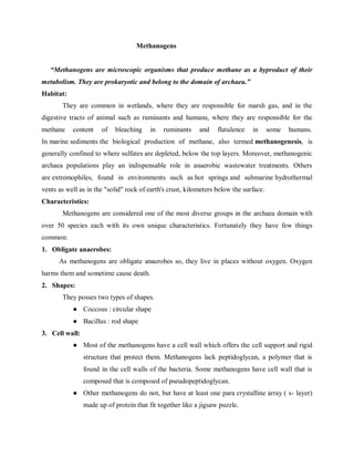
- 1. Methanogens “Methanogens are microscopic organisms that produce methane as a byproduct of their metabolism. They are prokaryotic and belong to the domain of archaea.” Habitat: They are common in wetlands, where they are responsible for marsh gas, and in the digestive tracts of animal such as ruminants and humans, where they are responsible for the methane content of bleaching in ruminants and flatulence in some humans. In marine sediments the biological production of methane, also termed methanogenesis, is generally confined to where sulfates are depleted, below the top layers. Moreover, methanogenic archaea populations play an indispensable role in anaerobic wastewater treatments. Others are extremophiles, found in environments such as hot springs and submarine hydrothermal vents as well as in the "solid" rock of earth's crust, kilometers below the surface. Characteristics: Methanogens are considered one of the most diverse groups in the archaea domain with over 50 species each with its own unique characteristics. Fortunately they have few things common: 1. Obligate anaerobes: As methanogens are obligate anaerobes so, they live in places without oxygen. Oxygen harms them and sometime cause death. 2. Shapes: They posses two types of shapes. ● Coccous : circular shape ● Bacillus : rod shape 3. Cell wall: ● Most of the methanogens have a cell wall which offers the cell support and rigid structure that protect them. Methanogens lack peptidoglycan, a polymer that is found in the cell walls of the bacteria. Some methanogens have cell wall that is composed that is composed of pseudopeptidoglycan. ● Other methanogens do not, but have at least one para crystalline array ( s- layer) made up of protein that fit together like a jigsaw puzzle.
- 2. 4. Consumption: Methanogens produce methane from substrates such as h2/co2, acetate, formate, methanol and methyl amines by process called methanogenesis. Most methanogens consume carbon dioxide, hydrogen and release methane gas. CO2 + 2H2O → CH4 + 2O2 Methanogenic bacteria are considered a critical group of bacteria, because of their phylogenetic diversity and the only producers of a hydrocarbon, methane. They require anoxic and highly reduced conditions for growth. These bacteria use co2 or methyl group as the terminal electron acceptor and produce methane as their catabolic end product. Example: ● Methanococcus jannaschii ● Methanobacterium paludis sp. Application: ● Methane production ● Biogas production ● Wastewater treatment Figure 2 Methanobacterium paludis sp. Figure 1Methanococcus jannaschii
- 3. CHYTRIDS Chytridiomycota are a division of zoosporic organisms in the kingdom fungi, informally known as chytrids. The name is derived from the greek chytridion, meaning "little pot", describing the structure containing unreleased zoospores. Chytrids are one of the early diverging fungal lineages, and their membership in kingdom fungi is demonstrated with chitin cell walls, a posterior whiplash flagellum, absorptive nutrition, use of glycogen as an energy storage compound, and synthesis of lysine by the α-amino adipic acid (aaa) pathway. Chytrids are unique among all fungi in having motile stages in their life cycle. No other fungi have this trait. These moltile stages take the form of 200 spores single cell with a single posterior flagellum. Chytrid have originated from a separate lineage/ from different ancestors than those of its higher fungi. Though the ancestors of ascomycetes and basidiomycetes are same but chytrids have separate ancestors. Chytrids are saprobic, degrading refractory materials such as chitin and keratin, and sometimes act as parasites. Habitat Chytrids are aquatic fungi, though those that thrive in the capillary network around soil particles are typically considered terrestrial. The zoospore is primarily a means of thoroughly exploring a small volume of water for a suitable substrate rather than a means of long-range dispersal. Chytrids have been isolated from a variety of aquatic habitats, including peats, bogs, rivers, ponds, springs, and ditches, and terrestrial habitats, such as acidic soils, alkaline soils, temperate forest soils, rainforest soils, arctic and antarctic soils. This has led to the belief that many chytrid species are ubiquitous and cosmopolitan. Ecological functions: Batrachochytrium dendrobatidis The chytrid batrachochytrium dendrobatidis is responsible for chytridiomycosis, a disease of amphibians. The process leading to frog mortality is thought to be the loss of essential ions through pores made in the epidermal cells by the chytrid during its replication. Other parasites: Chytrids mainly infect algae and other eukaryotic and prokaryotic microbes. The infection can be so severe as to control primary production within the lake. Chytrids may
- 4. also infect plant species; in particular, synchytrium endobioticum is an important potato pathogens Saprobes The most important ecological function chytrids perform is decomposition. These ubiquitous and cosmopolitan organisms are responsible for decomposition of refractory materials, such as pollen, cellulose, chitin, and keratin. Life cycle: Chytrids have a life cycle much like many of the other fungi's. For sexual reproduction, once the fungi starts meiosis the sporangium starts to germinate which then releases haploid zoospores which then germinate into a young gametophyte. Figure 3 life cycle of chytridiomycota (chytrids)
- 5. MICROBES AND INSECTS Insects are the most diverse group of animals with over a million different species found almost in every habitat, except the sea. Due to their widespread distribution, insects are inevitably associated with an extremely large variety of microscopic life forms, including viruses, bacteria, fungi, protozoa, nematodes and multicellular parasites. Although some of these microorganisms exhibit a rather wide host range, many associations are highly specialized and involve not only certain insect species but also particular life stages of the insect host. Interaction between microbes and insects All insects are colonized by microorganisms on the insect exoskeleton, in the gut and hemocoel, and within insect cells. The insect microbiota is generally different from microorganisms in the external environment, including ingested food. Specifically, certain microbial taxa are favored by the conditions and resources in the insect habitat, by their tolerance of insect immunity, and by specific mechanisms for their transmission. The resident microorganisms can promote insect fitness by contributing to nutrition, especially by providing essential amino acids, B vitamins, and, for fungal partners, sterols. Some microorganisms protect their insect hosts against pathogens, parasitoids, and other parasites by synthesizing specific toxins or modifying the insect immune system. Priorities for future research include elucidation of microbial contributions to detoxification, especially of plant allelochemicals in phytophagous insects, and resistance to pathogens; as well as their role in among-insect communication; and the potential value of manipulation of the microbiota to control insect pests. Mutualistic intereaction Microbes on the cuticle surface Although the insect exoskeleton is correctly recognized as a vitally important physical barrier against microbial infections, it is also a substrate that can be colonized by various microorganisms. Up to 1,000 culturable bacterial cells are associated with the body surface of Drosophila melanogaster. Factors limiting microbial populations on the insect cuticle can include physical disturbance (e.g., ecdysis and grooming behavior) as well as antimicrobial secretions (e.g., from the meta-pleural glands of ants, Hymenoptera). The extent to which cuticle-associated bacteria can proliferate and form stable communities, as occurs on human skin, is largely unknown.
- 6. Cuticular structures that promote colonization by specific microorganisms have evolved in many insects. In particular, the mycangia, i.e., cuticular invaginations housing fungi in adult insects, can be considered as culture vessels in which fungi required by the insect's offspring are stored and protected against abiotic factors and contamination by other microorganisms. The defining feature of mycangia—that they house fungi—is somewhat artificial because at least some mycangia additionally bear bacteria. Some cuticular modifications house bacteria exclusively. For example, solitary digger wasps of the tribe Philanthini retain Streptomyces spp. in cuticle-lined glandular reservoirs, in each of 5–6 antennal segments; and attine ants house actinobacteria of the genus Pseudonocardia in similar glandular invaginations, known as crypts or foveae, on the thorax, legs, or other locations of the body, varying with ant species. Gut microbes Gut microbiota of insects is composed both of prokaryotes and eukaryotes that live outside the gut cells. They usually inhabit the hind part of insect’s gut (hindgut), either moving freely in its lumen or remaining attached to its walls. In some phytophagous insects, likes termites and cockroaches, the hindgut is a chamber without oxygen (anaerobic) where fermentation of cellulose and other complex sugars takes place. Worker termite gut; the green part corresponds to the hindgut without oxygen. In termites, this anaerobic chamber contains facultative anaerobic prokaryotes (they can develop either with or without oxygen) and obligate anaerobic prokaryotes (they can only develop
- 7. without oxygen), such as spirochetes and methanogens, which aid in digestion. In addition, in some worker termites, this chamber also contains protozoans that play a major role in the digestion of wood cellulose Unlike other endosymbionts, gut microbes are horizontally transmitted between insects; that is, insects don’t inherit gut microbes from their parents, but they should acquire them throughout their lives. In termites, acquisition of gut microbes takes place through a process called trophallaxis: the workers, which are the only able to feed by themselves, digest the food and transmit the resulting product mixed with gut microorganisms to the rest of the colony members through their mouthparts. Moreover, microorganisms are removed during molting processes, so termites (and other insects performing trophollaxis) can acquire them again through trophollaxis. Endoparasites Parasites that live and/or develop inside an organism are known as endoparasites. They are also horizontally transmitted between insects. The most relevant endoparasitic relationship between insects and microorganisms, and the only one we are going to explain here, are vectors: the insect (or vector) serve as a container to the parasite until it reaches the definitive host. Parasites transported by vector usually are pathogenic protozoans harmful to vertebrates, like Trypanosoma, Leishmania or Plasmodium (Malaria). THE TSETSE FLY Multiple symbionts have coevolved with the tsetse fly, vector of Trypanosoma brucei (and other african trypanosomes) which is the causative agent of sleeping sickness. The fly feeds only on blood, which is rich in proteins but poor in nutrients. The symbionts supply vitamins and other nutrients that cannot be synthesized by the fly. At least two known symbionts with different ultra-structural characteristics are known and they can be found in different tissues within the insect: the primary and secondary endosymbionts are present in the gut tissue, while the third organism was identified in reproductive tissue. The interaction of the fly with the primary p-endosymbiont, wigglesworthia glossinidia, which forms a distinct lineage of γ-proteobacteria, can be characterized as an essential symbiosis. Sequencing of w. Glossinidia genome revealed the presence of several cofactor
- 8. biosynthetic pathways, including over 60 genes involved in the synthesis of vitamins and nutrients that are necessary for fly fertility. Anopheles mosquitoes Malaria is caused by a one-celled parasite called a Plasmodium. Female Anopheles mosquitoes pick up the parasite from infected people when they bite to obtain blood needed to nurture their eggs. Inside the mosquito the parasites reproduce and develop. When the mosquito bites again, the parasites contained in the salivary gland are injected and pass into the blood of the person being bitten. Malaria parasites multiply rapidly in the liver and then in red blood cells of the infected person. One to two weeks after a person is infected the first symptoms of malaria appear: usually fever, headache, chills and vomiting. If not treated promptly with effective medicines, malaria can kill by infecting and destroying red blood cells and by clogging the capillaries that carry blood to the brain or other vital organs. PATHOGENIC INTERACTION Just like humans and other vertebrates, insects are susceptible to many disease-causing organisms known as pathogens. Thousands of species of bacteria, fungi, viruses, protozoa and nematodes can sicken or kill insects. Even if the insects survive, the pathogens’ “sub-lethal” effects can keep their victims from feeding or reproducing. 1. Bacteria Most bacteria infect specific insect orders. Some naturally occurring insect-pathogenic bacteria have been isolated and mass-produced for commercial use. One of these, Bacillus thuringiensis or Bt, is the world’s most widely applied biological control agent. It exerts its toxicity only after plant-eating insects actually consume it. A highly dense protein crystal, the Bt toxin kills victims by first paralyzing their mid-gut, then their entire bodies. Like most other bacterial pathogens, Bt is specific to certain insect orders. Its short residual period also makes it an ideal candidate for pest management in fruits and vegetables. 2. Fungi Although an estimated 700-plus species of fungi can infect insects, fewer than 20 have been developed for insect management. Most insect-pathogenic fungi need cool, moist environments to germinate. Compared to most other insect pathogens, they have an extensive
- 9. host range. Beauveria bassiana, for example, can help manage beetles, ants, termites, true bugs, grasshoppers, mosquitoes and mites as well as other arthropod pests. It unleashes a toxin that weakens its host’s immune system, then overwhelms its dead host’s intestinal bacteria with an antibiotic. The tell-tale sign of B. bassiana’s carnage is its victim’s “white bloom” of fungal spores. Fungi can invade their insect host through natural openings in its cuticle. Thus, hosts need not consume pathogens but only come into direct contact with them. Although some fungi can take up to several weeks to kill their hosts, most infected insects die within three to seven days. 3. Viruses Most viruses that attack insects belong to a group called nuclear polyhedrosis viruses or NPVs. Their victims are usually young larvae of butterflies and moths, which become infected by eating NPV particles and typically die within several weeks. Some infected larvae hang limply from the tops of crop canopies, prompting the common name “caterpillar wilt” or “tree top” disease. Prevailing environmental factors heavily influence the insect-killing efficiencies of viruses. For example, they are adversely affected by sunlight, while the relatively slow speed at which they kill has also hindered their widespread acceptance for biocontrol. 4. Nematodes Nearly 40 known families of nematodes parasitize and consume insects and other arthropods. Some are hunter-cruisers while others are ambushers. The most beneficial of these “entomopathogenic” nematodes belong to the Heterorhabditidae and Steinernematidae families. Both families are “obligate” parasites: their survival depends on their hosts and on the symbiotic relationships the nematodes have evolved with disease-causing Xenorhabdus and Photorhabdus bacteria. Parasitic nematodes transport bacteria inside their host, penetrating the host via the mouth, anus, spiracles or cuticle. Once inside, the nematodes release the bacteria, which quickly multiply and kill the host. In turn, the nematode uses the bacteria and insect cadaver for food and shelter, maturing, mating and reproducing inside it. Infective-stage juvenile nematodes eventually emerge from the cadaver and seek out another host. Because they are highly mobile and can locate and destroy new victims in just a few days, entomopathogenic nematodes make outstanding candidates for all kinds of biological control. Some are applied to soils to successfully manage the underground life stages of insect pests.