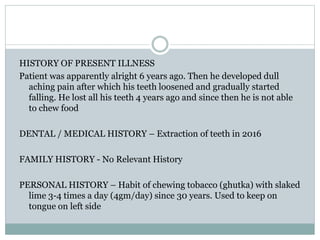
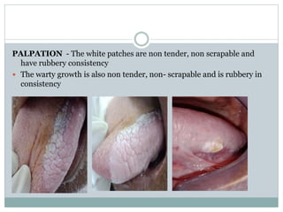
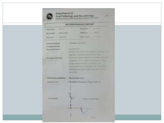
P R E S E N T E D B Y -
D R . H U M A I R A H U S S A I N
G U I D E D B Y -
D R . P R A S H A N T J A J U
CASE PRESENTATION OF
VERRUCOUS LEUKOPLAKIA
CONTENTS
 PERSONAL DETAILS
 CHIEF COMPLAINT
 HISTORY OF PRESENTING ILLNESS
 DENTAL/MEDICAL HISTORY
 PERSONAL HISTORY
 FAMILY HISTORY
 GENERAL PHYSICAL EXAMINATION
 EXTRAORAL EXAMINATION
 INTRAORAL EXAMINATION
 LOCAL EXAMINATION OF THE CONDITION
 PROVISIONAL DIAGNOSIS
 DIFFERENTIAL DIAGNOSIS
 INVESTIGATIONS
 FINAL DIAGNOSIS
 TREATMENT PLAN
PERSONAL DATA:
O.P.D. No: 34150/21
Name: Shanti Lal
Age: 64 years
Occupation: Farmer
Date: 26/10/20
Address:Harniyakala Sawan, Tehseel Kala Peepal, Zila - Shajapur
CHIEF COMPLAINT
 Patient complaints of inability to chew food since 4 years
HISTORY OF PRESENT ILLNESS
Patient was apparently alright 6 years ago. Then he developed dull
aching pain after which his teeth loosened and gradually started
falling. He lost all his teeth 4 years ago and since then he is not able
to chew food
DENTAL / MEDICAL HISTORY – Extraction of teeth in 2016
FAMILY HISTORY - No Relevant History
PERSONAL HISTORY – Habit of chewing tobacco (ghutka) with slaked
lime 3-4 times a day (4gm/day) since 30 years. Used to keep on
tongue on left side
 EDUCATION STATUS: Till class 7
GENERAL PHYSICAL EXAMINATION:
Patient was conscious and well oriented to time, place and person.
Built: Average
Nutritional status: Well nourished
Height & weight:
Gait & posture:
Speech:
Pallor: No abnormality detected
Icterus:
Cyanosis:
Clubbing:
Oedema:
Vital signs:
 Temperature – Afebrile
 Pulse – 74 beats/minute
 Respiration – 18 breaths/minute
 Blood pressure – 130/90 mm Hg
Extra Oral Examination :
Head Form:
Facial form:
Skin:
Hair:
Eyes:
Ears: No abnormality detected
Nose:
Paranasal sinuses:
Lymph nodes:
TMJ:Bilaterally symmetrical with no clicking or popping sound heard
Salivary glands:
Saliva:
Muscles of mastication:
Muscles of facial expression: No abnormality detected
Thyroid gland:
Trachea:
Carotid artery:
Others:
INTRA-ORAL EXAMINATION
Labial Mucosa –
Vermillion Border of Lip – No abnormality detected
Hard Palate –
Buccal Mucosa – Grayish white homogenous patch seen on left buccal
mucosa
Tongue – Grayish white homogenous patch seen on left dorsal, ventral
surfaces and lateral border of tongue
Floor of the mouth –
Palate –
Uvula – No abnormality detected
Vestibule –
Gingiva –
HARD TISSUE EXAMINATION:
Teeth present: Completely edentulous
Teeth missing:
11,12,13,14,15,16,17,21,22,23,24,25,26,27,31,32,33,34,35,36,37,41,42,43,
44,45,46,47
LOCAL EXAMINATION OF THE LESION
SOFT TISSUE EXAMINATION
 INSPECTION – Well defined, homogenous, grayish white patch
seen extending from tip of tongue till posterior 2/3rd surface of
tongue on left lateral border of about 5 cm anteroposteriorly and 1
cm mediolaterally (involving dorsal, lateral and ventral surfaces)
in dimensions
 There is whitish warty growth of 1× 1cm in dimensions in middle
third portion of ventral tongue on left side
 Well defined, homogenous, grayish white patch seen extending
from left retro commissure till buccal mucosa adjacent to alveolar
bone of 34 and 2 × 1 cm in dimensions
PALPATION - The white patches are non tender, non scrapable and
have rubbery consistency
 The warty growth is also non tender, non- scrapable and is rubbery in
consistency
PROVISIONAL DIAGNOSIS:
 Verrucous hyperplasia on left ventral surface of tongue
 Homogenous leukoplakia on dorsum of tongue
DIFFERENTIAL DIAGNOSIS
Verrucous leukoplakia
Verrucous Carcinoma
Chronic hyperplastic candidiasis
INVESTIGATIONS
 BLOOD INVESTIGATIONS
Complete blood picture
Bleeding and clotting time
 BIOPSY
Excisional biopsy of verrucous growth was done
 Haemoglobin : 12.4 gm%
 WBC Count : 8200/cmm
 Differential Count :
 Neutrophil :46.2%
 Lymphocytes : 47.4%
 Monocytes :6.4%
 RBC count : 4.47 mil./cu.mm
 RBC INDICES
MCV – 99.3
MCH- 35.9
MCHC- 36
 Platelet Count : 357000/ cumm
 Bleeding time : 1 min
 Clotting time : 3 min
TREATMENT PLAN
 Emergency Phase
Capsule Lycopene 8 mg once a day for 15 days
Topical retinoic acid 0.01% twice a day for 15 days
 Phase 1 – Quit the habit
 Preliminary impression taken
 Phase 3 – Removable prosthesis given
 Phase 4 – Patient recalled after 15 days
 Histopathological findings: The H and E stained section on
microscopic examination reveals hyperkeratotic filliform papilla
 Histopathological diagnosis: Normal Tongue tissue
FINAL DIAGNOSIS
 Homogenous Leukoplakia with left dorso-lateral surface of tongue
 Verrucous leukoplakia on left lateral border of tongue
DISCUSSION
White lesions are relatively frequent in the oral cavity with prevalence of
approximately 24.8%
Among them oral leukoplakia (OL) is quite prevalent (0.2-3.6%)
In a retrospective study, Hansen et al., reported that 26 of the 30 lesions
initially diagnosed as OL became oral carcinomas in patients followed
for 1-20 years (average, 6.1 years). After this study, these lesions were
named oral proliferative verrucous leukoplakia (OPVL)
According to the latest World Health Organization nomenclature, OPVL
conforms to the new terminology of “potentially malignant disorders”
given that it is neither a delimited lesion nor a condition
It is best-defined as a continuum of oral epithelial disease with
hyperkeratosis at one end of a clinical and microscopic spectrum and
verrucous carcinoma or squamous cell carcinoma at the other
Etiopathogenesis
 Many potential etiologies have been hypothesized, but little has been
proved about the origin of this disease process
 The disease seems to be idiopathic
 Tobacco is frequently absent as a known risk factor as OPVL occurs
both in smokers and non-smokers
 An association has been reported between human papillomavirus
(HPV) and OPVL
 Between 0% and 89% of OPVL are reported to be HPV positive,
especially for HPV types 16 and 18
 Apparently, there is no unequivocal pathogenetic link between HPV
and OPVL and it has also been reported in association with Epstein-
Barr virus or candida infection
 Despite such extensive works, the etiology of OPVL is still as enigmatic
as the disease itself
Clinical features
 Two of the largest studies of OPVL patients reported a predilection for
this lesion in elderly women, with a ratio as high as 4:1 for women to
men unlike other forms of OL. The mean age at the time of diagnosis is
slightly over 60 years
 It has been shown that almost all lesions occur bilaterally, mainly
affecting the lower alveolar ridge and buccal mucosa
 Clinically, it generally presents as a simple benign form, which tends to
spread and become diffuse
 In time, OPVL develops exophytic, wart-like or erythroplakic areas that
become oral carcinomas
 Histopathological features
 The microscopic findings associated with OPVL are dependent on the
stage of the disease and the adequacy of the biopsy
 Hansen et al., suggested histologic stages in the continuum of OPVL
with intermediates
 Grade 0: Normal mucosa
 Grade 2: Hyperkeratosis (clinical leukoplakia)
 Grade 4: Verrucous hyperplasia
 Grade 6: Verrucous carcinoma
 Grade 8: Papillary squamous cell carcinoma
 Grade 10: Less well-differentiated squamous cell carcinoma
Batsakis et al., reduced the number of histologic stages to four with
intermediates:
 Grade 0: Clinical flat leukoplakia without dysplasia
 Grade 2: Verrucous hyperplasia
 Grade 4: Verrucous carcinoma
 Grade 6: Conventional squamous cell carcinoma with intermediates
 It is of interest that the early phase of these lesions usually exhibits an
interface lymphocytic infiltrate that may have a pronounced lichenoid
pattern characterized by basal vacuolar degeneration containing
apoptotic cells and eosinophilic bodies, similar to types of oral
lichenoid stomatitis such as lichen planus
 Therefore, OPVL has no single defining histopathologic feature
 Diagnosis
 Because of the lack of specific histological criteria, the diagnosis of
OPVL is based on combined clinical and histopathologic evidence of
progression
 In previously published series, diagnosis of OPVL was made according
to Hansen's et al., definition
 There are few studies that apply a set of diagnostic criteria that are
mentioned as follows
 Ghazali et al., established the following criteria:
 The lesion starts as homogenous leukoplakia without evidence of
dysplasia at the first visit
 With time, some areas of leukoplakia become verrucous
 The disease progresses to the development of multiple isolated or
confluent lesions at the same or a different site
 With time, the disease progresses through the different
histopathological stages reported by Hansen et al.[2]
 The appearance of new lesions after treatment
 A follow-up period of no less than 1 year.
 Gandolfo et al., establish the following criteria:
 An initially innocuous lesion characterized by a homogenous plaque
that progresses over time to an exophytic, diffuse, usually multifocal,
lesion with a verrucous epithelial growth pattern
 Histopathologically, proliferative verrucous leukoplakia (PVL)
changes gradually from a simple plaque of hyperkeratosis without
dysplasia to verrucous hyperplasia, verrucous carcinoma or oral
squamous cell carcinoma (OSCC).
 Cerero-Lapiedra et al., established the following major and minor
criteria:
 Major criteria
 A leukoplakia lesion with more than two different oral sites, which is
most frequently found in the gingiva, alveolar processes and palate
 The existence of a verrucous area
 That the lesions have spread or engrossed during development of the
disease
 That there has been a recurrence in a previously treated area
 Histopathologically, there can be from simple epithelial hyperkeratosis
to verrucous hyperplasia, verrucous carcinoma or OSCC, whether in
situ or infiltrating.
Minor criteria
 An OL lesion that occupies at least 3 cm when adding all the affected
areas
 That the patient be female
 That patient (male or female) be a non-smoker
 A disease evolution higher than 5 years
 In order to make the diagnosis of PVL, it was suggested that one of the
two following combinations of the criteria mentioned before were met.
 Three major criteria (being E among them) or
 Two major criteria (being E among them) + two minor criteria.
 Nevertheless, at present, there is no criterion that will allow for the
early diagnosis of the disease
Treatment
 Advise patients with OPVL to avoid other known factors associated
with development of oral carcinoma, such as tobacco, alcohol and betel.
 Medical care
 Owing to the progressive nature of OPVL, many forms of therapy used
for the management of traditional leukoplakia have been disappointing.
Carbon dioxide laser, radiation, topical bleomycin solution, oral
retinoids, beta-carotene and systemic chemotherapy have all failed at
achieving permanent cure. Methisoprinol is a synthetic agent capable of
inhibiting viral ribonucleic acid synthesis and replication and of
stimulating antiviral cell–mediated reactions that has been shown to
have some clinical efficacy in HPV-induced lesions. Although
improvements have been noted with some of these modalities,
recurrence rates after cessation of therapy are high, often within
months of discontinuation of treatment
Surgical care
 This lesion is resistant to the presently available treatment modalities;
therefore, total excision with free surgical margins is critical combined
with a lifelong follow-up
 Malignant transformation and recurrences
 OPVL is known for its aggressive pathology, given its multifocal
involvement, high malignant transformation rates (60-100%), frequent
recurrences (87-100%) and high mortality rates (30-50%)
 The gingiva and palate represented the areas with the highest frequency
of these multiple malignant tumors
 Given the high tendency for (OSCCs) to appear in these patients, they
should be checked for life at least once every 6 months
REFERENCES
 Issrani R, Prabhu N, Keluskar V. Oral proliferative verrucous
leukoplakia: A case report with an update. Contemp Clin Dent.
2013;4(2):258-262. doi:10.4103/0976-237X.114887

Verrucous leukoplakia.pptx

  • 1.
    P R ES E N T E D B Y - D R . H U M A I R A H U S S A I N G U I D E D B Y - D R . P R A S H A N T J A J U CASE PRESENTATION OF VERRUCOUS LEUKOPLAKIA
  • 2.
    CONTENTS  PERSONAL DETAILS CHIEF COMPLAINT  HISTORY OF PRESENTING ILLNESS  DENTAL/MEDICAL HISTORY  PERSONAL HISTORY  FAMILY HISTORY  GENERAL PHYSICAL EXAMINATION  EXTRAORAL EXAMINATION  INTRAORAL EXAMINATION  LOCAL EXAMINATION OF THE CONDITION  PROVISIONAL DIAGNOSIS  DIFFERENTIAL DIAGNOSIS  INVESTIGATIONS  FINAL DIAGNOSIS  TREATMENT PLAN
  • 3.
    PERSONAL DATA: O.P.D. No:34150/21 Name: Shanti Lal Age: 64 years Occupation: Farmer Date: 26/10/20 Address:Harniyakala Sawan, Tehseel Kala Peepal, Zila - Shajapur
  • 4.
    CHIEF COMPLAINT  Patientcomplaints of inability to chew food since 4 years
  • 5.
    HISTORY OF PRESENTILLNESS Patient was apparently alright 6 years ago. Then he developed dull aching pain after which his teeth loosened and gradually started falling. He lost all his teeth 4 years ago and since then he is not able to chew food DENTAL / MEDICAL HISTORY – Extraction of teeth in 2016 FAMILY HISTORY - No Relevant History PERSONAL HISTORY – Habit of chewing tobacco (ghutka) with slaked lime 3-4 times a day (4gm/day) since 30 years. Used to keep on tongue on left side
  • 6.
  • 7.
    GENERAL PHYSICAL EXAMINATION: Patientwas conscious and well oriented to time, place and person. Built: Average Nutritional status: Well nourished Height & weight: Gait & posture: Speech: Pallor: No abnormality detected Icterus: Cyanosis: Clubbing: Oedema:
  • 8.
    Vital signs:  Temperature– Afebrile  Pulse – 74 beats/minute  Respiration – 18 breaths/minute  Blood pressure – 130/90 mm Hg
  • 9.
    Extra Oral Examination: Head Form: Facial form: Skin: Hair: Eyes: Ears: No abnormality detected Nose: Paranasal sinuses: Lymph nodes: TMJ:Bilaterally symmetrical with no clicking or popping sound heard
  • 10.
    Salivary glands: Saliva: Muscles ofmastication: Muscles of facial expression: No abnormality detected Thyroid gland: Trachea: Carotid artery: Others:
  • 11.
    INTRA-ORAL EXAMINATION Labial Mucosa– Vermillion Border of Lip – No abnormality detected Hard Palate – Buccal Mucosa – Grayish white homogenous patch seen on left buccal mucosa Tongue – Grayish white homogenous patch seen on left dorsal, ventral surfaces and lateral border of tongue Floor of the mouth – Palate – Uvula – No abnormality detected Vestibule – Gingiva –
  • 12.
    HARD TISSUE EXAMINATION: Teethpresent: Completely edentulous Teeth missing: 11,12,13,14,15,16,17,21,22,23,24,25,26,27,31,32,33,34,35,36,37,41,42,43, 44,45,46,47
  • 13.
    LOCAL EXAMINATION OFTHE LESION SOFT TISSUE EXAMINATION  INSPECTION – Well defined, homogenous, grayish white patch seen extending from tip of tongue till posterior 2/3rd surface of tongue on left lateral border of about 5 cm anteroposteriorly and 1 cm mediolaterally (involving dorsal, lateral and ventral surfaces) in dimensions  There is whitish warty growth of 1× 1cm in dimensions in middle third portion of ventral tongue on left side  Well defined, homogenous, grayish white patch seen extending from left retro commissure till buccal mucosa adjacent to alveolar bone of 34 and 2 × 1 cm in dimensions
  • 14.
    PALPATION - Thewhite patches are non tender, non scrapable and have rubbery consistency  The warty growth is also non tender, non- scrapable and is rubbery in consistency
  • 15.
    PROVISIONAL DIAGNOSIS:  Verrucoushyperplasia on left ventral surface of tongue  Homogenous leukoplakia on dorsum of tongue
  • 16.
    DIFFERENTIAL DIAGNOSIS Verrucous leukoplakia VerrucousCarcinoma Chronic hyperplastic candidiasis
  • 17.
    INVESTIGATIONS  BLOOD INVESTIGATIONS Completeblood picture Bleeding and clotting time  BIOPSY Excisional biopsy of verrucous growth was done
  • 18.
     Haemoglobin :12.4 gm%  WBC Count : 8200/cmm  Differential Count :  Neutrophil :46.2%  Lymphocytes : 47.4%  Monocytes :6.4%  RBC count : 4.47 mil./cu.mm  RBC INDICES MCV – 99.3 MCH- 35.9 MCHC- 36  Platelet Count : 357000/ cumm  Bleeding time : 1 min  Clotting time : 3 min
  • 20.
    TREATMENT PLAN  EmergencyPhase Capsule Lycopene 8 mg once a day for 15 days Topical retinoic acid 0.01% twice a day for 15 days  Phase 1 – Quit the habit  Preliminary impression taken  Phase 3 – Removable prosthesis given  Phase 4 – Patient recalled after 15 days
  • 21.
     Histopathological findings:The H and E stained section on microscopic examination reveals hyperkeratotic filliform papilla  Histopathological diagnosis: Normal Tongue tissue FINAL DIAGNOSIS  Homogenous Leukoplakia with left dorso-lateral surface of tongue  Verrucous leukoplakia on left lateral border of tongue
  • 23.
    DISCUSSION White lesions arerelatively frequent in the oral cavity with prevalence of approximately 24.8% Among them oral leukoplakia (OL) is quite prevalent (0.2-3.6%) In a retrospective study, Hansen et al., reported that 26 of the 30 lesions initially diagnosed as OL became oral carcinomas in patients followed for 1-20 years (average, 6.1 years). After this study, these lesions were named oral proliferative verrucous leukoplakia (OPVL) According to the latest World Health Organization nomenclature, OPVL conforms to the new terminology of “potentially malignant disorders” given that it is neither a delimited lesion nor a condition It is best-defined as a continuum of oral epithelial disease with hyperkeratosis at one end of a clinical and microscopic spectrum and verrucous carcinoma or squamous cell carcinoma at the other
  • 24.
    Etiopathogenesis  Many potentialetiologies have been hypothesized, but little has been proved about the origin of this disease process  The disease seems to be idiopathic  Tobacco is frequently absent as a known risk factor as OPVL occurs both in smokers and non-smokers  An association has been reported between human papillomavirus (HPV) and OPVL  Between 0% and 89% of OPVL are reported to be HPV positive, especially for HPV types 16 and 18  Apparently, there is no unequivocal pathogenetic link between HPV and OPVL and it has also been reported in association with Epstein- Barr virus or candida infection  Despite such extensive works, the etiology of OPVL is still as enigmatic as the disease itself
  • 25.
    Clinical features  Twoof the largest studies of OPVL patients reported a predilection for this lesion in elderly women, with a ratio as high as 4:1 for women to men unlike other forms of OL. The mean age at the time of diagnosis is slightly over 60 years  It has been shown that almost all lesions occur bilaterally, mainly affecting the lower alveolar ridge and buccal mucosa  Clinically, it generally presents as a simple benign form, which tends to spread and become diffuse  In time, OPVL develops exophytic, wart-like or erythroplakic areas that become oral carcinomas
  • 26.
     Histopathological features The microscopic findings associated with OPVL are dependent on the stage of the disease and the adequacy of the biopsy  Hansen et al., suggested histologic stages in the continuum of OPVL with intermediates  Grade 0: Normal mucosa  Grade 2: Hyperkeratosis (clinical leukoplakia)  Grade 4: Verrucous hyperplasia  Grade 6: Verrucous carcinoma  Grade 8: Papillary squamous cell carcinoma  Grade 10: Less well-differentiated squamous cell carcinoma Batsakis et al., reduced the number of histologic stages to four with intermediates:  Grade 0: Clinical flat leukoplakia without dysplasia  Grade 2: Verrucous hyperplasia  Grade 4: Verrucous carcinoma  Grade 6: Conventional squamous cell carcinoma with intermediates
  • 27.
     It isof interest that the early phase of these lesions usually exhibits an interface lymphocytic infiltrate that may have a pronounced lichenoid pattern characterized by basal vacuolar degeneration containing apoptotic cells and eosinophilic bodies, similar to types of oral lichenoid stomatitis such as lichen planus  Therefore, OPVL has no single defining histopathologic feature  Diagnosis  Because of the lack of specific histological criteria, the diagnosis of OPVL is based on combined clinical and histopathologic evidence of progression  In previously published series, diagnosis of OPVL was made according to Hansen's et al., definition  There are few studies that apply a set of diagnostic criteria that are mentioned as follows
  • 28.
     Ghazali etal., established the following criteria:  The lesion starts as homogenous leukoplakia without evidence of dysplasia at the first visit  With time, some areas of leukoplakia become verrucous  The disease progresses to the development of multiple isolated or confluent lesions at the same or a different site  With time, the disease progresses through the different histopathological stages reported by Hansen et al.[2]  The appearance of new lesions after treatment  A follow-up period of no less than 1 year.  Gandolfo et al., establish the following criteria:  An initially innocuous lesion characterized by a homogenous plaque that progresses over time to an exophytic, diffuse, usually multifocal, lesion with a verrucous epithelial growth pattern  Histopathologically, proliferative verrucous leukoplakia (PVL) changes gradually from a simple plaque of hyperkeratosis without dysplasia to verrucous hyperplasia, verrucous carcinoma or oral squamous cell carcinoma (OSCC).
  • 29.
     Cerero-Lapiedra etal., established the following major and minor criteria:  Major criteria  A leukoplakia lesion with more than two different oral sites, which is most frequently found in the gingiva, alveolar processes and palate  The existence of a verrucous area  That the lesions have spread or engrossed during development of the disease  That there has been a recurrence in a previously treated area  Histopathologically, there can be from simple epithelial hyperkeratosis to verrucous hyperplasia, verrucous carcinoma or OSCC, whether in situ or infiltrating. Minor criteria  An OL lesion that occupies at least 3 cm when adding all the affected areas  That the patient be female  That patient (male or female) be a non-smoker  A disease evolution higher than 5 years
  • 30.
     In orderto make the diagnosis of PVL, it was suggested that one of the two following combinations of the criteria mentioned before were met.  Three major criteria (being E among them) or  Two major criteria (being E among them) + two minor criteria.  Nevertheless, at present, there is no criterion that will allow for the early diagnosis of the disease Treatment  Advise patients with OPVL to avoid other known factors associated with development of oral carcinoma, such as tobacco, alcohol and betel.
  • 31.
     Medical care Owing to the progressive nature of OPVL, many forms of therapy used for the management of traditional leukoplakia have been disappointing. Carbon dioxide laser, radiation, topical bleomycin solution, oral retinoids, beta-carotene and systemic chemotherapy have all failed at achieving permanent cure. Methisoprinol is a synthetic agent capable of inhibiting viral ribonucleic acid synthesis and replication and of stimulating antiviral cell–mediated reactions that has been shown to have some clinical efficacy in HPV-induced lesions. Although improvements have been noted with some of these modalities, recurrence rates after cessation of therapy are high, often within months of discontinuation of treatment
  • 32.
    Surgical care  Thislesion is resistant to the presently available treatment modalities; therefore, total excision with free surgical margins is critical combined with a lifelong follow-up  Malignant transformation and recurrences  OPVL is known for its aggressive pathology, given its multifocal involvement, high malignant transformation rates (60-100%), frequent recurrences (87-100%) and high mortality rates (30-50%)  The gingiva and palate represented the areas with the highest frequency of these multiple malignant tumors  Given the high tendency for (OSCCs) to appear in these patients, they should be checked for life at least once every 6 months
  • 33.
    REFERENCES  Issrani R,Prabhu N, Keluskar V. Oral proliferative verrucous leukoplakia: A case report with an update. Contemp Clin Dent. 2013;4(2):258-262. doi:10.4103/0976-237X.114887