Downloaded 176 times

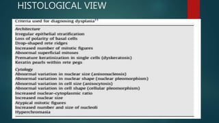


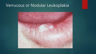


The document discusses premalignant lesions and biopsy, defining premalignant lesions and conditions, and describing various types of precancerous lesions such as leukoplakia and erythroplakia. It details their etiology, clinical features, histopathology, diagnosis, differential diagnoses, and treatment. Additionally, it covers the types of biopsy and procedures involved in diagnosing and treating such lesions.


