Downloaded 272 times


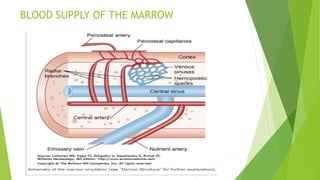


The bone marrow contains red and yellow marrow and produces around 6 billion new blood cells per kilogram of body weight daily. It consists of a hematopoietic component containing stem and progenitor cells embedded in a stromal component. The stroma contains sinusoids, adipocytes, extracellular matrix, endothelial cells, adventitial reticular cells, and CXCL12-abundant reticular cells. These stromal cells secrete growth factors and cytokines that regulate hematopoiesis. Hematopoietic cells adhere in specific niches in relation to sinusoids and extracellular matrix proteins via cellular adhesion molecules, which provides an environment suitable for cell survival, proliferation, differentiation, and maturation.