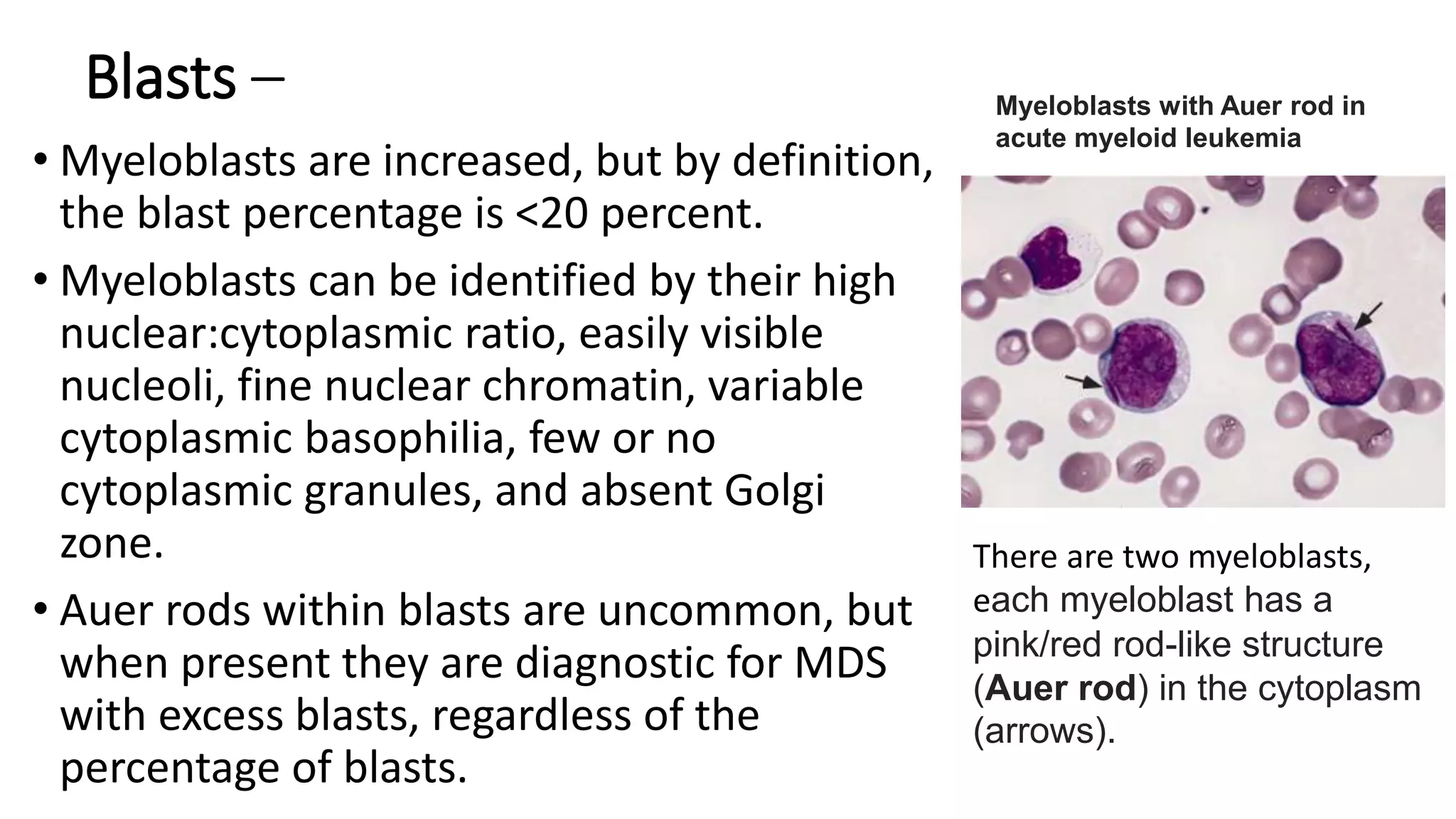
2. Introduction
⢠In these disorders, there is uncontrolled clonal proliferation
of one or more of the cell lines in the bone marrow, namely
erythroid, myeloid and megakaryocyte lines.
⢠Myeloproliferative disorders include:
1. Polycythaemia vera (PV),
2. Essential thrombocythaemia (ET),
3. Myelofibrosis and
4. Chronic myeloid leukaemia (CML)
3. ⢠These disorders are grouped together as there can be
transition from one disease to another; e.g. PV can
lead to myelofibrosis.
⢠They may also transform to acute myeloblastic
leukaemia.
4. Polycythaemia
⢠Polycythemia vera (PV, polycythemia rubra vera, maladie de
Vaquez) is one of the chronic myeloproliferative neoplasms
(MPNs), which are collectively characterized by clonal
proliferation of myeloid cells with variable morphologic
maturity and hematopoietic efficiency.
5. ⢠PV is distinguished clinically from the other MPNs by
the presence of an elevated red blood cell mass.
⢠However, an increased red blood cell mass alone is
insufficient to establish the diagnosis, since this is also
observed in conditions associated with chronic
hypoxia and with erythropoietin-secreting tumors
6. ⢠Polycythaemia (or erythrocytosis) is defined as an increase in
haemoglobin, PCV and red cell count.
⢠PCV is a more reliable indicator of polycythaemia than is Hb,
which may be disproportionately low in iron deficiency.
⢠Polycythaemia can be divided into absolute erythrocytosis
where there is a true increase in red cell volume, or relative
erythrocytosis where the red cell volume is normal but there
is a decrease in the plasma volume.
7. ⢠Absolute erythrocytosis is due to primary
polycythaemia (PV) or secondary polycythaemia.
⢠Secondary polycythaemia is due to either an
appropriate increase in red cells in response to
anoxia, or an inappropriate increase associated with
tumours, such as a renal carcinoma.
8.
9.
10. Primary polycythaemia: polycythaemia vera
(PV)
⢠PV is a clonal stem cell disorder in which there is an
alteration in the pluripotent progenitor cell leading to
excessive proliferation of erythroid, myeloid and
megakaryocytic progenitor cells.
⢠Over 95% of patients with PV have acquired mutations of
the gene Janus Kinase 2 (JAK2).
⢠JAK2 is a cytoplasmic tyrosine kinase that transduces signals,
especially those triggered by haematopoietic growth factors
such as erythropoietin, in normal and neoplastic cells.
11. Clinical features
⢠The onset is insidious.
⢠It usually presents in patients aged over 60 years with
tiredness, depression, vertigo, tinnitus and visual
disturbance.
⢠It should be noted that these symptoms are also common in
the normal population over the age of 60 and consequently,
PV is easily missed.
⢠These features, together with hypertension, angina,
intermittent claudication and a tendency to bleed, are
suggestive of PV.
12. ⢠Severe itching after a hot bath or when the patient is
warm is common.
⢠Gout due to increased cell turnover may be a feature,
and peptic ulceration occurs in a minority of patients.
⢠Thrombosis and haemorrhage are the major
complications of PV.
13. ⢠The patient is usually plethoric and has a
deep dusky cyanosis.
⢠Injection of the conjunctivae is
commonly seen.
⢠The spleen is palpable in 70% and is
useful in distinguishing PV from
secondary polycythaemia.
⢠The liver is enlarged in 50% of patients.
14. Diagnosis
⢠The measurement of
red cell and plasma
volume is not
necessary.
⢠There may be a raised
serum uric acid,
leucocyte alkaline
phosphatase and a
raised serum vitamin
B12 and vitamin B12
binding protein
(transcobalamin 1).
15. Management and prognosis
â˘Treatment is designed to maintain a normal
blood count and to prevent the complications
of the disease, particularly thromboses and
haemorrhage.
â˘Treatment is aimed at keeping the PCV below
0.45 L/L and the platelet count below 400 Ă
10^9/L.
16. â˘There are three types of specific treatment:
1.Venesection.
2.Chemotherapy.
3.Low-dose aspirin
17. Venesection.
⢠The removal of 400â500 mL weekly will successfully
relieve many of the symptoms of PV.
⢠Iron deficiency limits erythropoiesis.
⢠Venesection is often used as the sole treatment and
other therapy is reserved to control the
thrombocytosis.
⢠The aim is to maintain a packed cell volume (PVC) of
<0.45 L/L.
18. Chemotherapy
â˘Continuous or intermittent treatment with
hydroxycarbamide (hydroxyurea) is used frequently
because of the ease of controlling thrombocytosis.
â˘Low-dose intermittent busulfan may be more convenient
for elderly people, and this must be weighed against the
potential risk of long-term complications.
19. â˘Low-dose aspirin 100 mg daily with the
chemotherapy is used for patients with
recurrent thrombotic episodes.
â˘Anagrelide inhibits megakaryocyte
differentiation and is useful for thrombolysis.
20. Polycythaemia VS Surgery
⢠Polycythaemia should be controlled before surgery.
⢠Patients with uncontrolled PV have a high operative
risk; 75% of patients have severe haemorrhage
following surgery and 30% of these patients die.
⢠In an emergency, reduction of the haematocrit by
venesection and appropriate fluid replacement must
be carried out.
21. Prognosis
â˘PV develops into myelofibrosis in 30% of cases
and into acute myeloblastic leukaemia in 5% as
part of the natural history of the disease.
22. Essential thrombocythaemia
⢠Essential thrombocythaemia (ET) is a myeloproliferative disorder
closely related to PV.
⢠Patients have normal Hb levels and WBC but elevated platelet
counts.
⢠At diagnosis the platelet count will usually be >600 à 10^9/L, and
may be as high as 2000 Ă 10^9/L or rarely even higher.
⢠ET presents either symptomatically with thromboembolic or less
commonly bleeding problems or incidentally (e.g. at a routine
medical check).
23. ⢠The diagnosis of ET is not straightforward as there is no global gold
standard test.
⢠The JAK2 mutation tests are useful in that the gene is mutated in
about half of all cases of ET, confirming a myeloproliferative disorder.
⢠For the remaining 50% of patient with a normal JAK2 gene, clinical
assessment and observation over a period of time are required.
⢠As a generalization a person with a very high platelet count (>1000 Ă
10^9/L) who is clinically normal with good health will most likely
prove to have ET.
24. ⢠In a patient with a lower platelet count, e.g. 600 à 10^9/L,
and in poor health the diagnosis can be more difficult.
⢠Other disorders which may give rise to reactive high platelet
counts include autoimmune rheumatic disorders and
malignancy.
⢠Individuals who have been splenectomized (for any reason,
including trauma) sometimes have high platelet counts.
25. Major criteria
1.Platelet count âĽ450 Ă 10
9
/L
2.Bone marrow biopsy showing proliferation mainly of the megakaryocyte lineage with
increased numbers of enlarged, mature megakaryocytes with hyperlobulated nuclei. No
significant increase or left shift in neutrophil granulopoiesis or erythropoiesis and very rarely
minor (grade 1) increase in reticulin fibers.
3.Not meeting WHO criteria for BCR-ABL1
+
CML, PV, PMF, myelodysplastic syndromes, or
other myeloid neoplasms
4.Presence of JAK2, CALR, or MPL mutation
Minor criterion
1.Presence of a clonal marker or absence of evidence for reactive thrombocytosis
Diagnosis of ET requires meeting all 4 major criteria or the first 3 major criteria and the
minor criterion
WHO diagnostic criteria essential thrombocythemia
26. CLINICAL FEATURES
⢠Overview â Up to one-half of patients with ET are discovered
incidentally when thrombocytosis is noted on a complete blood count
obtained for some other reason.
⢠Others present with disease-related symptoms (eg, headache,
dizziness, visual changes) or complications (eg, thrombosis,
bleeding, first trimester fetal loss).
⢠Unlike in polycythemia vera, pruritus is very uncommon in ET,
occurring in less than 5 percent of patients.
27. Vasomotor symptoms â
⢠In patients with ET, "vasomotor symptoms" refer to a constellation of
symptoms that are thought to be related to microvascular
disturbances.
28. ⢠Vasomotor manifestations include:
âHeadache
âLightheadedness
âSyncope
âAtypical chest pain
âAcral paresthesia
âLivedo reticularis
âErythromelalgia
âTransient visual disturbance
Erythromelalgia involving the hands in essential
thrombocythemia
This photograph shows the presence of erythromelalgia of
the hands in a woman with essential thrombocythemia.
This condition is associated with burning pain in the feet
or hands accompanied by erythema, pallor, or cyanosis, in
the presence of palpable pulses, and is common in both
essential thrombocythemia and polycythemia vera.
29. Treatment
⢠Treatment is with hydroxycarbamide (hydroxyurea),
anagrelide or busulfan to control the platelet count to less
than 400 Ă 10^9/L.
â˘Îą-Interferon is also effective; it is administered by
subcutaneous injection.
⢠ET may eventually transform into PV, myelofibrosis or acute
leukaemia, but the disease may not progress for many years.
30. Myelofibrosis
⢠Myelofibrosis is a very debilitating chronic myeloproliferative
neoplasm.
⢠It may be primary or develop late in the course of essential
thrombocythaemia or polycythaemia vera.
⢠There is clonal proliferation of stem cells and myeloid metaplasia in
the liver, spleen and other organs.
⢠Increased fibrosis in the bone marrow is caused by hyperplasia of
abnormal megakaryocytes which release fibroblast-stimulating
factors such as platelet-derived growth factor.
31. Clinical features
⢠The disease presents insidiously with lethargy, weakness and weight
loss.
⢠Patients often complain of a âfullnessâ in the upper abdomen due to
splenomegaly.
⢠Severe pain related to respiration may indicate perisplenitis secondary
to splenic infarction, and bone pain and attacks of gout can complicate
the illness.
⢠Bruising and bleeding occur because of thrombocytopenia or abnormal
platelet function.
⢠Other physical signs include anaemia, fever and massive splenomegaly
32. Major criteria
1.Presence of megakaryocytic proliferation and atypia, accompanied by either reticulin and/or collagen
fibrosis grades 2 or 3
2.Not meeting WHO criteria for ET, PV, BCR-ABL1
+
CML, myelodysplastic syndromes, or other myeloid
neoplasms
3.Presence of JAK2, CALR, or MPL mutation or, in the absence of these mutations, presence of another clonal
marker,* or absence of reactive myelofibrosis
Âś
Minor criteria
Presence of at least 1 of the following, confirmed in 2 consecutive determinations:
1.Anemia not attributed to a comorbid condition
2.Leukocytosis âĽ11 Ă 10
9
/L
3.Palpable splenomegaly
4.LDH increased to above upper normal limit of institutional reference range
5.Leukoerythroblastosis
Diagnosis of overt PMF requires meeting all 3 major criteria, and at least 1 minor criterion
WHO diagnostic criteria overt primary myelofibrosis
33. Investigations
⢠Anaemia with leucoerythroblastic
features is present
⢠Poikilocytes and red cells with
characteristic tear-drop forms are seen.
⢠The WBC count may be over 100 Ă
10^9/L, and the differential WBC count
may be very similar to that seen in chronic
myeloid leukaemia (CML); later
leucopenia may develop.
⢠The platelet count may be very high, but
in later stages, thrombocytopenia occurs.
Teardrop-shaped red blood cells
(dacrocytes)
34. ⢠Bone marrow aspiration is often unsuccessful
and this gives a clue to the presence of the
condition.
⢠A bone marrow trephine is necessary to show the
markedly increased fibrosis.
⢠Increased numbers of megakaryocytes may be
seen.
⢠The Philadelphia chromosome is absent; this
helps to distinguish myelofibrosis from most
cases of CML.
⢠JAK2 mutation is present in approximately half of
the cases.
35. Treatment
⢠This consists of general supportive measures such as blood
transfusion, folic acid, analgesics and allopurinol.
⢠If the spleen becomes very large and painful, and transfusion
requirements are high, it may be advisable to perform splenectomy.
⢠Splenectomy may also result in relief of severe thrombocytopenia.
⢠Treatment for myelofibrosis is often difficult but an estimation of
prognosis from a prognostic scoring system is a good basis to start
planning a treatment strategy for the individual patient.
36. Prognosis
⢠Patients may survive for âĽ10 years; median survival is 3
years.
⢠Death may occur in 10â20% of cases from transformation to
acute myeloblastic leukaemia.
⢠The most common causes of death are cardiovascular
disease, infection and gastrointestinal bleeding.
37. Myelodysplasia
⢠Myelodysplasia (MDS) describes a group of acquired bone marrow
disorders that are due to a defect in stem cells.
⢠They are characterized by increasing bone marrow failure with
quantitative and qualitative abnormalities of all three myeloid cell
lines (red cells, granulocyte/monocytes and platelets).
38. ⢠The natural history of MDS is variable, but there is a
high morbidity and mortality owing to bone marrow
failure, and transformation into acute myeloblastic
leukaemia occurs in about 30% of cases.
39.
40. Clinical and laboratory features
⢠MDS occurs mainly in the elderly, and presents with
symptoms of anaemia, infection or bleeding due to
pancytopenia.
⢠Serial blood counts show evidence of increasing bone
marrow failure with anaemia, neutropenia, monocytosis
and thrombocytopenia, either alone or in combination.
⢠By contrast, in chronic myelomonocytic leukaemias (CMML),
monocytes are >1 Ă 10^9/L and the WBC count may be >100
Ă 10^9/L.
41. â˘The bone marrow usually shows increased cellularity
despite the pancytopenia.
â˘Dyserythropoiesis is present, and granulocyte
precursors and megakaryocytes also have abnormal
morphology.
â˘Ring sideroblasts are present in some types.
Ring sideroblasts in refractory anemia with
ring sideroblasts (RARS)
43. History and physical examination â
⢠The history should elicit details regarding consequences
or complications of cytopenias (eg, fatigue, infections,
bruising).
⢠It should also evaluate other potential causes for
cytopenias and/or dysplasia, including nutritional
status, alcohol and drug use, medications, exposure to
toxic chemicals, prior treatment with antineoplastic
agents or radiotherapy, and risk factors for HIV
infection.
44. ⢠Physical examination may reveal findings related to
cytopenias, including pallor, dyspnea, tachycardia;
manifestations of infections or mucosal ulceration;
and/or bleeding or bruising.
⢠Some patients may have splenomegaly, but
adenopathy is uncommon.
45. Complete blood count â
⢠Complete blood count (CBC) with leukocyte differential usually
demonstrates anemia, but neutropenia and thrombocytopenia are
more variable.
⢠Red blood cells â Anemia is almost uniformly present and is generally
associated with an inappropriately low reticulocyte response.
⢠Leukocytes â Approximately half of patients have a reduced total
white blood cell count (ie, leukopenia), usually resulting from
absolute neutropenia.
⢠Platelets â Variable levels of thrombocytopenia are present in
approximately one-quarter of patients, but isolated
thrombocytopenia is not a common early manifestation of MDS
46. Bone marrow examination â
⢠Bone marrow examination is an essential component
of the evaluation, diagnosis, and classification of MDS.
47. Blasts â
⢠Myeloblasts are increased, but by definition,
the blast percentage is <20 percent.
⢠Myeloblasts can be identified by their high
nuclear:cytoplasmic ratio, easily visible
nucleoli, fine nuclear chromatin, variable
cytoplasmic basophilia, few or no
cytoplasmic granules, and absent Golgi
zone.
⢠Auer rods within blasts are uncommon, but
when present they are diagnostic for MDS
with excess blasts, regardless of the
percentage of blasts.
Myeloblasts with Auer rod in
acute myeloid leukemia
There are two myeloblasts,
each myeloblast has a
pink/red rod-like structure
(Auer rod) in the cytoplasm
(arrows).
48. Myeloid cells â
⢠Impaired myeloid maturation is often readily apparent;
there may be a variable percentage of granulocytic
precursors and a relative maturation arrest at the
myelocyte stage.
⢠Maturation of the cytoplasm may progress more rapidly
than the nucleus and granulocytic precursors;
abnormally large size, abnormal nuclear shape, and
increased or decreased cytoplasmic granularity is
common.
49. Erythroid cells â
⢠Although erythroid hyperplasia (associated with
ineffective erythropoiesis) is usually seen, red cell
aplasia and/or hypoplasia also rarely occur.
⢠Morphologic abnormalities in the erythroid
precursors include large size, nuclear multilobation,
nuclear budding, and other abnormal forms.
50. Megakaryocytes â
⢠Megakaryocytes are usually normal or
increased in number and sometimes occur
in clusters.
⢠Abnormal megakaryocytes, including large
or very small mononuclear forms
(micromegakaryocytes or "dwarf
megakaryocytes"), megakaryocytes with
multiple dispersed nuclei ("pawn ball
megakaryocytes"), and hypogranular
megakaryocytes, are common findings.
Myelodysplastic syndrome with abnormal
megakaryocytic maturation
Bone marrow aspirate from a patient with
myelodysplastic syndrome. The megakaryocytes are
abnormal, with multiple small lobes seemingly
disconnected from each other ("pawn ball" changes,
arrows). (Wright-Giemsa).
51. Management
⢠Patients with <5% blasts in the bone marrow are usually
managed conservatively with red cell and platelet
transfusions and antibiotics for infections, as they are
needed.
⢠Haemopoietic growth factors (e.g. erythropoietin, G-CSF)
may be useful in some patients.
⢠Patients with >5% blasts have a less favourable prognosis,
and a number of treatment options are available:
52. ⢠Supportive care only is suitable for elderly patients with
other medical problems.
⢠âGentleâ chemotherapy (low-dose or single-agent, e.g.
azacytidine) may be useful in patients with high WBC counts.
⢠Intensive chemotherapy schedules used for acute
myeloblastic leukaemia may be tried in patients under the
age of 60, but the remission rate is less, and prolonged
pancytopenia may occur owing to poor haemopoietic
regeneration because of the defect in stem cells.
53. ⢠Lenalidomide (a thalidomide analogue) has been proven to
be remarkably successful in the treatment of early stage
myelodysplasia with a chromosome 5q deletion (the 5qâ
syndrome).
⢠Avoid use in women of childbearing age.
⢠Bone marrow transplantation offers the hope of cure in the
small proportion of MDS patients who are under the age of
50 and who have an HLA-identical sibling or an unrelated
HLA-matched donor.
Editor's Notes
myeloproliferative neoplasms (MPNs)
CML: chronic myeloid leukemia; ET: essential thrombocythemia; PMF: primary myelofibrosis; PV: polycythemia vera; WHO: World Health Organization
Perisplenitis is acute inflammation of the splenic capsule and its peritoneal covering.
CML: chronic myeloid leukemia; ET: essential thrombocythemia; LDH: lactate dehydrogenase; PMF: primary myelofibrosis; PV: polycythemia vera* In the absence of any of the 3 major clonal mutations, the search for the most frequent accompanying mutations (eg, ASXL1, EZH2, TET2, IDH1/IDH2, SRSF2, SF3B1) are of help in determining the clonal nature of the disease.œ Bone marrow fibrosis secondary to infection, autoimmune disorder, or other chronic inflammatory conditions, hairy cell leukemia or other lymphoid neoplasm, metastatic malignancy, or toxic (chronic) myelopathies.
View all
The Philadelphia (Ph) chromosome is an abbreviated chromosome 22 that was shortchanged in a reciprocal exchange of material with chromosome 9.Â
ing sideroblasts are erythroblasts with iron-loaded mitochondria visualized by Prussian blue staining (Perls' reaction) as a perinuclear ring of blue granules
Blasts: Immature blood cells. Leukemic blasts do not grow and age normally; they proliferate