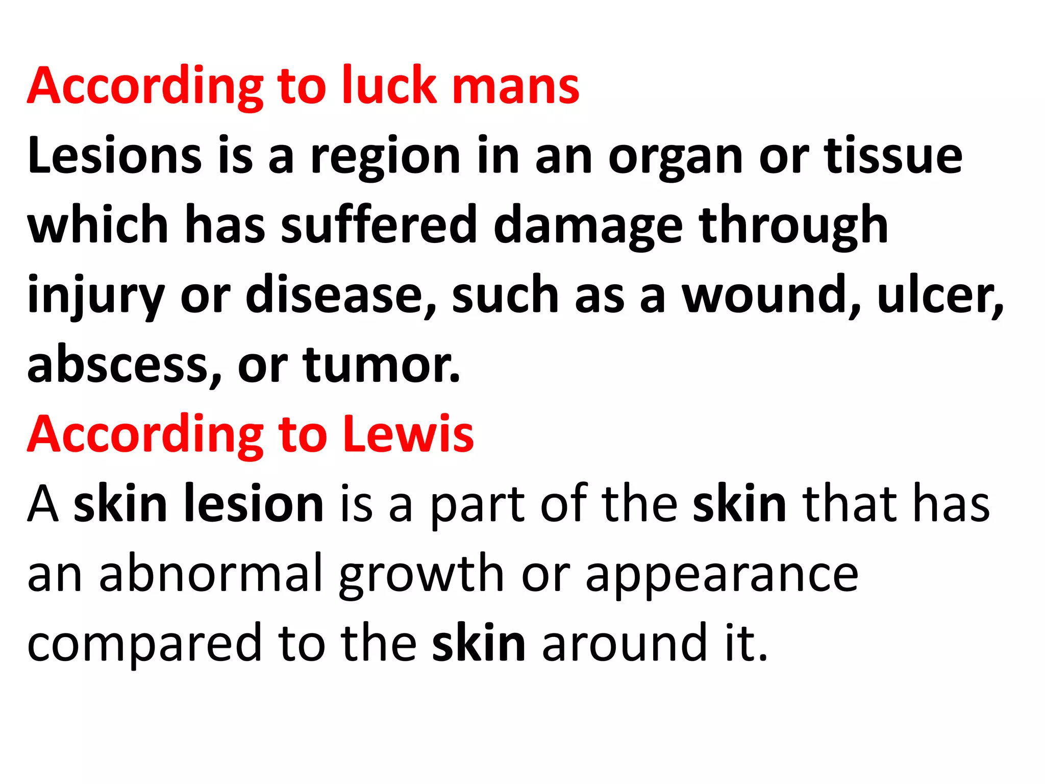
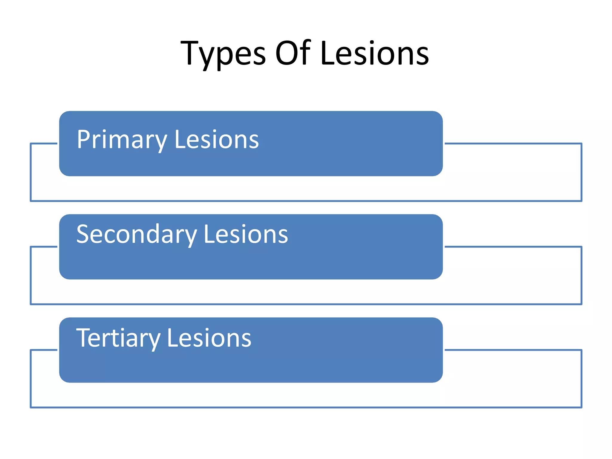
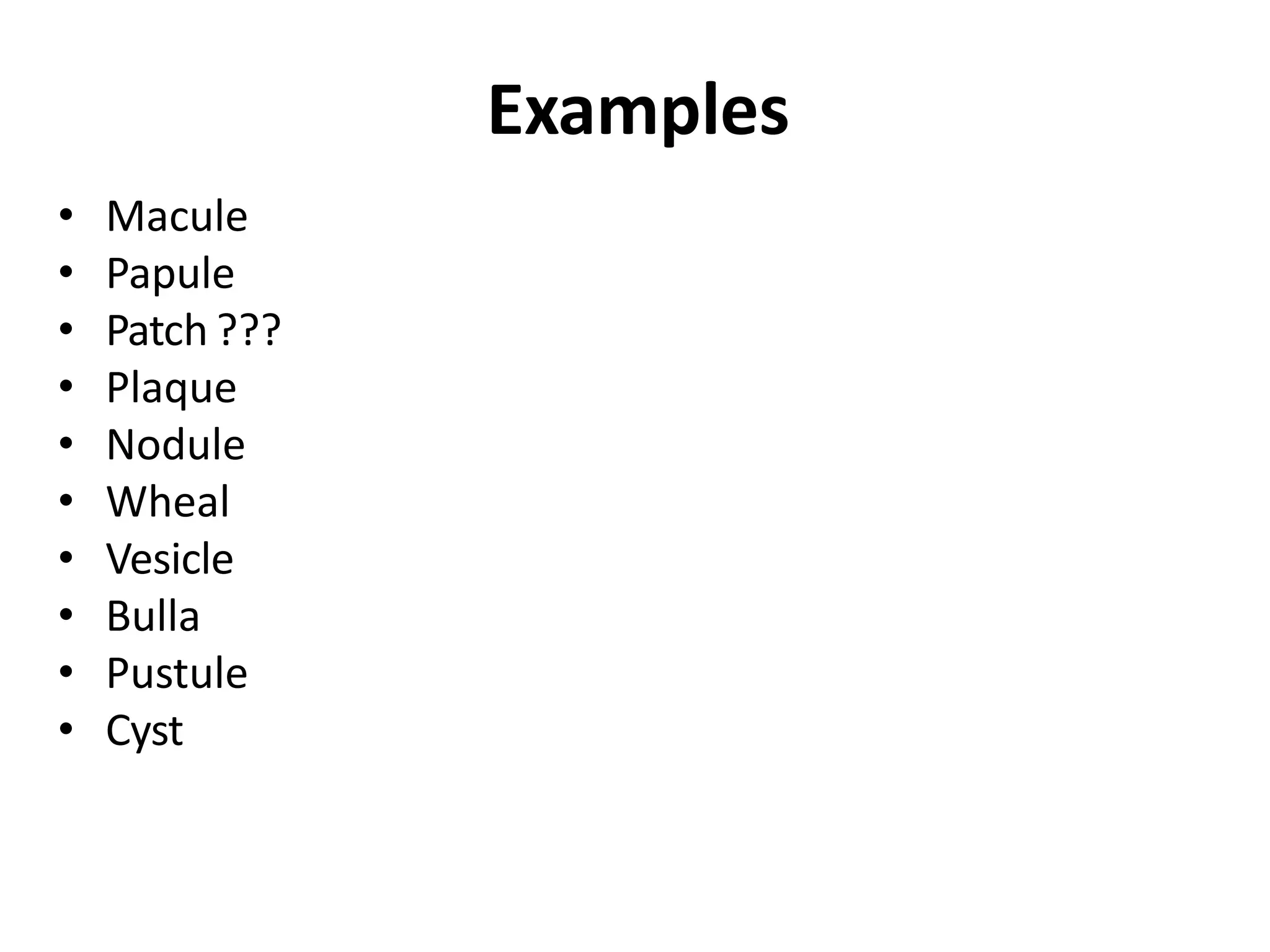
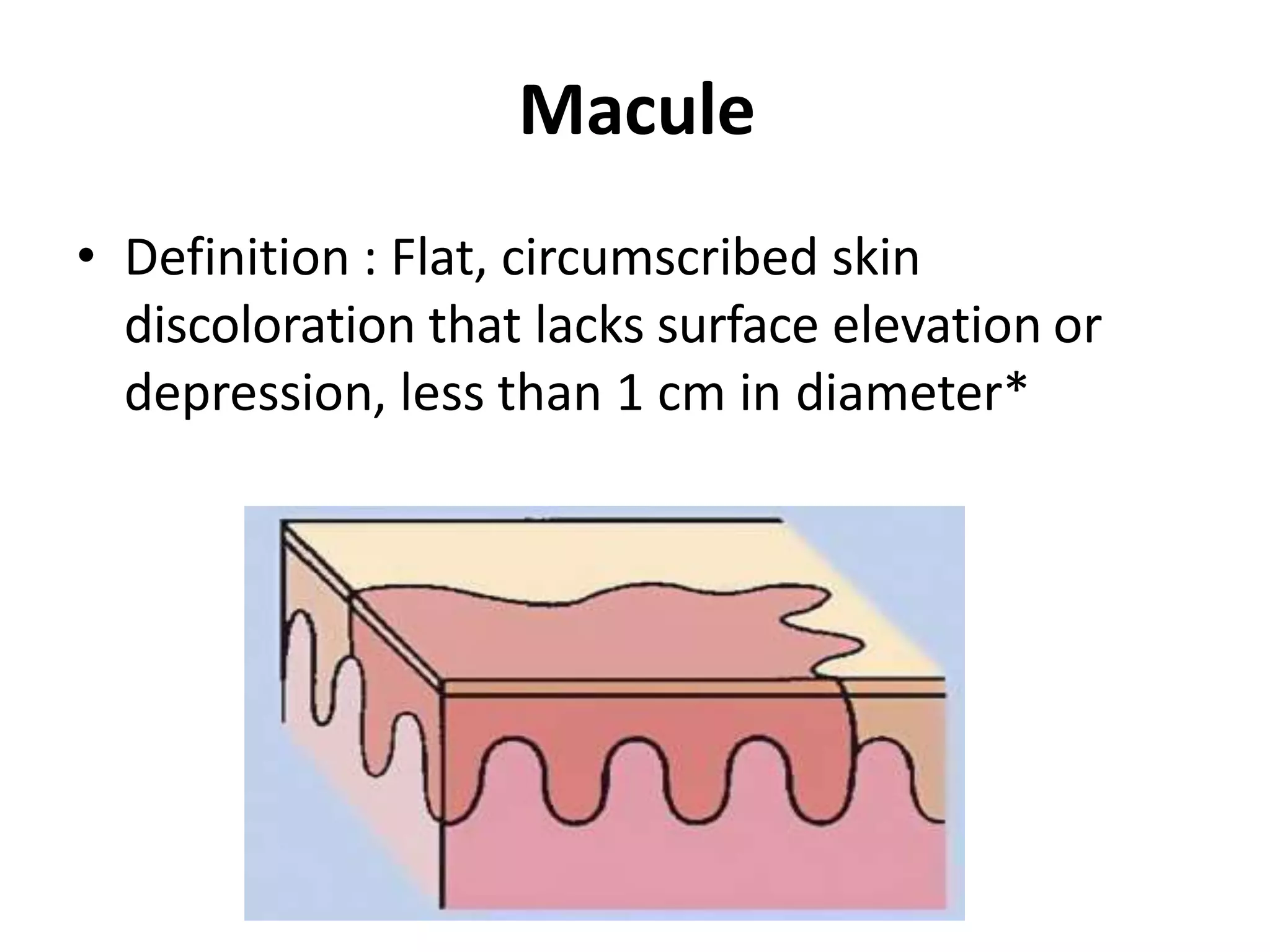
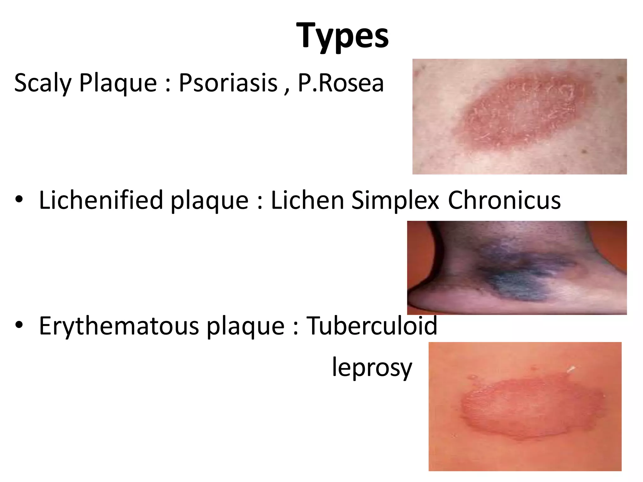
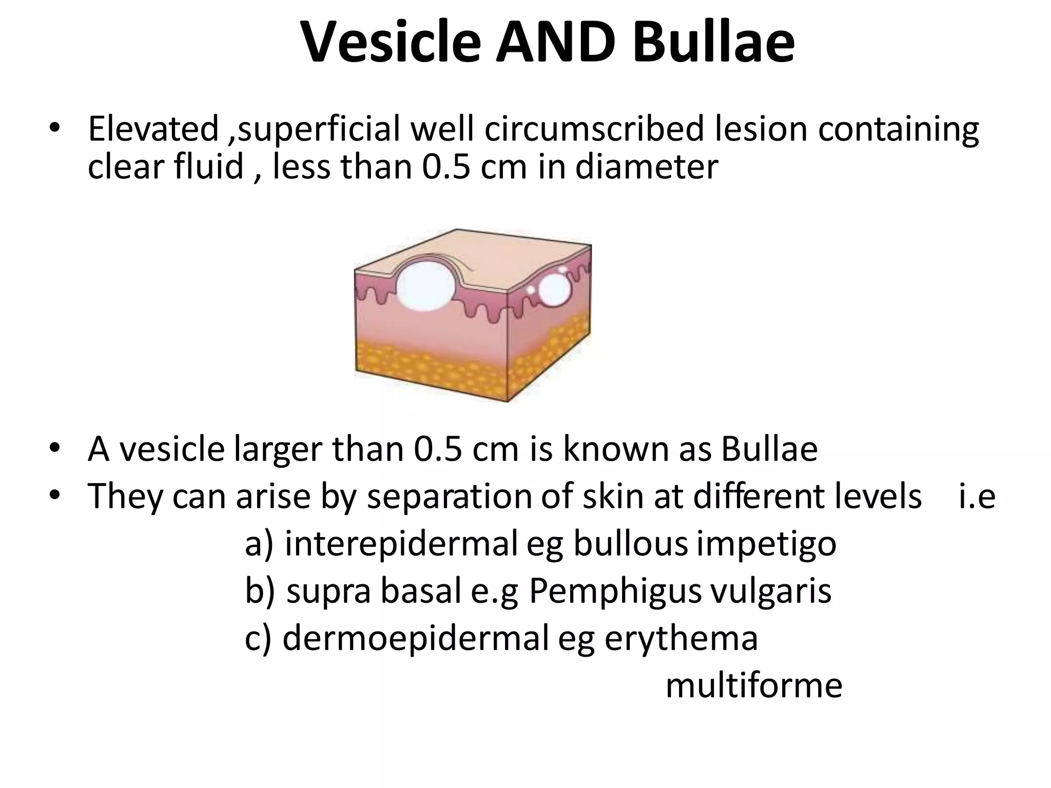
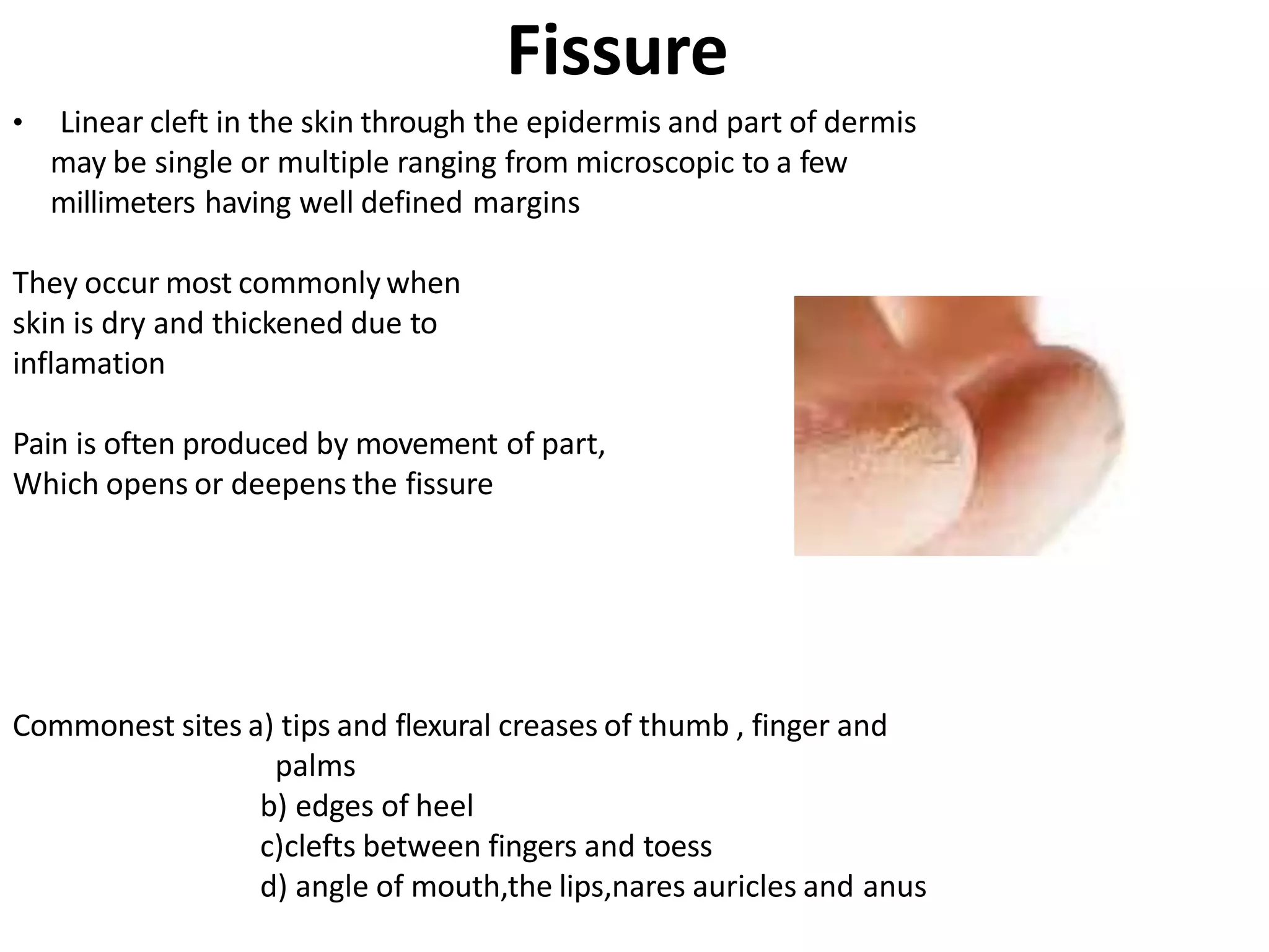
LESIONS AND
ABRASIONS OF SKIN
PRESENTED BY
According to luck mans
Lesions is a region in an organ or tissue
which has suffered damage through
injury or disease, such as a wound, ulcer,
abscess, or tumor.
According to Lewis
A skin lesion is a part of the skin that has
an abnormal growth or appearance
compared to the skin around it.
Types Of Lesions
Primary Lesions
Secondary Lesions
Tertiary Lesions
Primary Lesions
Definition : lesion occurring on non pathological
skin which have not been altered by trauma,
manipulation (scratching, scrubbing), or natural
regression over time.
Examples
• Macule
• Papule
• Patch ???
• Plaque
• Nodule
• Wheal
• Vesicle
• Bulla
• Pustule
• Cyst
Macule
• Definition : Flat, circumscribed skin
discoloration that lacks surface elevation or
depression, less than 1 cm in diameter*
• TYPES
1)Hypopigmented : due to decrease in number of
melanocytes or the amount of pigment they produce
E.g. : tuberous sclerosis , nevus achromicus , etc
nevus achromicus
2)Depigmented : due to complete loss of
melanocytes E.g.: vitiligo , halonaevus
3)Hyperpigmented : due to excess of melanin in skin
A)brown or black : due to excess of melanin in epidermis
E.g.: freckles or chloasma
B)Bluish-grey : due to increase melanin in dermis (
tyndall effect )
E.g : Mongolian spot, nevus of Ota
C)Erythema: increased blood flow through the skin
causing capillary dilatation
E.g : macular viral and
drug rash
D)Purpura/Ecchymosis: extravasation of RBCs in
Dermis
(large purpura : ecchymosis)
E)g : thrombocytic purpura
Schamberg’s purpura
Papule
Solid , well circumscribed elevated lesion, less than one cm
in diameter
Formed by
1)hyperplasia of epidermis , dermis or both
E.g : verruca vulgaris
2)Metabolic deposits or cellular infiltrates
E.g : xanthelasma
SHAPE
COLOUR
Shape
• Pointed : Follicular Lichen planus
• Papulosquamous : Guttate Psoriasis
• Dome : Molluscum contagiosum
• Flat topped : lichen plannus
Colour
• Red : Psoriasis
• Pearly white : molluscum contagiosum
• Violaceous : Lichen planus
• Coppery : seccondary syphilis
• Yellowish : Xanthelasma
• Blue/Black : Mallignant Melanoma
• Purpuric : henoch-schönlein purpura
Plaque
•Elevated well circumscribed more than 1 cm in
diameter ,occupying relatively large surface area
in comparison with its height above the skin
surface
Types
Scaly Plaque : Psoriasis , P.Rosea
• Lichenified plaque : Lichen Simplex Chronicus
• Erythematous plaque : Tuberculoid
leprosy
Patch
• Patch / Large macule ???
Nodule
• Palpable solid round lesion ,usually larger than
1cm in diameter .occupying relatively larger
vertical diameter as compared to suface
diameter.
• PAPULE
• SEEN BETTER
NODULE
FELT BETTER
• E.g : erythema nodosum,Lipoma,Sebaceous cyst
kerato acanthoma,xanthelasma,
Wheals
• Evanescent , edematous , platue-like
elevations of various sizes
• Usually oval or arcuate , pink to red ,
surrounded by a flare of macular erythema
• Caused by transient vascular reaction in the
upper dermis mainly due to vasodilatation and
increased permeability of capillaries
• It is the characteristic lesion of urticaria
Wheals
Vesicle AND Bullae
• Elevated ,superficial well circumscribed lesion containing
clear fluid , less than 0.5 cm in diameter
• A vesicle larger than 0.5 cm is known as Bullae
• They can arise by separation of skin at different levels i.e
a) interepidermal eg bullous impetigo
b) supra basal e.g Pemphigus vulgaris
c) dermoepidermal eg erythema
multiforme
• Lesions may be
• A) tense : Pompholyx
• Flaccid : Pemphigus Vulgaris
• Umblicated : varicella zoster
Pustule
• Well-circumscribed, elevated lesion containing
visible purulent exudate
• Pustules are characteristic of pustular
psoriasis, pyoderma, rosacea
Cyst
• A sac that contains liquid or semisolid material
in a well-defined cavity
• 2 most common type-
• A) Epidermal cyst: lined with squamous
epithelium and produce keratinous material
• B) pilar cyst: originate from hair follicle lined
with multilayered epithelium
Abscess
• An abscess is a collection of pus below the
skin
• Pus in an abscess is invisible but clinically be
interpreted as sign of inflammation in the
overlying skin
•
• Abscess cavities do not have well-defined
lining as cyst
Secondary lesions: modification of primary skin lesions
that result from traumatic injury , evolution from
primary lesion , or other external factors
• Crust
• Scale
• Erosion
• Ulcer
• Fissure
• Scar
• Atrophy
• Telangiectasia
Crust
• A collection of cellular debris
,dried serum,pus or blood and
sometimes bacterial debries
• antecedent primary lesion is
usually a vesicle,bulla or
pustule
Types
• Golden yellow,soft friable : Impetigo
• Yellowish : Flavus
• Thick hard and tough : third degree burns
• Lamillated,elevated, black or green : syphillus
(oyster shell)
Erosion
• Partial focal loss of the epidermis alone. Heals
without a scar
• Eg herpes zooster,TEN,Erosion interdigitale
Scale
• Abnormal shedding or accumulation of the
stratum corneum in visible flakes is called scaling
• Formed due to - a) formation of epidermal cells is
rapid
OR
b) process of normal keratinisation is
interferred with
Types
• Fine and delicate : P.versicolor
• Coarse : eczema
• Stratified : Psoriasis
Ulcer
• A full-thickness, focal loss
of epidermis along with
parts of dermis ,heals
with scarring
• E.g : bed sores , Syphillus
Diabetic foot
Fissure
• Linear cleft in the skin through the epidermis and part of dermis
may be single or multiple ranging from microscopic to a few
millimeters having well defined margins
They occur most commonly when
skin is dry and thickened due to
inflamation
Pain is often produced by movement of part,
Which opens or deepens the fissure
Commonest sites a) tips and flexural creases of thumb , finger and
palms
b) edges of heel
c)clefts between fingers and toess
d) angle of mouth,the lips,nares auricles and anus
Scar
• A collection of new connective tissue, that
replaces lost substances in the demis or deep
dermal tissues
• They may be atrophic or hypertrophic
• Hypertrophic scars / Keloid develop when
fibrous components predominate
An abrasion is a type of open wound that's caused
by the skin rubbing against a rough surface. It may
be called a scrape or a graze. When an abrasion is
caused by the skin sliding across hard ground, it may
be called road rash. Abrasions are very common
injuries. They can range from mild to severe.
According to lippincott
An abrasion is a partial thickness wound
caused by damage to the skin and can be
superficial involving only the epidermis to
deep, involving the deep dermis.
According to luckmans
Abrasion
Punctate or linear abrasion produced by mechanical means
Usually involving only the epidermis
Caused by scratching with fingernails in a variety of disease
E.g : atopic dermatitis , Scabies
If the damage is due to constant
friction or mechanical trauma
the word Abrasion may be used
THANK YOU

lesions and abrasions.pptx

  • 1.
  • 2.
  • 4.
    According to luckmans Lesions is a region in an organ or tissue which has suffered damage through injury or disease, such as a wound, ulcer, abscess, or tumor. According to Lewis A skin lesion is a part of the skin that has an abnormal growth or appearance compared to the skin around it.
  • 5.
    Types Of Lesions PrimaryLesions Secondary Lesions Tertiary Lesions
  • 6.
    Primary Lesions Definition :lesion occurring on non pathological skin which have not been altered by trauma, manipulation (scratching, scrubbing), or natural regression over time.
  • 7.
    Examples • Macule • Papule •Patch ??? • Plaque • Nodule • Wheal • Vesicle • Bulla • Pustule • Cyst
  • 8.
    Macule • Definition :Flat, circumscribed skin discoloration that lacks surface elevation or depression, less than 1 cm in diameter*
  • 9.
    • TYPES 1)Hypopigmented :due to decrease in number of melanocytes or the amount of pigment they produce E.g. : tuberous sclerosis , nevus achromicus , etc nevus achromicus
  • 10.
    2)Depigmented : dueto complete loss of melanocytes E.g.: vitiligo , halonaevus 3)Hyperpigmented : due to excess of melanin in skin A)brown or black : due to excess of melanin in epidermis E.g.: freckles or chloasma
  • 11.
    B)Bluish-grey : dueto increase melanin in dermis ( tyndall effect ) E.g : Mongolian spot, nevus of Ota
  • 12.
    C)Erythema: increased bloodflow through the skin causing capillary dilatation E.g : macular viral and drug rash D)Purpura/Ecchymosis: extravasation of RBCs in Dermis (large purpura : ecchymosis) E)g : thrombocytic purpura Schamberg’s purpura
  • 13.
    Papule Solid , wellcircumscribed elevated lesion, less than one cm in diameter Formed by 1)hyperplasia of epidermis , dermis or both E.g : verruca vulgaris 2)Metabolic deposits or cellular infiltrates E.g : xanthelasma
  • 14.
  • 15.
    Shape • Pointed :Follicular Lichen planus • Papulosquamous : Guttate Psoriasis • Dome : Molluscum contagiosum • Flat topped : lichen plannus
  • 16.
    Colour • Red :Psoriasis • Pearly white : molluscum contagiosum • Violaceous : Lichen planus • Coppery : seccondary syphilis • Yellowish : Xanthelasma • Blue/Black : Mallignant Melanoma • Purpuric : henoch-schönlein purpura
  • 17.
    Plaque •Elevated well circumscribedmore than 1 cm in diameter ,occupying relatively large surface area in comparison with its height above the skin surface
  • 18.
    Types Scaly Plaque :Psoriasis , P.Rosea • Lichenified plaque : Lichen Simplex Chronicus • Erythematous plaque : Tuberculoid leprosy
  • 19.
    Patch • Patch /Large macule ???
  • 20.
    Nodule • Palpable solidround lesion ,usually larger than 1cm in diameter .occupying relatively larger vertical diameter as compared to suface diameter. • PAPULE • SEEN BETTER NODULE FELT BETTER
  • 21.
    • E.g :erythema nodosum,Lipoma,Sebaceous cyst kerato acanthoma,xanthelasma,
  • 22.
    Wheals • Evanescent ,edematous , platue-like elevations of various sizes • Usually oval or arcuate , pink to red , surrounded by a flare of macular erythema • Caused by transient vascular reaction in the upper dermis mainly due to vasodilatation and increased permeability of capillaries • It is the characteristic lesion of urticaria
  • 23.
  • 24.
    Vesicle AND Bullae •Elevated ,superficial well circumscribed lesion containing clear fluid , less than 0.5 cm in diameter • A vesicle larger than 0.5 cm is known as Bullae • They can arise by separation of skin at different levels i.e a) interepidermal eg bullous impetigo b) supra basal e.g Pemphigus vulgaris c) dermoepidermal eg erythema multiforme
  • 25.
    • Lesions maybe • A) tense : Pompholyx • Flaccid : Pemphigus Vulgaris • Umblicated : varicella zoster
  • 26.
    Pustule • Well-circumscribed, elevatedlesion containing visible purulent exudate • Pustules are characteristic of pustular psoriasis, pyoderma, rosacea
  • 27.
    Cyst • A sacthat contains liquid or semisolid material in a well-defined cavity • 2 most common type- • A) Epidermal cyst: lined with squamous epithelium and produce keratinous material • B) pilar cyst: originate from hair follicle lined with multilayered epithelium
  • 28.
    Abscess • An abscessis a collection of pus below the skin • Pus in an abscess is invisible but clinically be interpreted as sign of inflammation in the overlying skin • • Abscess cavities do not have well-defined lining as cyst
  • 29.
    Secondary lesions: modificationof primary skin lesions that result from traumatic injury , evolution from primary lesion , or other external factors • Crust • Scale • Erosion • Ulcer • Fissure • Scar • Atrophy • Telangiectasia
  • 30.
    Crust • A collectionof cellular debris ,dried serum,pus or blood and sometimes bacterial debries • antecedent primary lesion is usually a vesicle,bulla or pustule
  • 31.
    Types • Golden yellow,softfriable : Impetigo • Yellowish : Flavus • Thick hard and tough : third degree burns • Lamillated,elevated, black or green : syphillus (oyster shell)
  • 32.
    Erosion • Partial focalloss of the epidermis alone. Heals without a scar • Eg herpes zooster,TEN,Erosion interdigitale
  • 33.
    Scale • Abnormal sheddingor accumulation of the stratum corneum in visible flakes is called scaling • Formed due to - a) formation of epidermal cells is rapid OR b) process of normal keratinisation is interferred with
  • 34.
    Types • Fine anddelicate : P.versicolor • Coarse : eczema • Stratified : Psoriasis
  • 35.
    Ulcer • A full-thickness,focal loss of epidermis along with parts of dermis ,heals with scarring • E.g : bed sores , Syphillus Diabetic foot
  • 36.
    Fissure • Linear cleftin the skin through the epidermis and part of dermis may be single or multiple ranging from microscopic to a few millimeters having well defined margins They occur most commonly when skin is dry and thickened due to inflamation Pain is often produced by movement of part, Which opens or deepens the fissure Commonest sites a) tips and flexural creases of thumb , finger and palms b) edges of heel c)clefts between fingers and toess d) angle of mouth,the lips,nares auricles and anus
  • 37.
    Scar • A collectionof new connective tissue, that replaces lost substances in the demis or deep dermal tissues • They may be atrophic or hypertrophic • Hypertrophic scars / Keloid develop when fibrous components predominate
  • 40.
    An abrasion isa type of open wound that's caused by the skin rubbing against a rough surface. It may be called a scrape or a graze. When an abrasion is caused by the skin sliding across hard ground, it may be called road rash. Abrasions are very common injuries. They can range from mild to severe. According to lippincott
  • 41.
    An abrasion isa partial thickness wound caused by damage to the skin and can be superficial involving only the epidermis to deep, involving the deep dermis. According to luckmans
  • 42.
    Abrasion Punctate or linearabrasion produced by mechanical means Usually involving only the epidermis Caused by scratching with fingernails in a variety of disease E.g : atopic dermatitis , Scabies If the damage is due to constant friction or mechanical trauma the word Abrasion may be used
  • 53.