Downloaded 140 times


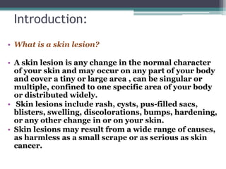
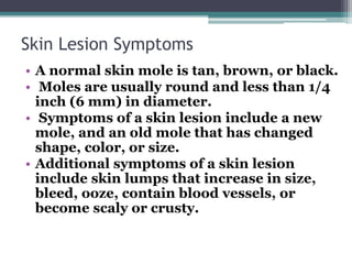
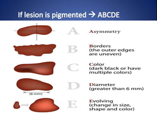
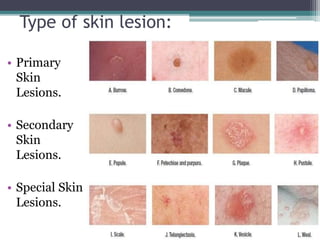

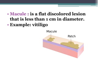
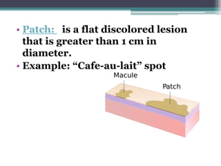
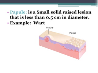
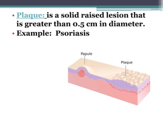
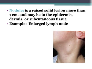

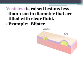
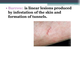
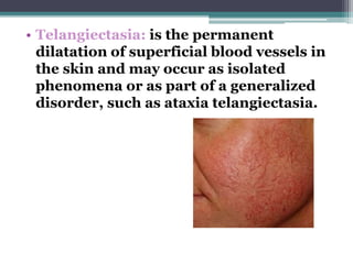
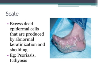
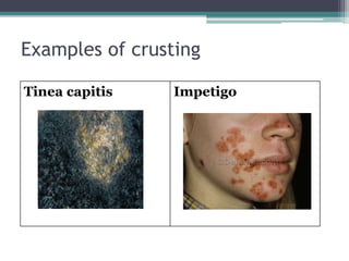
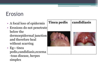
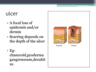
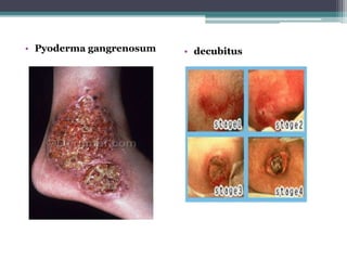

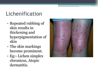
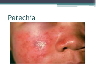

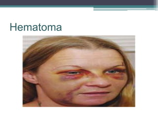


This document discusses assessing and classifying skin lesions. It begins by defining skin lesions and describing common symptoms. It then categorizes lesions as primary, secondary, or vascular. Primary lesions include macules, papules, plaques, nodules, vesicles, and pustules. Secondary lesions result from primary lesions, such as scales, crusts, erosions, and fissures. Vascular lesions involve changes to blood vessels. The document provides examples and characteristics of different lesion types and outlines how to assess lesions by visual inspection and palpation of characteristics like temperature, moisture, and texture.


![PERI-PROSTHETIC FRACTURE NAIL-PLATE CONSTRUCT [NPC].pptx](https://cdn.slidesharecdn.com/ss_thumbnails/drarunkumardrmohamedashrafperiprostheticfrasturenail-plateconstructnpc-260209164459-7e9d15a1-thumbnail.jpg?width=640&height=640&fit=bounds)


