FUNCTION OF SKIN
Protection:
•Physical and mechanical barrier to skin
• Barrier to chemical hazards
Regulation of body temperature:
• Regulate water loss and prevent dehydration
• Maintain body temperature
Photoprotection:
• Protect excessive ultraviolet exposure via melanin
14.
FUNCTION OF SKIN
Immunity:
•Antigen presentation by Langerhans cells
Metabolic function
• Production of vitamin D
Sensory and autonomic function
• Meissners & Pacinian corpuscles
• Pain, heat and cold receptors
Sociosexual & cosmetic function
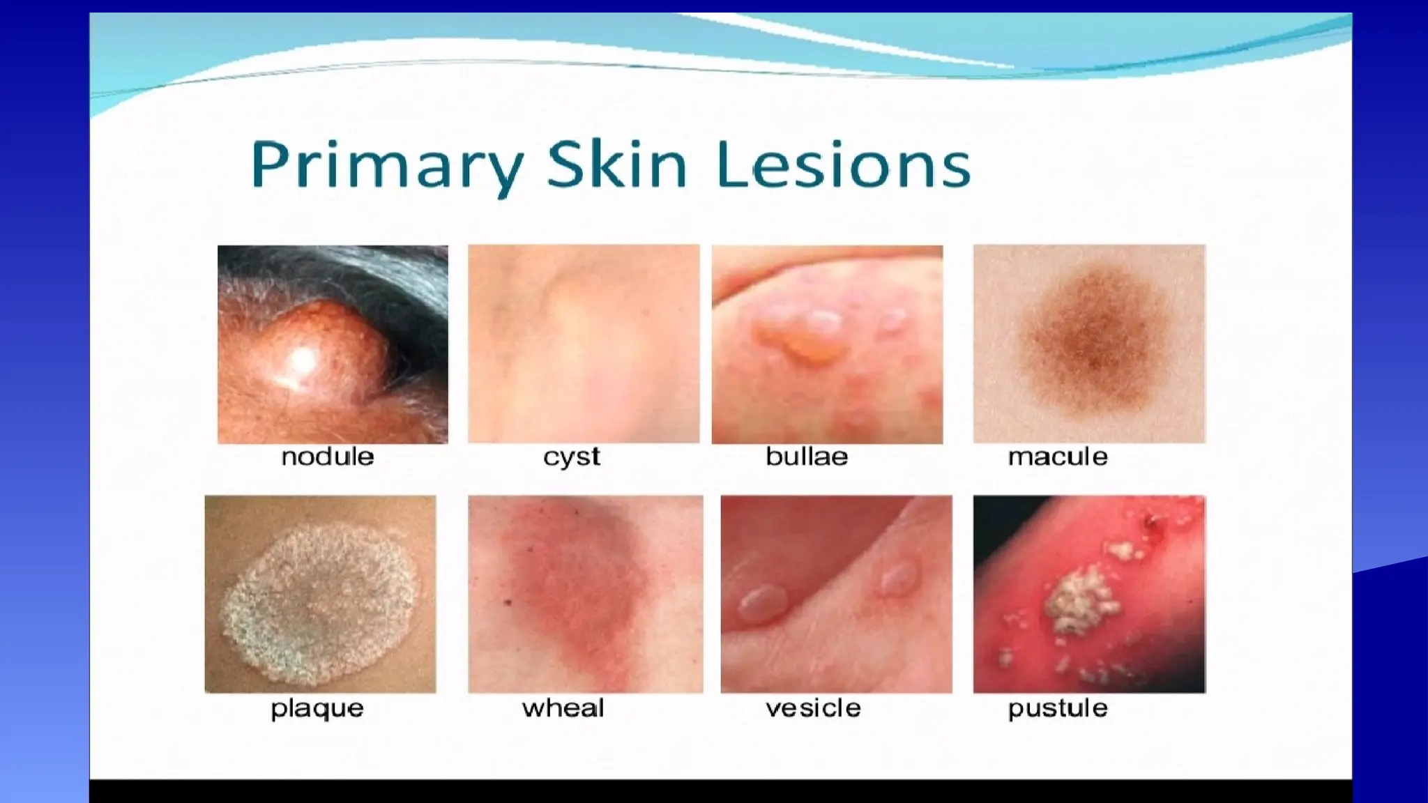
PRIMARY LESIONS
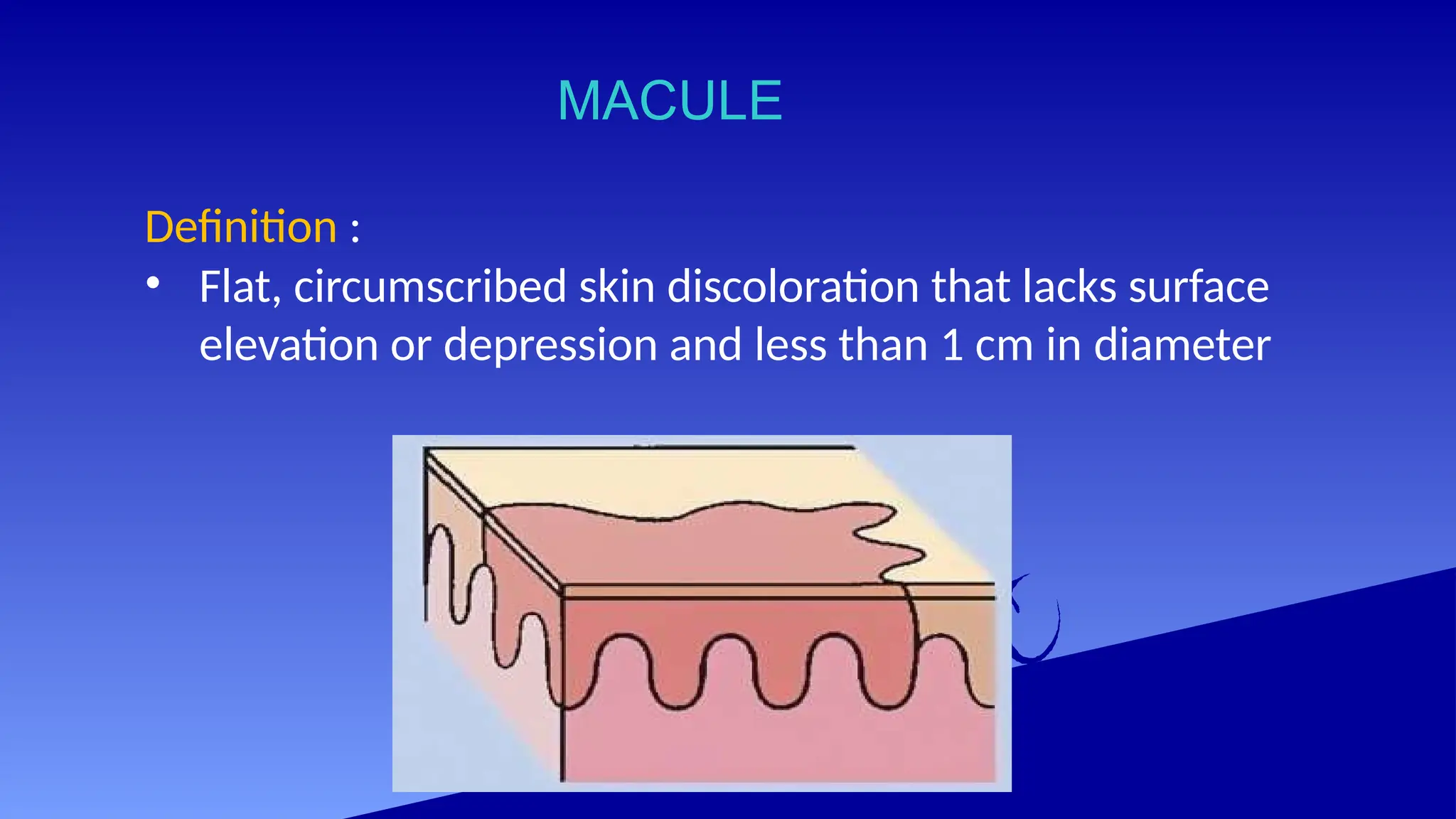
Definition :
•lesion occurring on non pathological skin
which have not been altered by trauma,
manipulation (scratching, scrubbing) or
natural regression over time
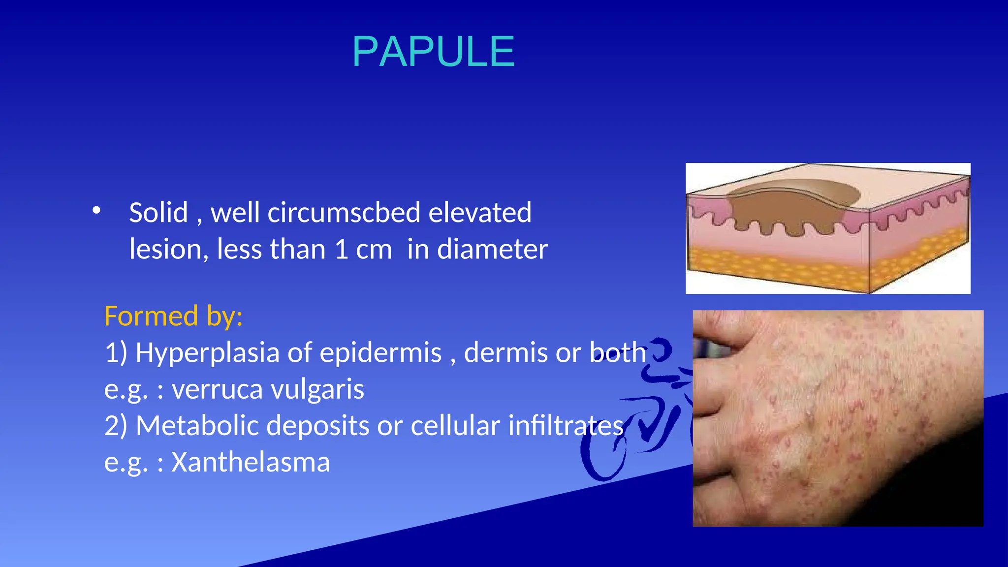
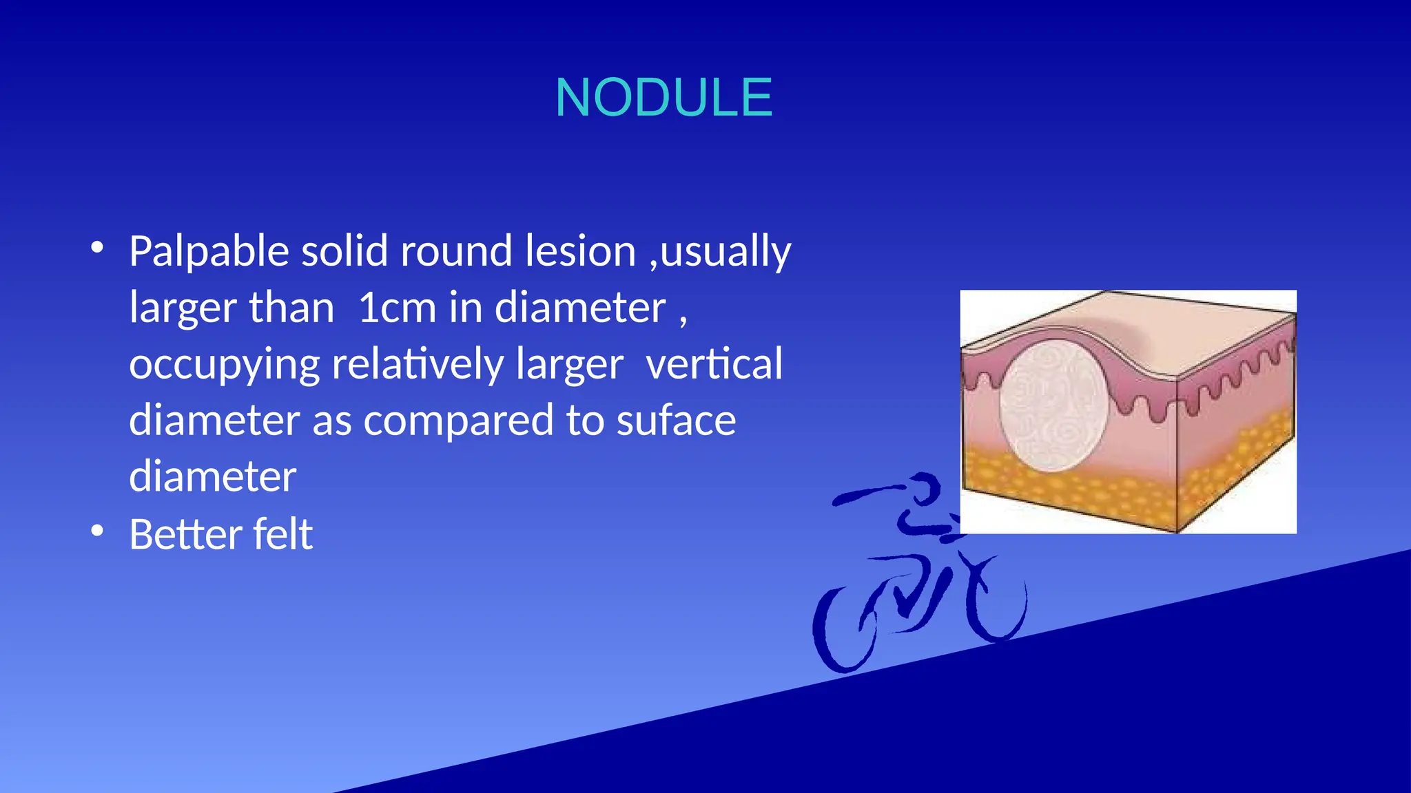
PAPULE
• Solid ,well circumscbed elevated
lesion, less than 1 cm in diameter
Formed by:
1) Hyperplasia of epidermis , dermis or both
e.g. : verruca vulgaris
2) Metabolic deposits or cellular infiltrates
e.g. : Xanthelasma
PLAQUE
•Elevated well circumscribed,more than 1 cm in
diameter ,occupying relatively large surface area
in comparison with its height above the skin
surface
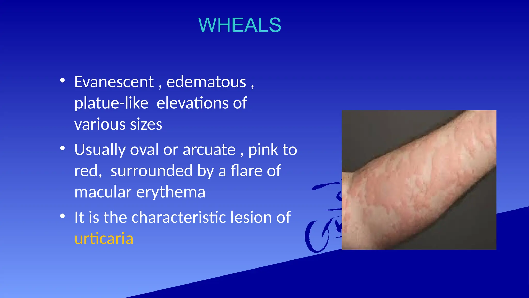
WHEALS
• Evanescent ,edematous ,
platue-like elevations of
various sizes
• Usually oval or arcuate , pink to
red, surrounded by a flare of
macular erythema
• It is the characteristic lesion of
urticaria
32.
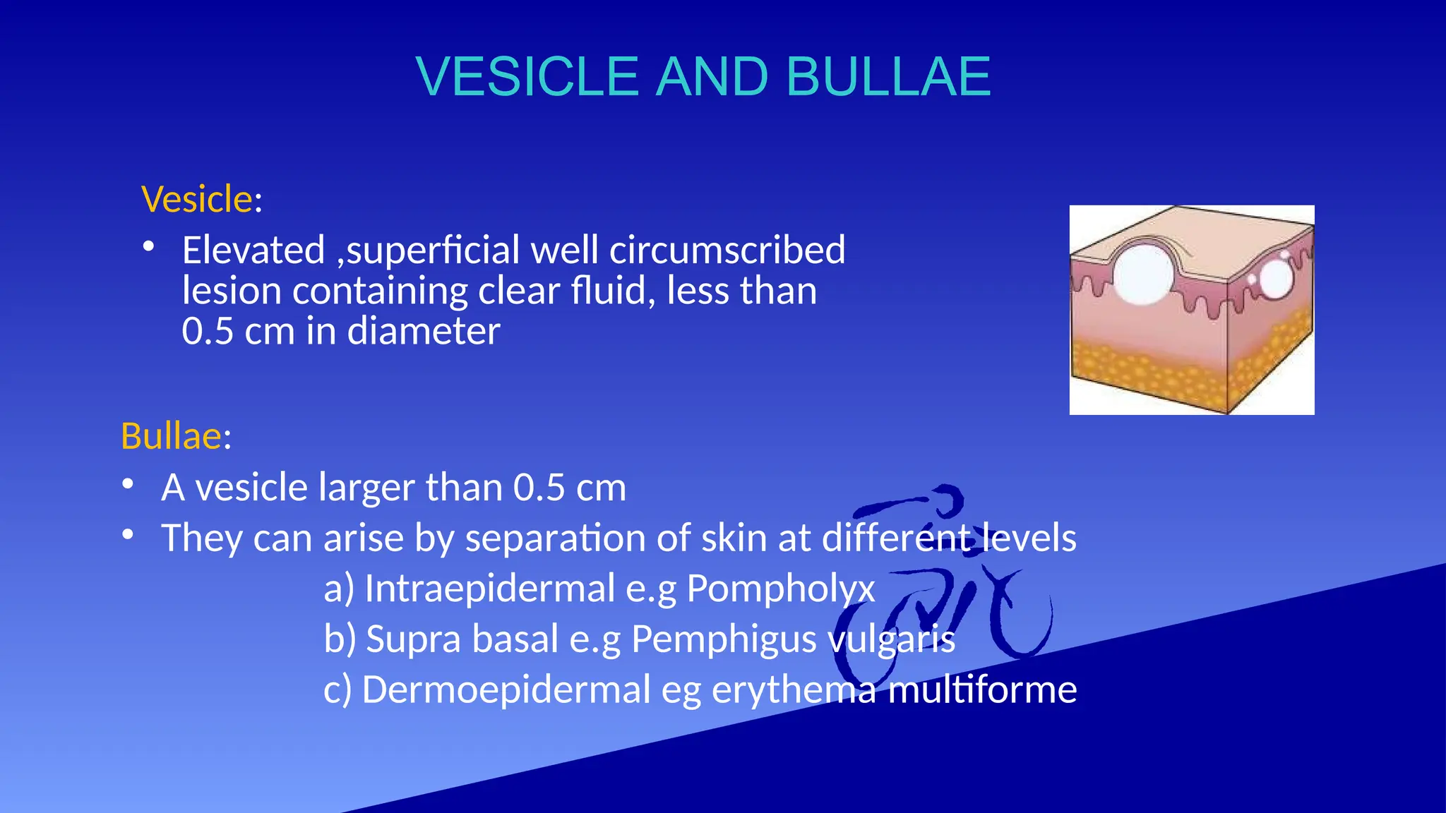
VESICLE AND BULLAE
Vesicle:
•Elevated ,superficial well circumscribed
lesion containing clear fluid, less than
0.5 cm in diameter
Bullae:
• A vesicle larger than 0.5 cm
• They can arise by separation of skin at different levels
a) Intraepidermal e.g Pompholyx
b) Supra basal e.g Pemphigus vulgaris
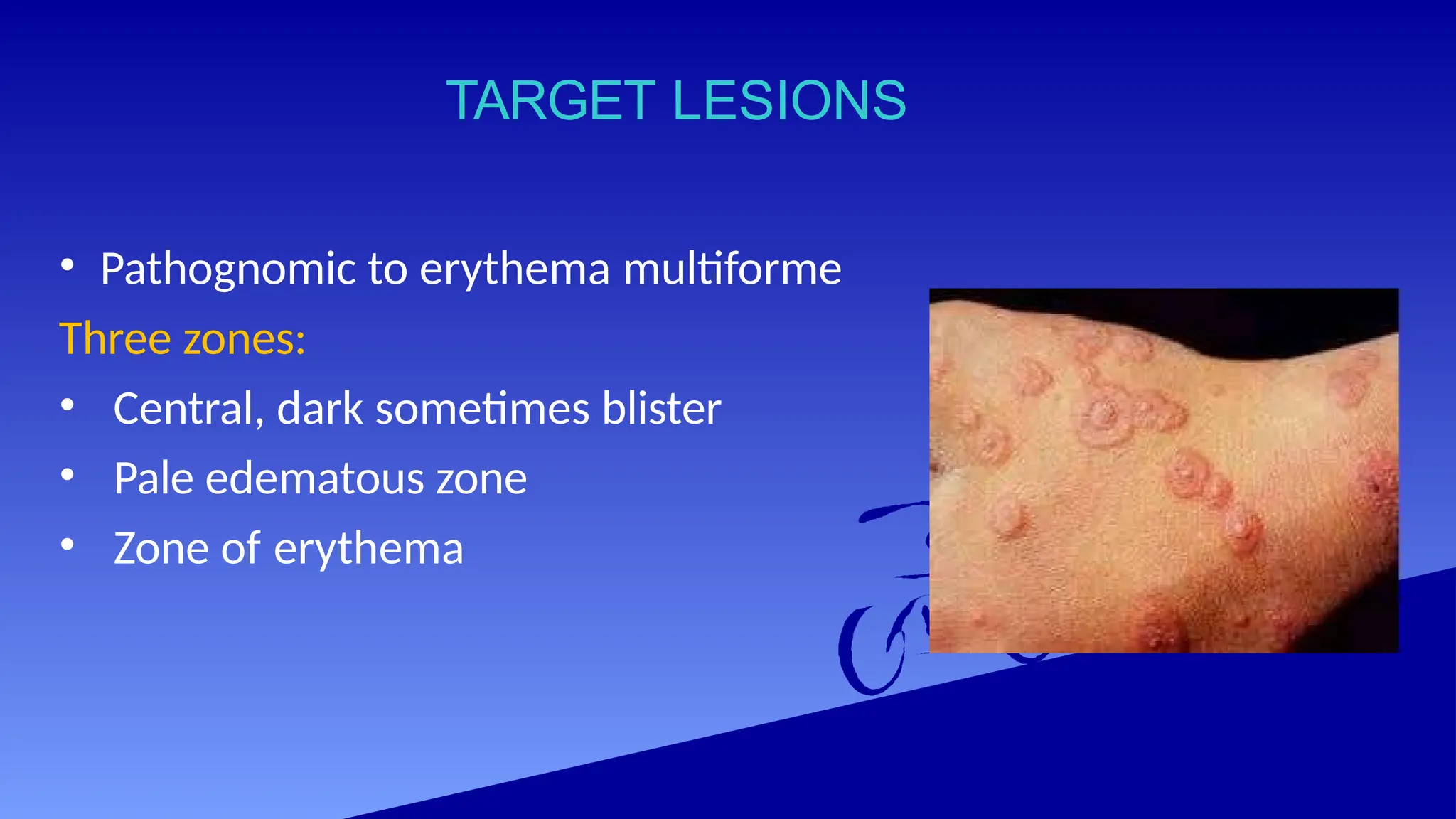
c) Dermoepidermal eg erythema multiforme
33.
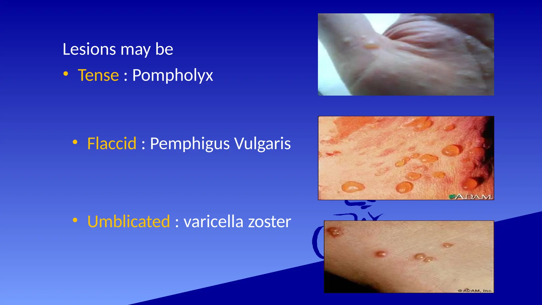
Lesions may be
•Tense : Pompholyx
• Flaccid : Pemphigus Vulgaris
• Umblicated : varicella zoster
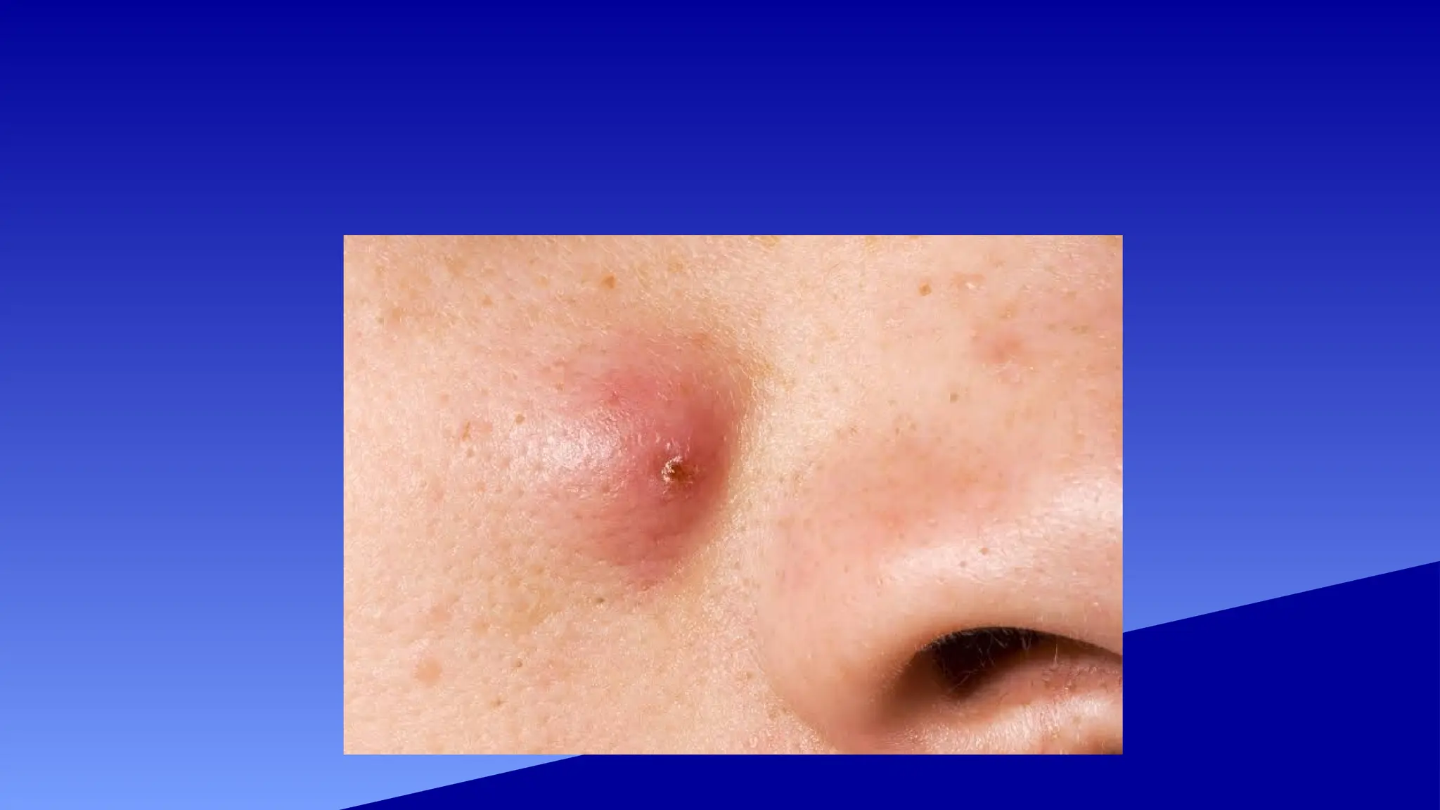
CYST
• A sacthat contains liquid or semisolid material
in a well-defined cavity
Types of cyst:
• Epidermal cyst: lined with squamous
epithelium and produce keratinous material
• Pilar cyst
37.
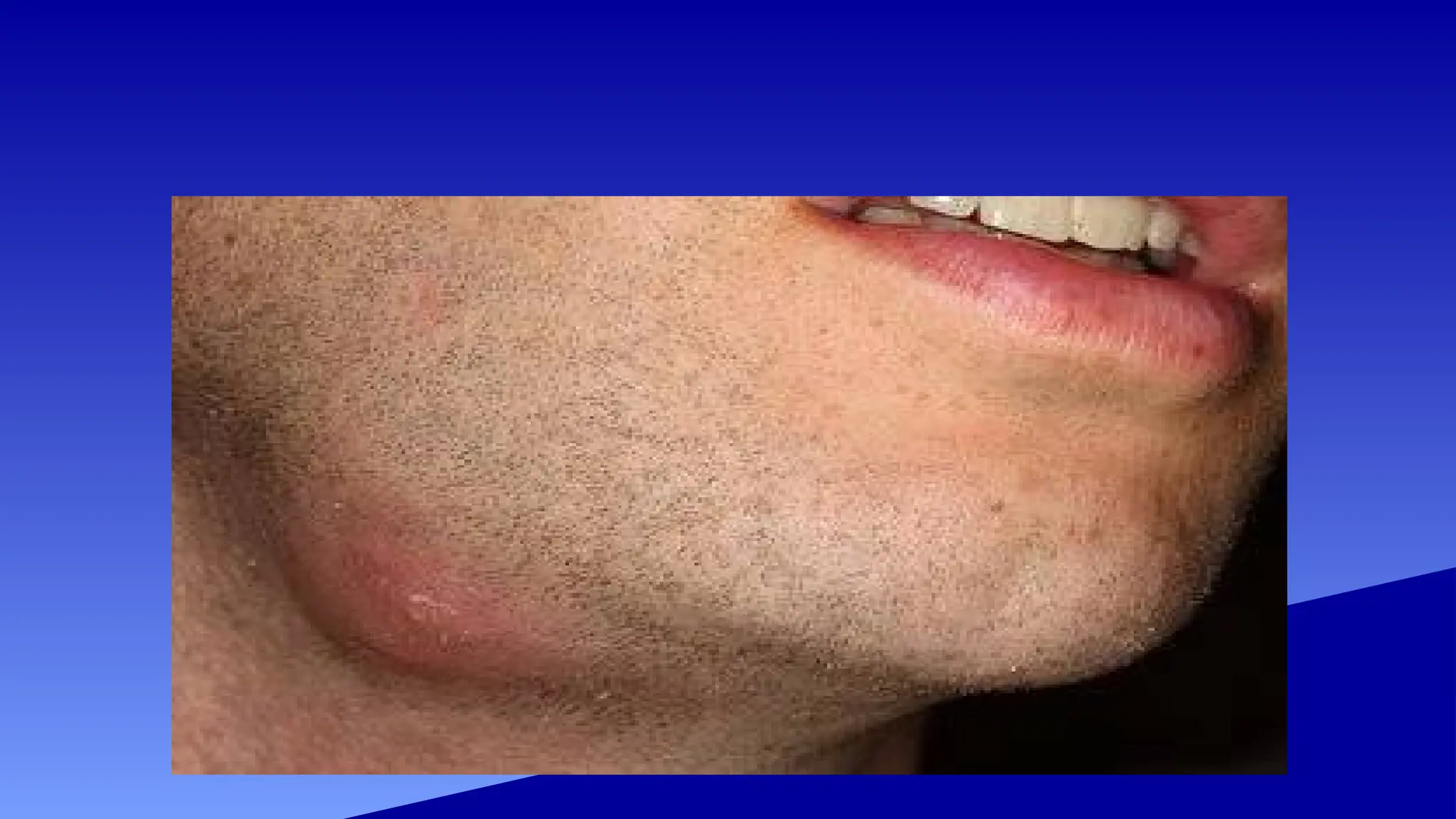
ABSCESS
• An abscessis a collection of pus below the
skin
• Pus in an abscess is invisible but clinically
be interpreted as sign of inflammation in
the overlying skin
• Abscess cavities do not have well-defined
lining as cyst
39.
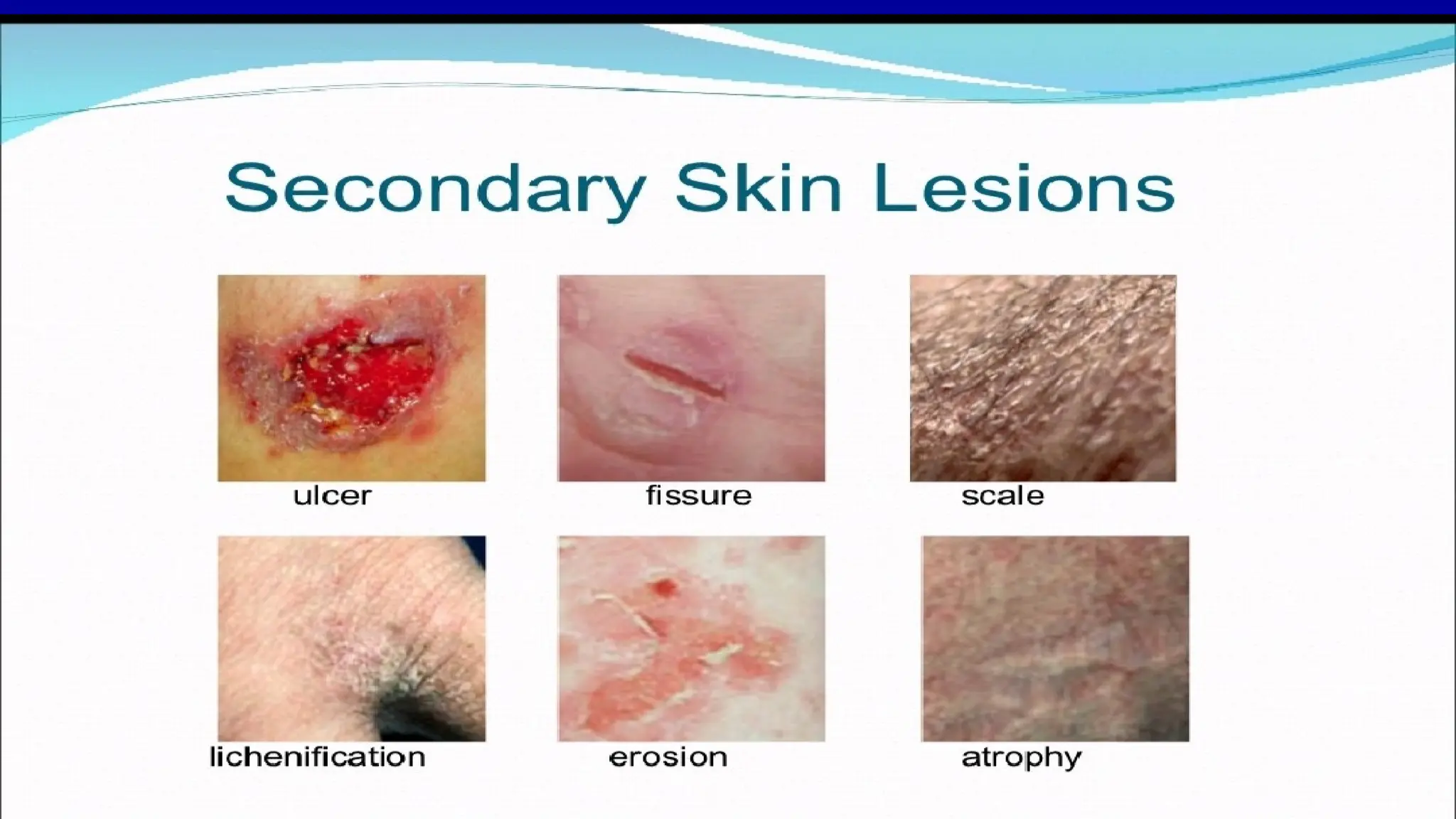
SECONDARY LESIONS
Modification ofprimary skin lesions that result from
traumatic injury , evolution from primary lesion , or other
external factors
• Crust
• Scale
• Erosion
• Ulcer
• Fissure
• Scar
• Atrophy
• Telangiectasia
41.
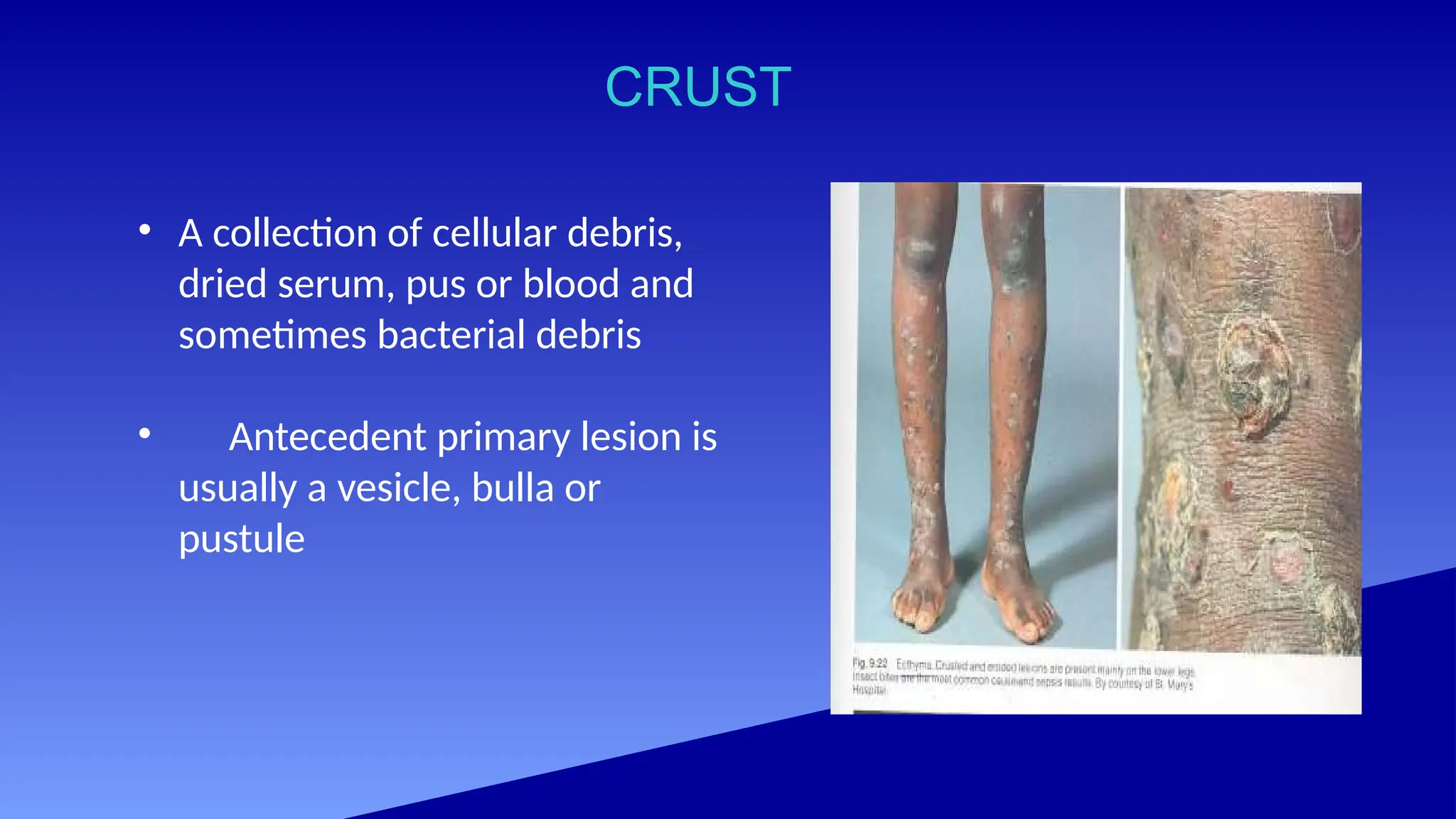
CRUST
• A collectionof cellular debris,
dried serum, pus or blood and
sometimes bacterial debris
• Antecedent primary lesion is
usually a vesicle, bulla or
pustule
42.
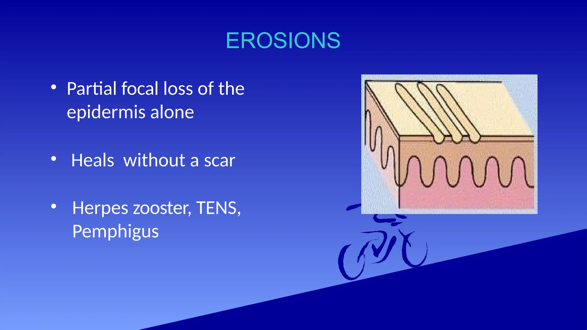
EROSIONS
• Partial focalloss of the
epidermis alone
• Heals without a scar
• Herpes zooster, TENS,
Pemphigus
44.
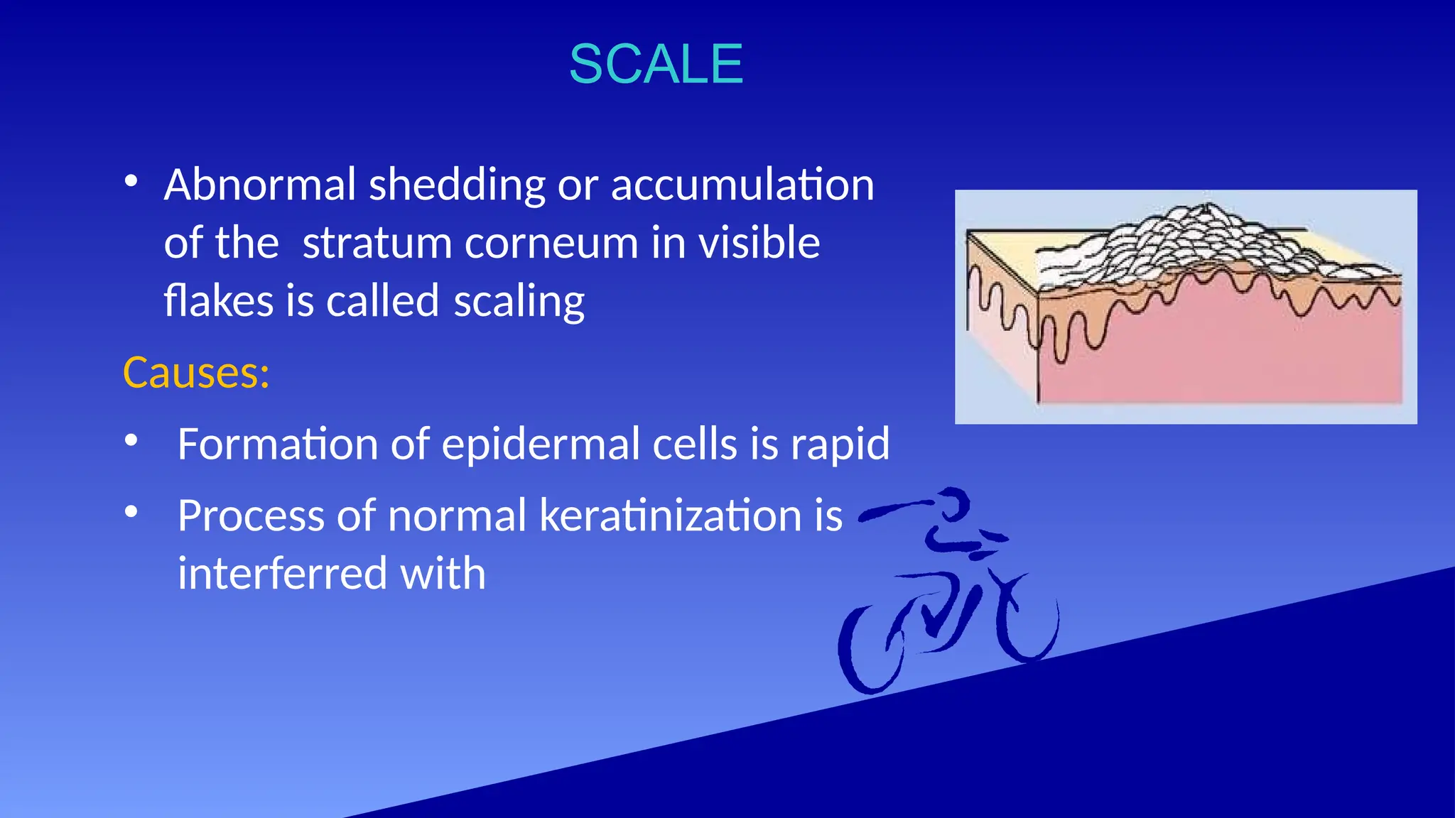
SCALE
• Abnormal sheddingor accumulation
of the stratum corneum in visible
flakes is called scaling
Causes:
• Formation of epidermal cells is rapid
• Process of normal keratinization is
interferred with
45.
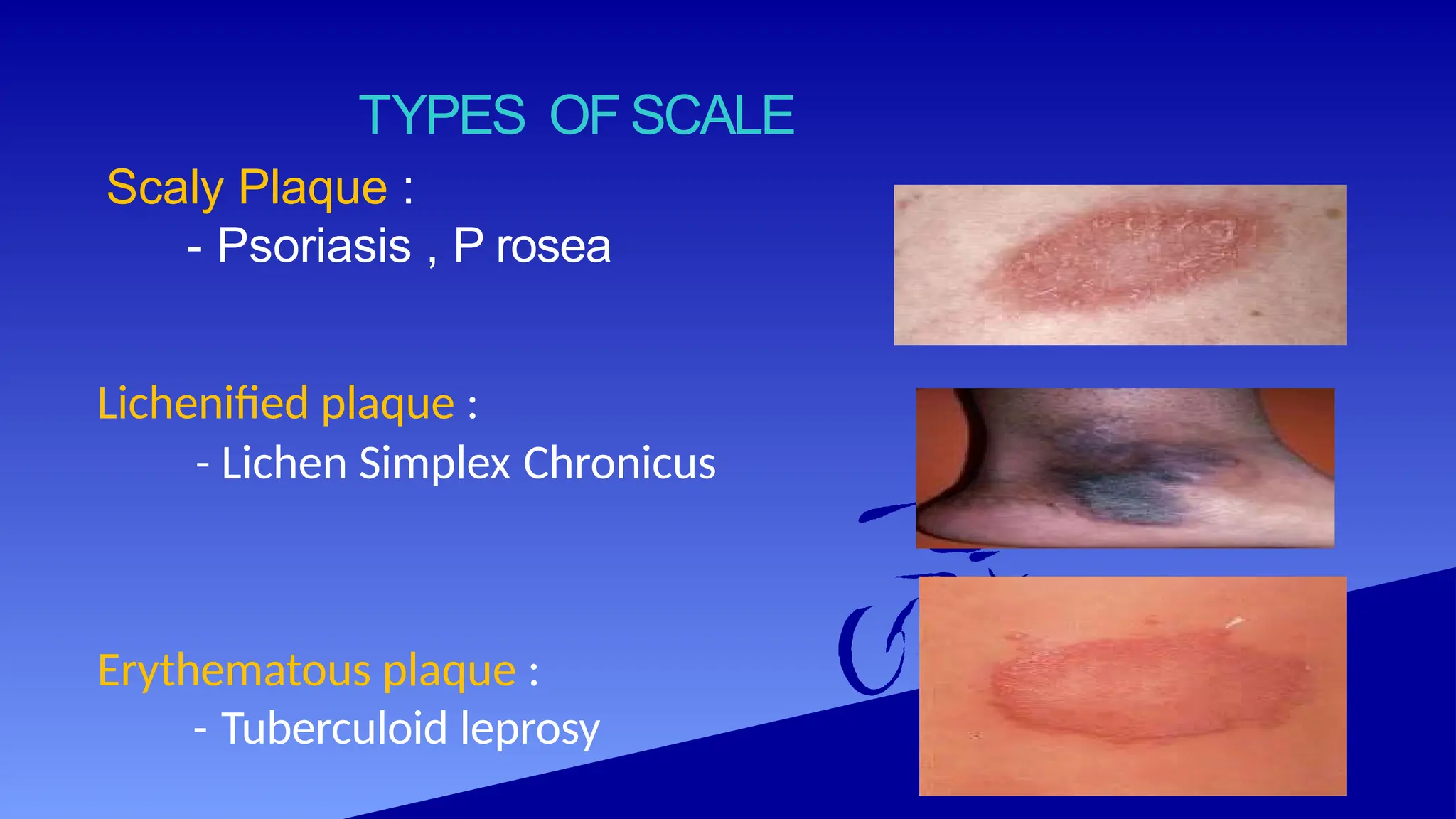
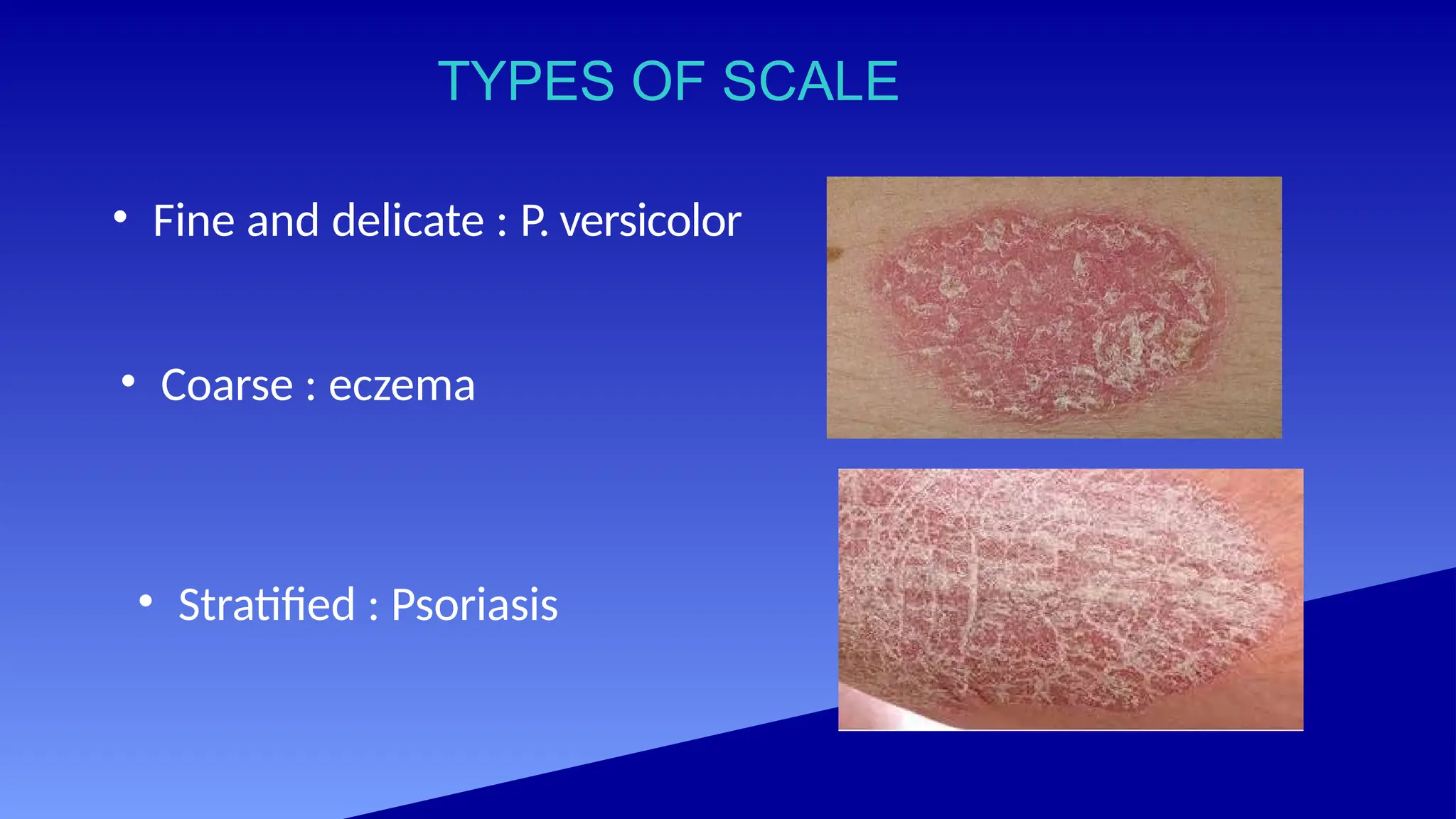
TYPES OF SCALE
•Fine and delicate : P. versicolor
• Coarse : eczema
• Stratified : Psoriasis
46.
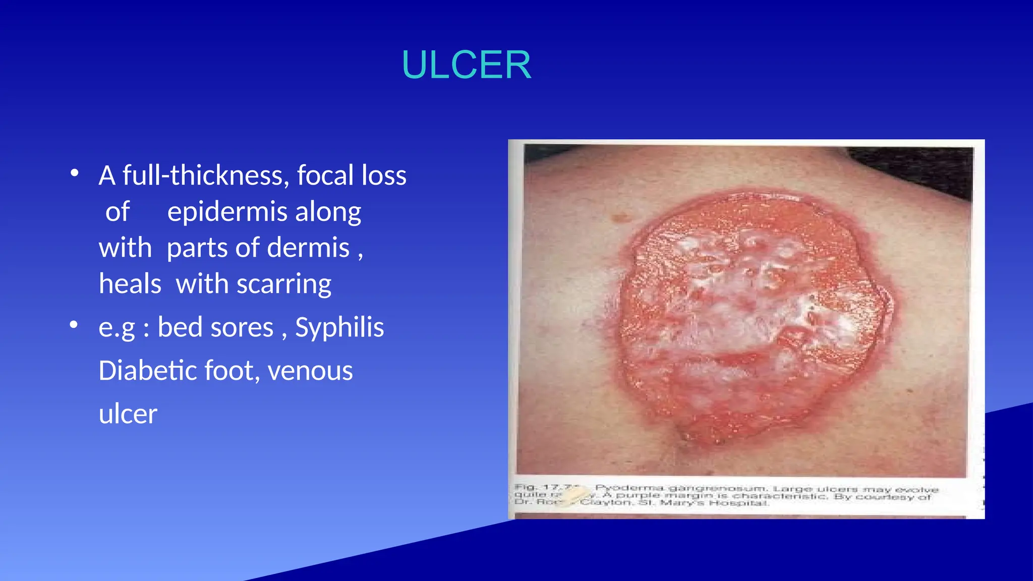
ULCER
• A full-thickness,focal loss
of epidermis along
with parts of dermis ,
heals with scarring
• e.g : bed sores , Syphilis
Diabetic foot, venous
ulcer
47.
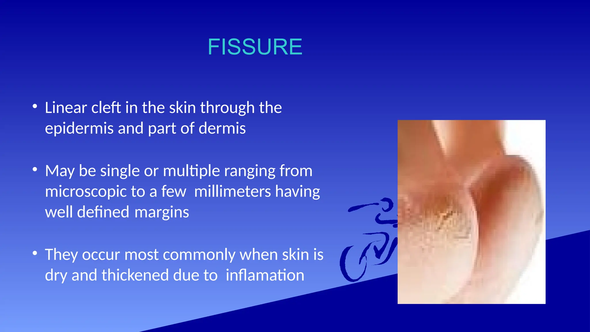
FISSURE
• Linear cleftin the skin through the
epidermis and part of dermis
• May be single or multiple ranging from
microscopic to a few millimeters having
well defined margins
• They occur most commonly when skin is
dry and thickened due to inflamation
48.
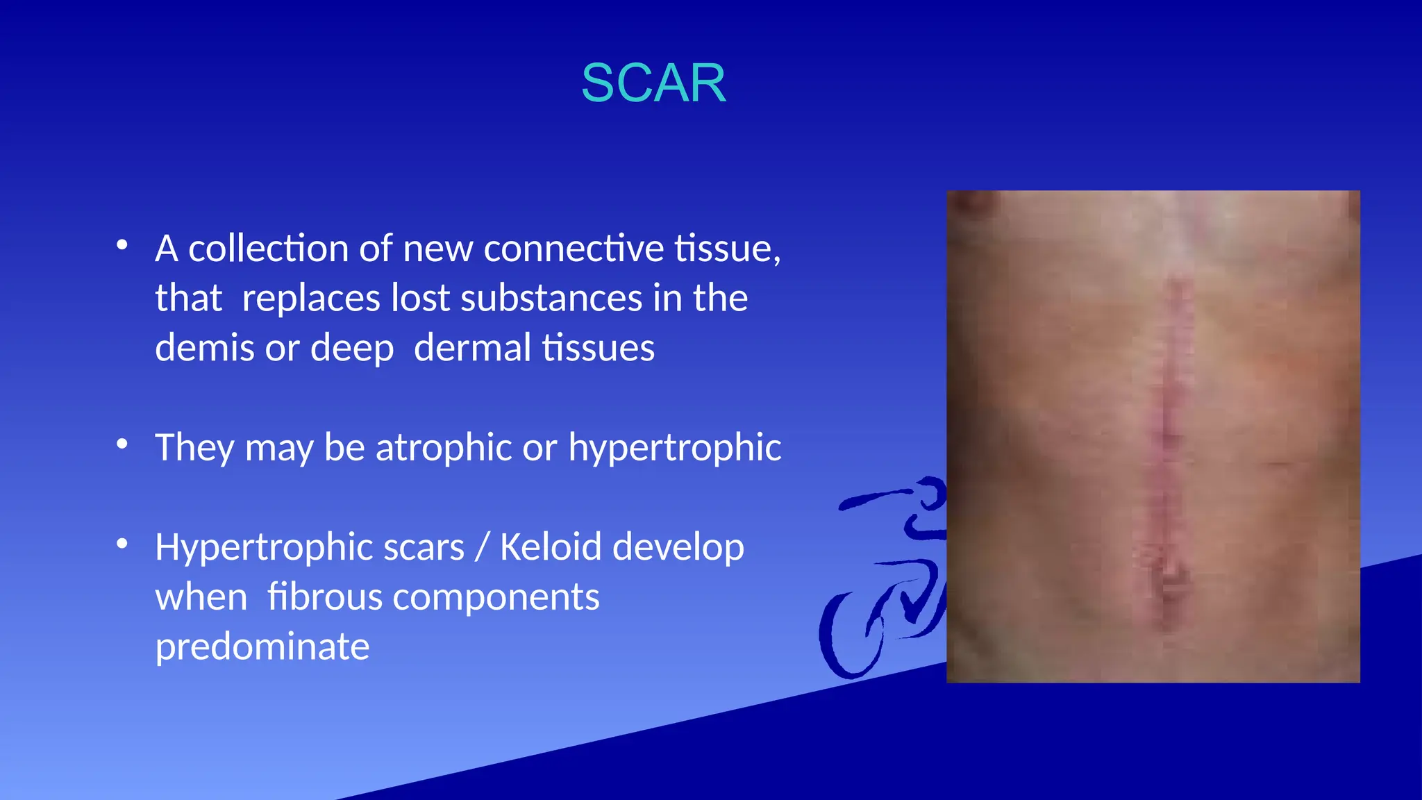
SCAR
• A collectionof new connective tissue,
that replaces lost substances in the
demis or deep dermal tissues
• They may be atrophic or hypertrophic
• Hypertrophic scars / Keloid develop
when fibrous components
predominate
49.
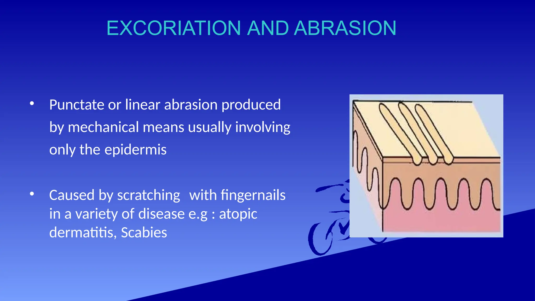
EXCORIATION AND ABRASION
•Punctate or linear abrasion produced
by mechanical means usually involving
only the epidermis
• Caused by scratching with fingernails
in a variety of disease e.g : atopic
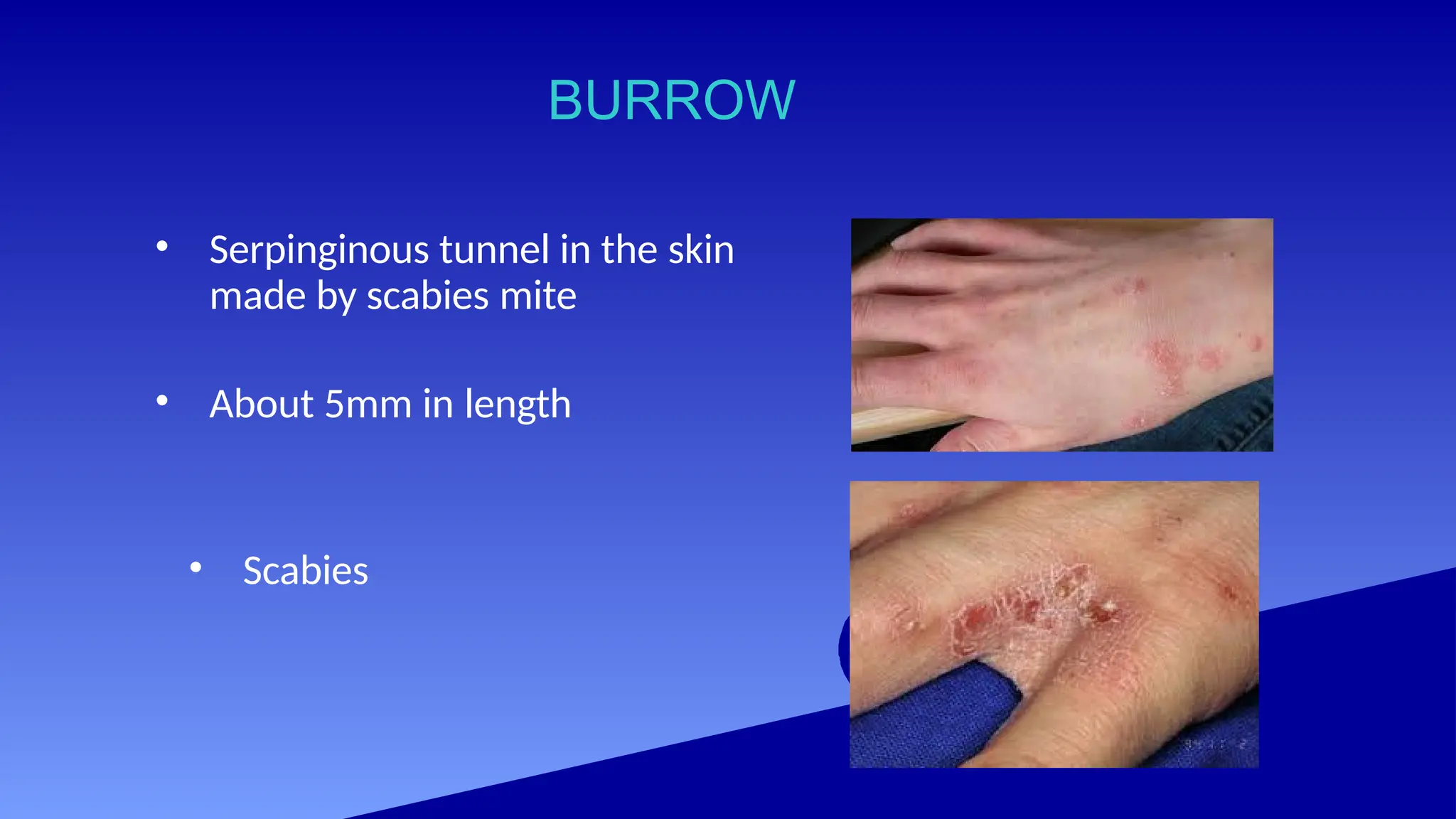
dermatitis, Scabies
50.
ATROPHY
Reduction in thecomponents of a tissue, Organ or
part of body. In the skin
1) Epidermal Atrophy: results from decrease in
epidermal cells .Gives rise to frequently transparent
epidermis and alteration of skin surface i.e. loss of
normal skin lines and fine wrinkling
2) Dermal Atrophy: results from decrease in the
reticular or papillary dermis. Clinically seen as
depression of skin
51.
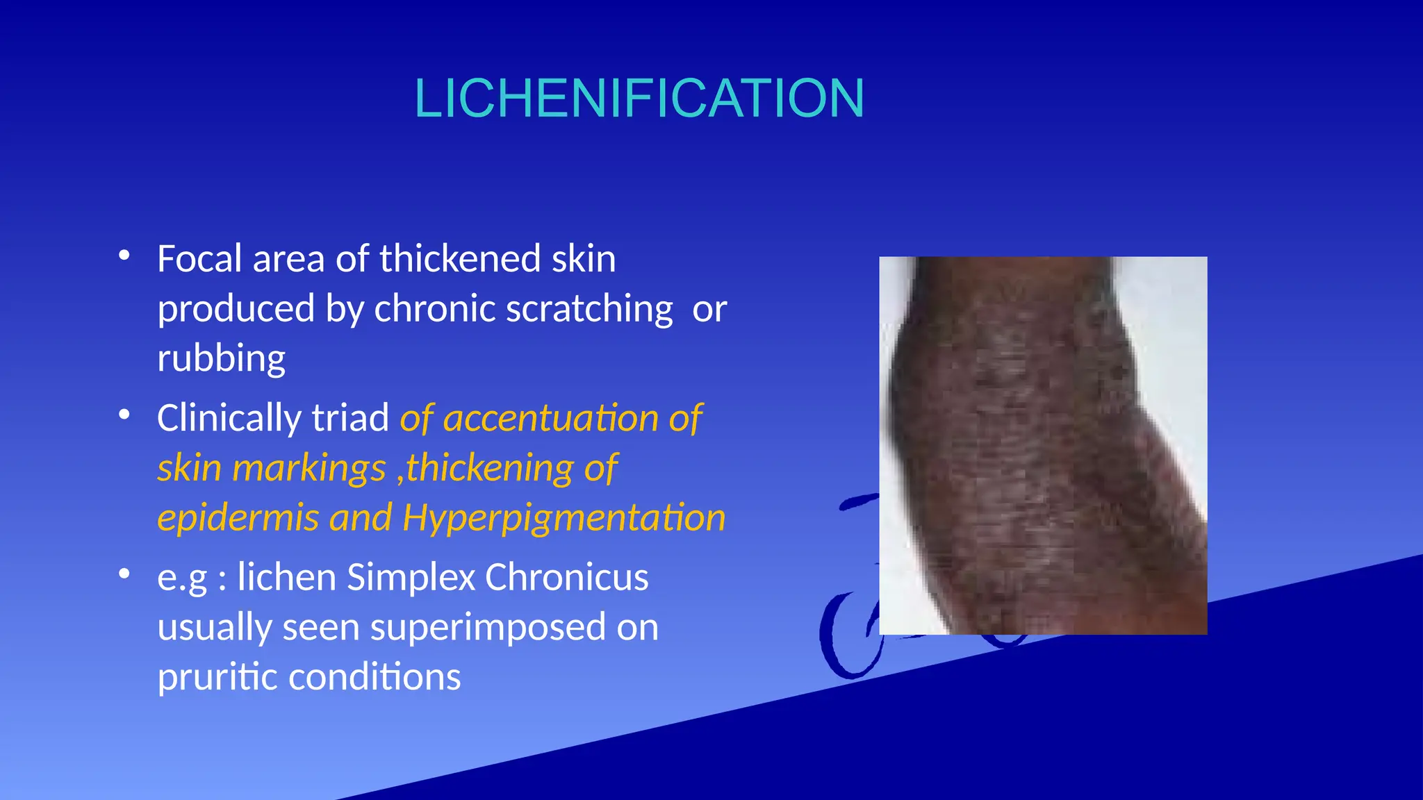
LICHENIFICATION
• Focal areaof thickened skin
produced by chronic scratching or
rubbing
• Clinically triad of accentuation of
skin markings ,thickening of
epidermis and Hyperpigmentation
• e.g : lichen Simplex Chronicus
usually seen superimposed on
pruritic conditions