INTRODUCTION
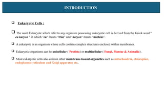
Eukaryotic Cells:
The word Eukaryote which refer to any organism possessing eukaryotic cell is derived from the Greek word "
eu karyon " in which "eu" means "true" and "karyon" means "nucleus".
A eukaryote is an organism whose cells contain complex structures enclosed within membranes.
Eukaryotic organisms can be unicellular ( Protista) or multicellular ( Fungi, Plantae & Animalia) .
Most eukaryotic cells also contain other membrane-bound organelles such as mitochondria, chloroplast,
endoplasmic reticulum and Golgi apparatus etc.
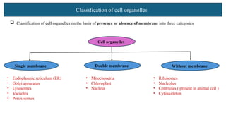
Classification of cellorganelles
Classification of cell organelles on the basis of presence or absence of membrane into three categories
Cell organelles
• Endoplasmic reticulum (ER)
• Golgi apparatus
• Lysosomes
• Vacuoles
• Peroxisomes
• Mitochondria
• Chloroplast
• Nucleus
• Ribosomes
• Nucleolus
• Centrioles ( present in animal cell )
• Cytoskeleton
Single membrane Double membrane Without membrane
5.
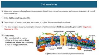
Cell membrane
Outermostboundary of cytoplasm which separates the cell from external environment and controls the entrance & exit of
molecules or ions.
It has highly selective permeable.
Several types of models have been put forward to explain the structure of cell membrane.
The most accepted model explaining the structure of cell membrane is fluid mosaic model, proposed by Singer and
Nicolson in 1972.
Functions:
- Play important role in various
cellular activities, such as signal
transduction, membrane trafficking
as well as energy conversion.
Figure 2: Fluid mosaic model of plasma membrane
6.
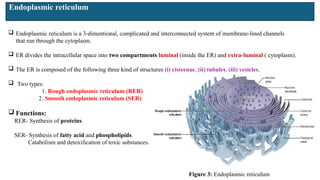
Endoplasmic reticulum
Endoplasmicreticulum is a 3-dimentional, complicated and interconnected system of membrane-lined channels
that run through the cytoplasm.
ER divides the intracellular space into two compartments luminal (inside the ER) and extra-luminal ( cytoplasm).
The ER is composed of the following three kind of structures (i) cisternae, (ii) tubules, (iii) vesicles.
Two types:
1. Rough endoplasmic reticulum (RER)
2. Smooth endoplasmic reticulum (SER)
Functions:
RER- Synthesis of proteins.
SER- Synthesis of fatty acid and phospholipids.
Catabolism and detoxification of toxic substances.
iii
Figure 3: Endoplasmic reticulum
7.
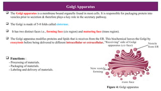
Golgi Apparatus
TheGolgi apparatus is a membrane bound organelle found in most cells. It is responsible for packaging protein into
vesicles prior to secretion & therefore plays a key role in the secretary pathway.
The Golgi is made of 5-8 folds called cisternae.
It has two distinct faces i.e., forming face (cis region) and maturing face (trans region).
The Golgi apparatus modifies proteins and lipids that is receives from the ER. This biochemical leaves the Golgi by
exocytosis before being delivered to different intracellular or extracellular.
Functions :
- Processing of materials.
- Packaging of materials.
- Labeling and delivery of materials.
Figure 4: Golgi apparatus
8.
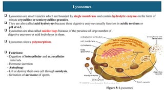
Lysosomes
Lysosomes aresmall vesicles which are bounded by single membrane and contain hydrolytic enzymes in the form of
minute crystalline or semicrystalline granules.
They are also called acid hydrolyses because these digestive enzymes usually function in acidic medium or
pH of 4-5.
Lysosomes are also called suicide bags because of the presence of large number of
digestive enzymes or acid hydrolyses in them.
Lysosomes shows polymorphism.
Functions:
- Digestion of intracellular and extracellular
materials
- Hormone secretion
- Autophagy
- kill or destroy their own cell through autolysis.
- formation of acrosome of sperm.
Figure 5: Lysosomes
9.
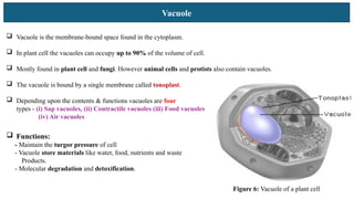
Vacuole
Figure 6: Vacuoleof a plant cell
Vacuole is the membrane-bound space found in the cytoplasm.
In plant cell the vacuoles can occupy up to 90% of the volume of cell.
Mostly found in plant cell and fungi. However animal cells and protists also contain vacuoles.
The vacuole is bound by a single membrane called tonoplast.
Depending upon the contents & functions vacuoles are four
types - (i) Sap vacuoles, (ii) Contractile vacuoles (iii) Food vacuoles
(iv) Air vacuoles
Functions:
- Maintain the turgor pressure of cell
- Vacuole store materials like water, food, nutrients and waste
Products.
- Molecular degradation and detoxification.
10.
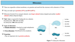
Ribosomes
These areorganelles without membrane, are granular and small dot like structure with a diameter of 15nm.
They are made up to proteins (15%) and RNA (65%).
Ribosome formed of two unequal subunits- one larger subunit (dome shaped) and another smaller
subunit (oblate-ellipsoid shape).
Mg2+ ions is required for binding the two subunits.
-In prokaryotes- 70S (50S and 30S)
-In eukaryotes- 80S (60S and 40S)
Types of ribosomes:
(I) Bound ribosomes- Attached to rough endoplasmic
reticulum.
(ii) Free ribosomes- Distributed in the cytoplasm.
Function:
- Play an important role in protein synthesis.
Figure 7: Eukaryotic ribosomes
11.
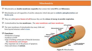
Mitochondria
Mitochondria aredouble-membrane organelles that contain their own DNA and Ribosomes.
Mitochondria are cell organelles of aerobic eukaryotes which take part in oxidative phosphorylation and
Krebs cycle.
They are called power houses of cell because they are the release of energy in aerobic respiration.
A mitochondrion has two membranes : The outer membrane and inner membrane.
The inner membrane of mitochondria has many folds and
form layered structure called cristae.
Functions:
- Production of energy.
- Synthesis of ATP.
- help in building certain part of blood & hormones
like testosterone and estrogen.
- Play important role in the process of apoptosis.
- Store and release of calcium.
Figure 8: Mitochondria
12.
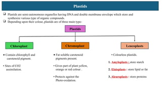
Plastids
Plastids aresemi-autonomous organelles having DNA and double membrane envelope which store and
synthesize various type of organic compounds.
Depending upon their colour, plastids are of three main type-
Plastids
Chloroplast Chromoplast Leucoplasts
• Contain chlorophyll and
carotenoid pigment.
• Sites of CO2
assimilation.
• Fat soluble carotenoid
pigments present.
• Gives part of plant yellow,
orange or red colour .
• Protects against the
Photo-oxidation.
• Colourless plastids.
1. Amyloplasts : store starch
2. Elaioplasts : store lipid or fat
3. Aleuroplasts : store proteins
13.
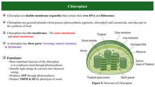
Chloroplast
Functions:
- Mostimportant function of the chloroplast
is to synthesize food through photosynthesis.
- Absorbs light energy & converts into chemical
energy .
- Produces ATP through photosynthesis.
- Produce NDPH & O2 by photolysis of water.
Chloroplast are double-membrane organelles that contain their own DNA and Ribosomes.
Chloroplasts are greenish plastids which possess photosynthetic pigments, chlorophyll and carotenoids, and take part in
the synthesis of food.
Chloroplasts has two membranes : The outer membrane
and inner membrane.
A chloroplast has three parts- Envelope, matrix (stroma),
& thylakoids.
Figure 9: Structure of Chloroplast
14.
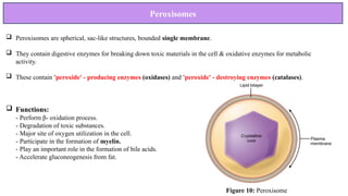
Peroxisomes
Peroxisomes arespherical, sac-like structures, bounded single membrane.
They contain digestive enzymes for breaking down toxic materials in the cell & oxidative enzymes for metabolic
activity.
These contain 'peroxide' - producing enzymes (oxidases) and 'peroxide' - destroying enzymes (catalases).
Functions:
- Perform β- oxidation process.
- Degradation of toxic substances.
- Major site of oxygen utilization in the cell.
- Participate in the formation of myelin.
- Play an important role in the formation of bile acids.
- Accelerate gluconeogenesis from fat.
Figure 10: Peroxisome
15.
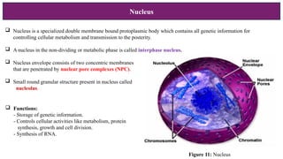
Nucleus
Nucleus isa specialized double membrane bound protoplasmic body which contains all genetic information for
controlling cellular metabolism and transmission to the posterity.
A nucleus in the non-dividing or metabolic phase is called interphase nucleus.
Nucleus envelope consists of two concentric membranes
that are penetrated by nuclear pore complexes (NPC).
Small round granular structure present in nucleus called
nucleolus.
Functions:
- Storage of genetic information.
- Controls cellular activities like metabolism, protein
synthesis, growth and cell division.
- Synthesis of RNA.
Figure 11: Nucleus
16.
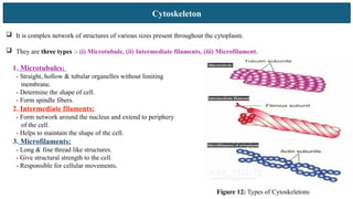
Cytoskeleton
It iscomplex network of structures of various sizes present throughout the cytoplasm.
They are three types :- (i) Microtubule, (ii) Intermediate filaments, (iii) Microfilament.
1. Microtubules:
- Straight, hollow & tubular organelles without limiting
membrane.
- Determine the shape of cell.
- Form spindle fibers.
2. Intermediate filaments:
- Form network around the nucleus and extend to periphery
of the cell.
- Helps to maintain the shape of the cell.
3. Microfilaments:
- Long & fine thread like structures.
- Give structural strength to the cell.
- Responsible for cellular movements.
Figure 12: Types of Cytoskeletons