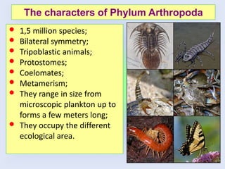
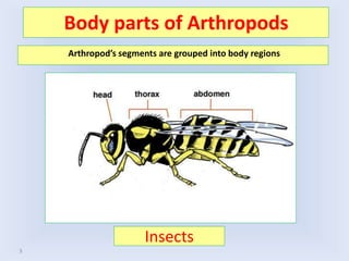
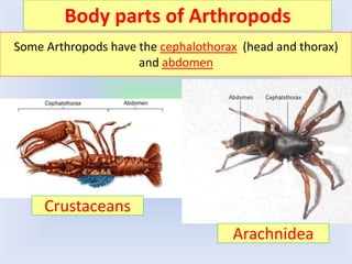
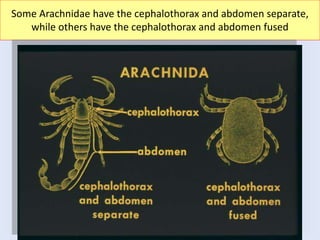
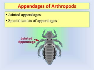
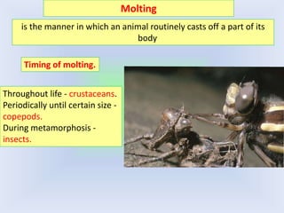
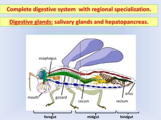
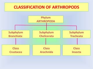
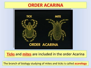
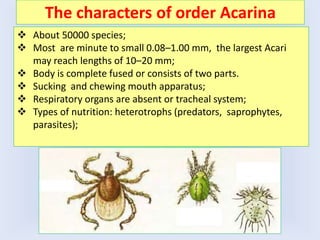
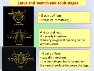
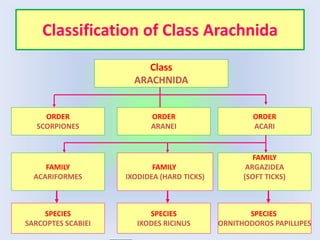
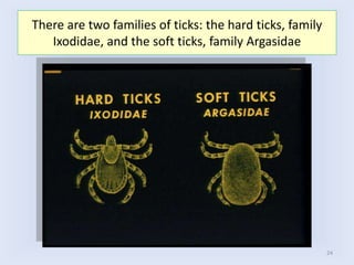
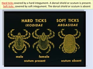
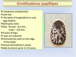
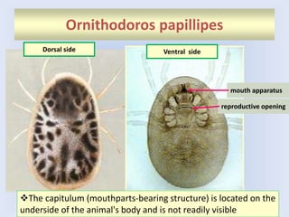
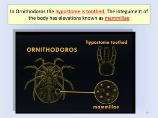
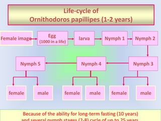
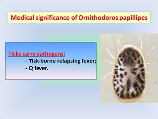
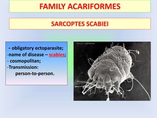
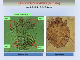
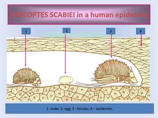
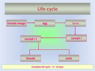
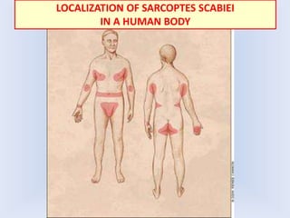
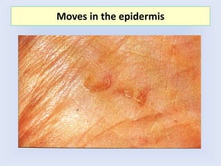
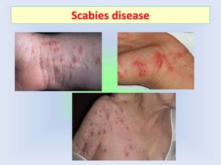
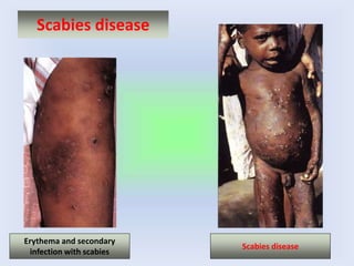
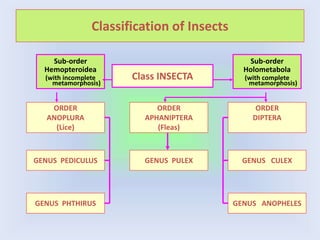
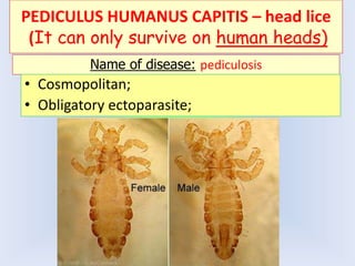
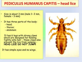
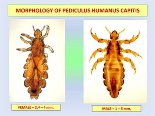
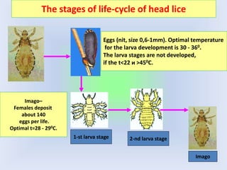
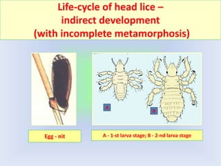
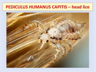
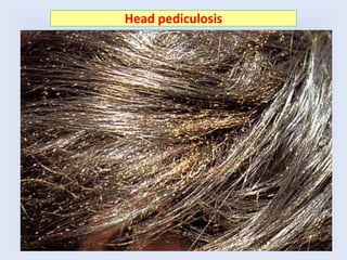
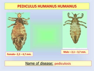
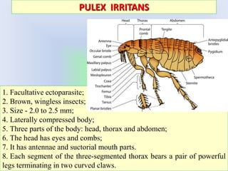
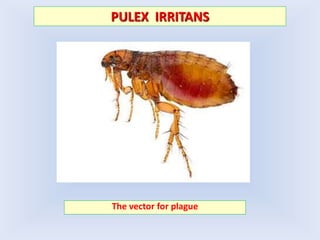
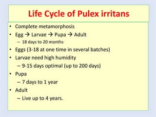
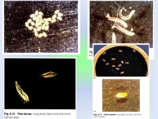
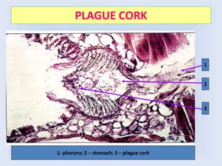
Kursk State Medical University
department of biology, medical genetics and ecology
Lecture 5
Medical Arachnoentomology.
Phylum Arthropoda.
Yelena Novikova, PhD of Biology,
Associate Professor
• 1,5 million species;
• Bilateral symmetry;
• Tripoblastic animals;
• Protostomes;
• Coelomates;
• Metamerism;
• They range in size from
microscopic plankton up to
forms a few meters long;
• They occupy the different
ecological area.
The characters of Phylum Arthropoda
Insects
3
Body parts of Arthropods
Arthropod’s segments are grouped into body regions
Body parts of Arthropods
Some Arthropods have the cephalothorax (head and thorax)
and abdomen
Crustaceans
Arachnidea
5
Some Arachnidae have the cephalothorax and abdomen separate,
while others have the cephalothorax and abdomen fused
Appendages of Arthropods
• Jointed appendages
• Specialization of appendages
Exoskeleton
• Exoskeleton- body covered with a hard external
skeleton (chitin)
• Exoskeleton good for small things, protects body
from damage (rainfall, falling, etc.).
• Striated muscles are attached to the exoskeleton
is the manner in which an animal routinely casts off a part of its
body
Molting
Timing of molting.
Throughout life - crustaceans.
Periodically until certain size -
copepods.
During metamorphosis -
insects.
Complete digestive system with regional specialization.
mouth
esophagus
gizzard
cecum rectum
anus
foregut midgut hindgut
Digestive glands: salivary glands and hepatopancreas.
Open Circulatory System
Open hemocoel as a result of hard exoskeleton and lack of internal
segmentation.
Muscular heart required since body movements can’t move blood.
Hemolymph has amebocytes, pigments, and some have clotting
factors.
RESPIRATORY SYSTEM
Respiratory structures depend on habitat.
Gills in aquatic animals.
Arachnid book lung.
Trachea in terrestrial insects.
EXCRETORY SYSTEM
Malpighian tubules in
arachnids and insects.
Blind tubes extend into
hemocoel and empty into
gut.
Malpighian tubules
Antennal and maxillary
glands in crustaceans.
Produce ammonia with
some urea and uric
acid.
NERVOUS SYSTEM OF ARTHROPODS
Primitive brain and Ventral Nerve Cord
General
• most arthropods are gonochoric
• fertilization may be external or internal in aquatic arthropods
• always internal in terrestrial species
Ontogenesis:
• Direct and indirect development (with complete and
incomplete metamorphosis).
REPRODUCTION AND ONTOGENESIS
CLASSIFICATION OF ARTHROPODS
Phylum
ARTHROPODA
Subphylum
Branchiata
Subphylum
Chelicerata
Subphylum
Tracheata
Class
Crustacea
Class
Insecta
Class
Arachnida
Medical Arachnology
ORDER ACARINA
The branch of biology studying of mites and ticks is called acarology
Ticks and mites are included in the order Acarina
The characters of order Acarina
 About 50000 species;
 Most are minute to small 0.08–1.00 mm, the largest Acari
may reach lengths of 10–20 mm;
 Body is complete fused or consists of two parts.
 Sucking and chewing mouth apparatus;
 Respiratory organs are absent or tracheal system;
 Types of nutrition: heterotrophs (predators, saprophytes,
parasites);
Sucking mouth apparatus
hypostome
It consists of a central hypostome, usually bearing recurved teeth, on
each side a chelicera with cutting digits, and palps.
chelicera
palps
Sucking mouth apparatus of tick
hypostome
21
There are four stages in the life history of a tick: eggs, larva,
nymph and adult (complete metamorphosis).
Some ticks have but one nymphal stage, while others have
several nymphal stages.
The stages of the life-cycle
22
- 3 pairs of legs;
- sexually immature;
Larva and nymph and adult stages
 4 pairs of legs;
 sexually immature;
 having no genital opening on the
ventral surface.
- 4 pairs of legs;
- sexually immature;
- the genital opening is located on
the ventral surface between the legs.
Classification of Class Arachnida
Class
ARACHNIDA
ORDER
SCORPIONES
ORDER
ARANEI
ORDER
ACARI
FAMILY
IXODIDEA (HARD TICKS)
FAMILY
ACARIFORMES
FAMILY
ARGAZIDEA
(SOFT TICKS)
SPECIES
SARCOPTES SCABIEI
SPECIES
IXODES RICINUS
SPECIES
ORNITHODOROS PAPILLIPES
24
There are two families of ticks: the hard ticks, family
Ixodidae, and the soft ticks, family Argasidae
25
Hard ticks covered by a hard integument. A dorsal shield or scutum is present.
Soft ticks covered by soft integument. The dorsal shield or scutum is absent
26
Hard ticks have the spiracular plates located behind the 4th leg.
Soft ticks have the spiracular plates located behind the 3rd leg.
FAMILY IXODIDEA (HARD TICKS)
Morphology of Ixodes ricinus
Female, 3-4 mm
Male, 2,5 mm
Scutum
Female of Ixodes ricinus after feeding
1
2
1- scutum; 2 – mouth apparatus.
LIFE-CYCLE OF IXODES RICINUS
Feeding of female
MEDICAL SIGNIFICANCE OF IXODES TICKS
Ticks vectors of pathogens:
A) rickettsioses:
-Marseilles fever;
-Q fever;
-borne typhus in Central Asia;
B) viral infections:
- Spring-summer encephalitis
- Hemorrhagic fever;
- Scottish sheep encephalitis;
B) bacterial infections:
-tularemia;
-brucellosis;
-plague.
FAMILY ARGASIDAE (SOFT TICKS)
Ornithodoros papillipes
 temporary ectoparasite;
soft tick;
 the body of longitudinal or oval,
egg-shaped;
dark-grey color;
Size : female - 8,2 mm,
male— 5,8 mm;
4 pairs of legs;
 eyes are reduced;
characteristic welt on the edge
of the body;
sexual dimorphism is weak;
able to starve up to 11-13 years;
Ornithodoros papillipes
mouth apparatus
reproductive opening
Dorsal side Ventral side
The capitulum (mouthparts-bearing structure) is located on the
underside of the animal's body and is not readily visible
36
In Ornithodoros the hypostome is toothed. The integument of
the body has elevations known as mammillae
Life-cycle of
Ornithodoros papillipes (1-2 years)
Female imago
Egg
(1000 in a life)
larva Nymph 1 Nymph 2
Nymph 3
Nymph 4
female male
Nymph 5
female
female male
male
Because of the ability for long-term fasting (10 years)
Ticks carry pathogens:
- Tick-borne relapsing fever;
- Q fever.
Medical significance of Ornithodoros papillipes
FAMILY ACARIFORMES
SARCOPTES SCABIEI
- obligatory ectoparasite;
-name of disease – scabies;
- cosmopolitan;
-Transmission:
person-to-person.
SARCOPTES SCABIEI (female)
Ventral side Dorsal side
size: 0,3 – 0,4 х 0,2 – 0,3 mm
legs legs
legs
legs
Mouth apparatus
SARCOPTES SCABIEI in a human epidermis
1 3
2 4
1- male; 2- egg; 3 - female; 4 – epidermis.
Life-cycle
Female imago
male
female
nymph I I
larva
nymph I
egg
Complete life-cycle - 9 – 14 days
LOCALIZATION OF SARCOPTES SCABIEI
IN A HUMAN BODY
Moves in the epidermis
Scabies disease
Scabies disease
Erythema and secondary
infection with scabies
Scabies disease
MEDICAL ENTOMOLOGY
Classification of Insects
Class INSECTA
ORDER
ANOPLURA
(Lice)
GENUS PULEX
GENUS PEDICULUS
ORDER
APHANIPTERA
(Fleas)
ORDER
DIPTERA
GENUS PHTHIRUS
GENUS CULEX
GENUS ANOPHELES
Sub-order
Hemopteroidea
(with incomplete
metamorphosis)
Sub-order
Holometabola
(with complete
metamorphosis)
ORDER
ANOPLURA
PEDICULUS HUMANUS CAPITIS – head lice
(It can only survive on human heads)
Name of disease: pediculosis
• Cosmopolitan;
• Obligatory ectoparasite;
• Size is about 3 mm (male 1– 2 mm,
female – 3 mm)
• It has three parts of the body:
- head;
- thorax;
- abdomen;
It has 6 legs with strong claws
which are designed for holding
tightly onto hair - these claws also
prevent the lice from jumping -
HEAD LICE DO NOT JUMP!!
It has simple eyes and no wings.
PEDICULUS HUMANUS CAPITIS – head lice
MORPHOLOGY OF PEDICULUS HUMANUS CAPITIS
FEMALE – 2,4 – 4 mm. MALE – 1 – 3 mm.
The stages of life-cycle of head lice
Imago–
Females deposit
about 140
eggs per life.
Optimal t=28 - 290С.
1-st larva stage 2-nd larva stage
Imago
Eggs (nit, size 0,6-1mm). Optimal temperature
for the larva development is 30 - 360.
The larva stages are not developed,
if the t<22 и >450С.
Life-cycle of head lice –
indirect development
(with incomplete metamorphosis)
Egg - nit A - 1-st larva stage; B - 2-nd larva stage
А
B
PEDICULUS HUMANUS CAPITIS – head lice
Head pediculosis
Pediculosis eyelashes
Head lice eggs on hair and head louse on fingernail
Photo courtesy of Foundation Studios
A head louse using its powerful claws to grasp a hair shaft
Source: Sally Speare and Rick Speare ©, School of Public Health and Tropical Medicine, James
Cook University, Townsville, 1998.
• Size is about 3,5 mm
(male 2-3 mm, female –
4 mm)
• It has 6 legs,
• It is tan to grayish-white.
• If separated from their
hosts, lice die at room
temperature.
PEDICULUS HUMANUS HUMANUS – body louse
PEDICULUS HUMANUS HUMANUS
Female– 2,2 – 4,7 mm.
Male – 2,1 – 3,7 mm.
Name of disease: pediculosis
Places of biting of P. humanus humanus
Phthirus pubis – pubic louse (crab louse)
Female – 1,5 mm; Male – 1,0 mm
1. The crab louse is found in the person's pubic hair.
2. An adult crab louse is about 1.3–2 mm long (slightly smaller than the body
louse and head louse).
3. It can be distinguished from those other species by its almost round body.
4. It has four pairs of the legs, two pairs of legs are much thicker than the front
legs and have large claws.
Disease - phthiriasis
Medical significance of lice
• The body louse and head louse cause the Pediculosis disease.
• The crab louse causes the Phthiriasis disease.
• The body louse is the vector of epidemic typhus,
relapsing fever, and trench fever.
• The head louse and the crab louse have never been
incriminated in disease transmission.
ORDER APHANIPTERA
PULEX IRRITANS
1. Facultative ectoparasite;
2. Brown, wingless insects;
3. Size - 2.0 to 2.5 mm;
4. Laterally compressed body;
5. Three parts of the body: head, thorax and abdomen;
6. The head has eyes and combs;
7. It has antennae and suctorial mouth parts.
8. Each segment of the three-segmented thorax bears a pair of powerful
legs terminating in two curved claws.
PULEX IRRITANS
The vector for plague
Life Cycle of Pulex irritans
• Complete metamorphosis
• Egg  Larvae  Pupa  Adult
– 18 days to 20 months
• Eggs (3-18 at one time in several batches)
• Larvae need high humidity
– 9-15 days optimal (up to 200 days)
• Pupa
– 7 days to 1 year
• Adult
– Live up to 4 years.
Life-cycle of Pulex irritance
imago
egg
imago
larva
pupa
PLAGUE CORK
1
2
3
1- pharynx; 2 – stomach; 3 – plague cork
ORDER - DIPTERA
FAMILY - CULICIDAE
Family
Anopheles and Culex
Anopheles Culex
Anopheles Culex
Landing of females
Anopheles и Culex
Heads of females ANOPHELES И CULEX
1
2
3
Anoph. Culex
1 – proboscis; 2 – antennae; 3 – palps; 4 - eyes
4
Heads of females ANOPHELES И CULEX
Anopheles Culex
EGGS OF ANOPHELES И CULEX
Anopheles Culex
-single;
- boat shaped;
- lateral floats;
-Small clusters - raft;
- cigar shaped;
- no lateral floats;
LARVAE OF ANOPHELES И CULEX
1 – head; 2 – thorax; 3 – abdomen;
4 - spiracles
1 – head; 2 – thorax; 3 – abdomen;
4 – respiratory siphon
Anopheles Culex
-free swimming;
- body consists of head, thorax, abdomen;
- it is vegetarian;
- floats horizontally surface feeder;
- very active – swift movement;
- no respiratory siphon;
- head downwards;
- slower with snake like movement;
- long narrow respiratory siphon;
Landing of larvae
Larva of Culex
Larva of Anopheles
Pupae of Culex и Anopheles
Culex Anopheles
1- spiracles; 2 – cephalothorax; 3 - abdomen
Culex
1
2
3
-Resting stage, does not feed, prefer stay quite at water surface;
- body consists of cephalothorax and narrow abdomen;
Respiratory spiracles are tubular shaped Respiratory spiracles are funnel shaped
FLIES
MUSKA DOMESTICA
Mechanical carrier of protozoan and helminthes
• Size – 6-9 mm
• Dark grey color
• The head of the adult fly has
reddish-eyes and sponging
mouthparts.
• The thorax bears four narrow
black stripes and there is a
sharp upward bend in the
fourth longitudinal wing
vein.
• The abdomen is grey or
yellowish with dark midline
and irregular dark markings
on the sides.
Morphology of Muska Domestica
Life-cycle of Muska Domestica
(complete metamorphosis)

Lecture 5. Arthropoda.2016.pptx

  • 1.
    Kursk State MedicalUniversity department of biology, medical genetics and ecology Lecture 5 Medical Arachnoentomology. Phylum Arthropoda. Yelena Novikova, PhD of Biology, Associate Professor
  • 2.
    • 1,5 millionspecies; • Bilateral symmetry; • Tripoblastic animals; • Protostomes; • Coelomates; • Metamerism; • They range in size from microscopic plankton up to forms a few meters long; • They occupy the different ecological area. The characters of Phylum Arthropoda
  • 3.
    Insects 3 Body parts ofArthropods Arthropod’s segments are grouped into body regions
  • 4.
    Body parts ofArthropods Some Arthropods have the cephalothorax (head and thorax) and abdomen Crustaceans Arachnidea
  • 5.
    5 Some Arachnidae havethe cephalothorax and abdomen separate, while others have the cephalothorax and abdomen fused
  • 6.
    Appendages of Arthropods •Jointed appendages • Specialization of appendages
  • 7.
    Exoskeleton • Exoskeleton- bodycovered with a hard external skeleton (chitin) • Exoskeleton good for small things, protects body from damage (rainfall, falling, etc.). • Striated muscles are attached to the exoskeleton
  • 8.
    is the mannerin which an animal routinely casts off a part of its body Molting Timing of molting. Throughout life - crustaceans. Periodically until certain size - copepods. During metamorphosis - insects.
  • 9.
    Complete digestive systemwith regional specialization. mouth esophagus gizzard cecum rectum anus foregut midgut hindgut Digestive glands: salivary glands and hepatopancreas.
  • 10.
    Open Circulatory System Openhemocoel as a result of hard exoskeleton and lack of internal segmentation. Muscular heart required since body movements can’t move blood. Hemolymph has amebocytes, pigments, and some have clotting factors.
  • 11.
    RESPIRATORY SYSTEM Respiratory structuresdepend on habitat. Gills in aquatic animals. Arachnid book lung. Trachea in terrestrial insects.
  • 12.
    EXCRETORY SYSTEM Malpighian tubulesin arachnids and insects. Blind tubes extend into hemocoel and empty into gut. Malpighian tubules Antennal and maxillary glands in crustaceans. Produce ammonia with some urea and uric acid.
  • 13.
    NERVOUS SYSTEM OFARTHROPODS Primitive brain and Ventral Nerve Cord
  • 14.
    General • most arthropodsare gonochoric • fertilization may be external or internal in aquatic arthropods • always internal in terrestrial species Ontogenesis: • Direct and indirect development (with complete and incomplete metamorphosis). REPRODUCTION AND ONTOGENESIS
  • 15.
  • 16.
  • 17.
    ORDER ACARINA The branchof biology studying of mites and ticks is called acarology Ticks and mites are included in the order Acarina
  • 18.
    The characters oforder Acarina  About 50000 species;  Most are minute to small 0.08–1.00 mm, the largest Acari may reach lengths of 10–20 mm;  Body is complete fused or consists of two parts.  Sucking and chewing mouth apparatus;  Respiratory organs are absent or tracheal system;  Types of nutrition: heterotrophs (predators, saprophytes, parasites);
  • 19.
    Sucking mouth apparatus hypostome Itconsists of a central hypostome, usually bearing recurved teeth, on each side a chelicera with cutting digits, and palps. chelicera palps
  • 20.
    Sucking mouth apparatusof tick hypostome
  • 21.
    21 There are fourstages in the life history of a tick: eggs, larva, nymph and adult (complete metamorphosis). Some ticks have but one nymphal stage, while others have several nymphal stages. The stages of the life-cycle
  • 22.
    22 - 3 pairsof legs; - sexually immature; Larva and nymph and adult stages  4 pairs of legs;  sexually immature;  having no genital opening on the ventral surface. - 4 pairs of legs; - sexually immature; - the genital opening is located on the ventral surface between the legs.
  • 23.
    Classification of ClassArachnida Class ARACHNIDA ORDER SCORPIONES ORDER ARANEI ORDER ACARI FAMILY IXODIDEA (HARD TICKS) FAMILY ACARIFORMES FAMILY ARGAZIDEA (SOFT TICKS) SPECIES SARCOPTES SCABIEI SPECIES IXODES RICINUS SPECIES ORNITHODOROS PAPILLIPES
  • 24.
    24 There are twofamilies of ticks: the hard ticks, family Ixodidae, and the soft ticks, family Argasidae
  • 25.
    25 Hard ticks coveredby a hard integument. A dorsal shield or scutum is present. Soft ticks covered by soft integument. The dorsal shield or scutum is absent
  • 26.
    26 Hard ticks havethe spiracular plates located behind the 4th leg. Soft ticks have the spiracular plates located behind the 3rd leg.
  • 27.
  • 28.
    Morphology of Ixodesricinus Female, 3-4 mm Male, 2,5 mm Scutum
  • 29.
    Female of Ixodesricinus after feeding 1 2 1- scutum; 2 – mouth apparatus.
  • 30.
  • 31.
  • 32.
    MEDICAL SIGNIFICANCE OFIXODES TICKS Ticks vectors of pathogens: A) rickettsioses: -Marseilles fever; -Q fever; -borne typhus in Central Asia; B) viral infections: - Spring-summer encephalitis - Hemorrhagic fever; - Scottish sheep encephalitis; B) bacterial infections: -tularemia; -brucellosis; -plague.
  • 33.
  • 34.
    Ornithodoros papillipes  temporaryectoparasite; soft tick;  the body of longitudinal or oval, egg-shaped; dark-grey color; Size : female - 8,2 mm, male— 5,8 mm; 4 pairs of legs;  eyes are reduced; characteristic welt on the edge of the body; sexual dimorphism is weak; able to starve up to 11-13 years;
  • 35.
    Ornithodoros papillipes mouth apparatus reproductiveopening Dorsal side Ventral side The capitulum (mouthparts-bearing structure) is located on the underside of the animal's body and is not readily visible
  • 36.
    36 In Ornithodoros thehypostome is toothed. The integument of the body has elevations known as mammillae
  • 37.
    Life-cycle of Ornithodoros papillipes(1-2 years) Female imago Egg (1000 in a life) larva Nymph 1 Nymph 2 Nymph 3 Nymph 4 female male Nymph 5 female female male male Because of the ability for long-term fasting (10 years)
  • 38.
    Ticks carry pathogens: -Tick-borne relapsing fever; - Q fever. Medical significance of Ornithodoros papillipes
  • 39.
    FAMILY ACARIFORMES SARCOPTES SCABIEI -obligatory ectoparasite; -name of disease – scabies; - cosmopolitan; -Transmission: person-to-person.
  • 40.
    SARCOPTES SCABIEI (female) Ventralside Dorsal side size: 0,3 – 0,4 х 0,2 – 0,3 mm legs legs legs legs Mouth apparatus
  • 41.
    SARCOPTES SCABIEI ina human epidermis 1 3 2 4 1- male; 2- egg; 3 - female; 4 – epidermis.
  • 42.
    Life-cycle Female imago male female nymph II larva nymph I egg Complete life-cycle - 9 – 14 days
  • 43.
    LOCALIZATION OF SARCOPTESSCABIEI IN A HUMAN BODY
  • 44.
    Moves in theepidermis
  • 45.
  • 46.
    Scabies disease Erythema andsecondary infection with scabies Scabies disease
  • 47.
  • 48.
    Classification of Insects ClassINSECTA ORDER ANOPLURA (Lice) GENUS PULEX GENUS PEDICULUS ORDER APHANIPTERA (Fleas) ORDER DIPTERA GENUS PHTHIRUS GENUS CULEX GENUS ANOPHELES Sub-order Hemopteroidea (with incomplete metamorphosis) Sub-order Holometabola (with complete metamorphosis)
  • 49.
  • 50.
    PEDICULUS HUMANUS CAPITIS– head lice (It can only survive on human heads) Name of disease: pediculosis • Cosmopolitan; • Obligatory ectoparasite;
  • 51.
    • Size isabout 3 mm (male 1– 2 mm, female – 3 mm) • It has three parts of the body: - head; - thorax; - abdomen; It has 6 legs with strong claws which are designed for holding tightly onto hair - these claws also prevent the lice from jumping - HEAD LICE DO NOT JUMP!! It has simple eyes and no wings. PEDICULUS HUMANUS CAPITIS – head lice
  • 52.
    MORPHOLOGY OF PEDICULUSHUMANUS CAPITIS FEMALE – 2,4 – 4 mm. MALE – 1 – 3 mm.
  • 53.
    The stages oflife-cycle of head lice Imago– Females deposit about 140 eggs per life. Optimal t=28 - 290С. 1-st larva stage 2-nd larva stage Imago Eggs (nit, size 0,6-1mm). Optimal temperature for the larva development is 30 - 360. The larva stages are not developed, if the t<22 и >450С.
  • 54.
    Life-cycle of headlice – indirect development (with incomplete metamorphosis) Egg - nit A - 1-st larva stage; B - 2-nd larva stage А B
  • 55.
  • 56.
  • 57.
  • 58.
    Head lice eggson hair and head louse on fingernail Photo courtesy of Foundation Studios
  • 59.
    A head louseusing its powerful claws to grasp a hair shaft Source: Sally Speare and Rick Speare ©, School of Public Health and Tropical Medicine, James Cook University, Townsville, 1998.
  • 60.
    • Size isabout 3,5 mm (male 2-3 mm, female – 4 mm) • It has 6 legs, • It is tan to grayish-white. • If separated from their hosts, lice die at room temperature. PEDICULUS HUMANUS HUMANUS – body louse
  • 61.
    PEDICULUS HUMANUS HUMANUS Female–2,2 – 4,7 mm. Male – 2,1 – 3,7 mm. Name of disease: pediculosis
  • 62.
    Places of bitingof P. humanus humanus
  • 63.
    Phthirus pubis –pubic louse (crab louse) Female – 1,5 mm; Male – 1,0 mm 1. The crab louse is found in the person's pubic hair. 2. An adult crab louse is about 1.3–2 mm long (slightly smaller than the body louse and head louse). 3. It can be distinguished from those other species by its almost round body. 4. It has four pairs of the legs, two pairs of legs are much thicker than the front legs and have large claws. Disease - phthiriasis
  • 65.
    Medical significance oflice • The body louse and head louse cause the Pediculosis disease. • The crab louse causes the Phthiriasis disease. • The body louse is the vector of epidemic typhus, relapsing fever, and trench fever. • The head louse and the crab louse have never been incriminated in disease transmission.
  • 66.
  • 67.
    PULEX IRRITANS 1. Facultativeectoparasite; 2. Brown, wingless insects; 3. Size - 2.0 to 2.5 mm; 4. Laterally compressed body; 5. Three parts of the body: head, thorax and abdomen; 6. The head has eyes and combs; 7. It has antennae and suctorial mouth parts. 8. Each segment of the three-segmented thorax bears a pair of powerful legs terminating in two curved claws.
  • 68.
  • 69.
    Life Cycle ofPulex irritans • Complete metamorphosis • Egg  Larvae  Pupa  Adult – 18 days to 20 months • Eggs (3-18 at one time in several batches) • Larvae need high humidity – 9-15 days optimal (up to 200 days) • Pupa – 7 days to 1 year • Adult – Live up to 4 years.
  • 70.
    Life-cycle of Pulexirritance imago egg imago larva pupa
  • 72.
    PLAGUE CORK 1 2 3 1- pharynx;2 – stomach; 3 – plague cork
  • 73.
  • 74.
  • 75.
    Anopheles Culex Landing offemales Anopheles и Culex
  • 76.
    Heads of femalesANOPHELES И CULEX 1 2 3 Anoph. Culex 1 – proboscis; 2 – antennae; 3 – palps; 4 - eyes 4
  • 77.
    Heads of femalesANOPHELES И CULEX Anopheles Culex
  • 78.
    EGGS OF ANOPHELESИ CULEX Anopheles Culex -single; - boat shaped; - lateral floats; -Small clusters - raft; - cigar shaped; - no lateral floats;
  • 79.
    LARVAE OF ANOPHELESИ CULEX 1 – head; 2 – thorax; 3 – abdomen; 4 - spiracles 1 – head; 2 – thorax; 3 – abdomen; 4 – respiratory siphon Anopheles Culex -free swimming; - body consists of head, thorax, abdomen; - it is vegetarian; - floats horizontally surface feeder; - very active – swift movement; - no respiratory siphon; - head downwards; - slower with snake like movement; - long narrow respiratory siphon;
  • 80.
    Landing of larvae Larvaof Culex Larva of Anopheles
  • 81.
    Pupae of Culexи Anopheles Culex Anopheles 1- spiracles; 2 – cephalothorax; 3 - abdomen Culex 1 2 3 -Resting stage, does not feed, prefer stay quite at water surface; - body consists of cephalothorax and narrow abdomen; Respiratory spiracles are tubular shaped Respiratory spiracles are funnel shaped
  • 82.
  • 83.
    MUSKA DOMESTICA Mechanical carrierof protozoan and helminthes • Size – 6-9 mm • Dark grey color • The head of the adult fly has reddish-eyes and sponging mouthparts. • The thorax bears four narrow black stripes and there is a sharp upward bend in the fourth longitudinal wing vein. • The abdomen is grey or yellowish with dark midline and irregular dark markings on the sides.
  • 85.
  • 86.
    Life-cycle of MuskaDomestica (complete metamorphosis)