Surgical anatomy of salivary glands
•Download as PPTX, PDF•
24 likes•2,913 views

The major salivary glands are the parotid, submandibular, and sublingual glands. The parotid gland is the largest and is located below and in front of the ear. The submandibular gland is beneath the jawbone and the sublingual gland is beneath the floor of the mouth. Each gland has specific blood supply, nerve innervation and ducts through which saliva passes. Imaging modalities like ultrasound and CT are useful for evaluating salivary gland disorders. Surgical procedures like gland excision require careful dissection to preserve structures like nerves and ducts.

Recommended
More Related Content
What's hot
What's hot (20)
Similar to Surgical anatomy of salivary glands
Similar to Surgical anatomy of salivary glands (20)
More from Shibani Sarangi
More from Shibani Sarangi (9)
Recently uploaded
Recently uploaded (20)
Surgical anatomy of salivary glands
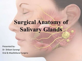
- 1. Surgical Anatomy of Salivary Glands Presented by- Dr Shibani Sarangi Oral & Maxillofacial Surgery
- 2. Introduction The salivary glands are the exocrine glands, glands with ducts, that produce saliva and pour their secretion in the oral cavity .They are classified as: 1) Major (Paired) -Parotid Submandibular Sublingual 2) Minor -Those in the Tongue, Palatine Tonsil, Palate Lips & Cheek
- 4. Development of Salivary glands • They originate from oral epithelium buds invading the underlying ectomesenchyme. • Origin of epithelial buds: Ectodermal –Parotid gland,Minor salivary glands Endodermal-Submandibular gland Sublingual gland
- 7. Functions of Saliva • The functions of saliva are: • Chemical digestion: breaks down starch by the function of “salivary amylase” • Helps chewing and swallowing. • Lubricating effect: moisturizes the inside of the mouth and creates smoother speech. • Solvent effect: dissolves food and allows the tongue to taste food.
- 9. PAROTID GLAND • Largest major Salivary gland • Average Wt - 25gm • Irregular lobulated mass lying mainly below the external acoustic meatus between mandible and sternomastoid. • On the surface of the masseter, small detached part lies b/w zygomatic arch and parotid duct- accessory parotid gland or ‘socia parotidis’
- 10. • Parotid Capsule : • Derived from investing layer of deep cervical fascia. • Superficial lamina-thick, closely adherent-sends fibrous septa into the gland. • Deep lamina-thin- attached to styloid process, mandible and tympanic plate. • Stylomandibular ligament
- 11. External Features -It resembles an inverted 3 sided pyramid having four surfaces – • Superior(Base of the Pyramid) • Superficial • Anteromedial • Posteromedial -Separated by three borders :- • Anterior • Posterior • Medial
- 12. Relations- • Superior Surface – • Concave • Related to : 1) Cartilaginous part of external acoustic meatus 2) Post. Aspect of temperomandibular joint 3) Auriculotemporal Nerve 4) Sup. Temporal vessels
- 13. • Apex – Overlaps posterior belly of digastric and adjoining part of carotid triangle. • Superficial Surface – • Covered by : Skin Superficial fascia containing facial branches of Great Auricular nerve Superficial parotid lymph nodes and post fibers of Platysma
- 14. Anteromedial Surface – • Grooved by posterior border of ramus of mandible • Related to - 1)Masseter 2)Lateral Surface of temporomandibular joint 3) Medial pterygoid muscles 4)Emerging branches of Facial Nerve
- 15. • Posteromedial Surface : • Related to- 1) Mastoid process with sternomastoid and posterior belly of digastric. 2) Styloid process with structures attached to it. 3) External Carotid artery which enters the gland through the surface 4) Internal Carotid artery which lies deep to styloid process
- 16. BORDERS • Anterior border • Separates superficial surface from anteromedial surface • Structures which emerge at this border -Parotid Duct -Terminal Branches of facial nerve -Transverse facial vessels
- 17. Posterior border • Separates superficial surface from posteromedial surface • Overlaps sternocleidomastoid muscule Medial Border- • Separates anteromedial surface from posteromedial surface • Related to lateral wall of pharynx
- 18. STRUCTURES WITHIN THE PAROTID GLAND
- 19. ARTERIES
- 20. VEINS
- 21. NERVE
- 22. • To expose the trunk of the facial nerve at the stylomastoid foramen the dissection passes down the avascular plane between the parotid gland and the external acoustic canal • The nerve lies about 9 mm from the posterior belly of the digastric muscle and 11 mm from the bony external meatus • The facial nerve then passes downward and forward over the styloid process and associated muscles for about 1.3 cm
- 24. Parotid duct • ductus parotideus; Stensen’s duct • 5 cm in length • Appears in the anterior border of the gland • Runs anteriorly and downwards on the masseter b/w the upper and lower buccal branches of Facial nerve.
- 25. • At the anterior border of masseter it pierces • Buccal pad of fat • Buccopharyngeal fascia • Buccinator Muscle • It opens into the vestibule of mouth opposite to the 2nd upper molar.
- 26. Blood supply • Arterial- Branch of External Carotid artery • Venous- Into the External Juglar vein • Lymphatic drainage- Upper deep Cervical nodes via Parotid nodes Nerve supply Parasympathetic –Auriculotemporal nerve Sympathetic- plexus around middle meningeal
- 27. Identifying Facial nerve • Tragus pointer :1.0cm-1.5cm deep and slightly anterior and inferior to the tip of the external auditory canal cartilage. • The nerve bisects the angle made by the diagastric and tympanic plate. • 1.0cm deep to attachment of the posterior belly of the diagastric groove of the mastoid bone. • The CN VII lies 6-8mm distal to the end point of the tympano mastoid fissure. • Nerve stimulator • Magnifying loops
- 28. Approaches • Pre auricular • Submandibular • Combination of both
- 29. • During surgical removal of parotid gland for any tumour the facial nerve is preserved by removing the glands in two parts superficial and deep lobe separately. • Retrograde approach to the trunk from either Mandibular branch,where it passes over Retromandibular vein. • Supravital staining of Parotid gland. Preventing injury to Facial nerve
- 30. Complications • Facial Nerve injury • Frey's syndrome-(Gustatory sweating)- It is a condition where sweating & sometimes flushing of skin in the area of distribution of Auriculotemporal nerve that occures;which is caused by stimulation to saliva secretion. • It is caused due to damage to postganglionic parasympathetic fibres from the otic ganglion,that became united to sympathetic fibres arising from Superior cervical ganglion.
- 31. SUBMANDIBULAR SALIVARY GLANDS • Irregular in shape • Weigh about 15 grams and contribute some 60–67% of unstimulated saliva secretion. • Large superficial and small deeper part continous with each other around the posterior border of mylohyoid.
- 32. Superficial part- • Situated in the digastric triangle • Wedged b/w body of mandible and mylohyoid • 3 surfaces – Inferior, Medial, Lateral
- 33. Capsule • Derived from deep cervical fascia . • Superficial Layer is attached to base of mandible • Deep layer attached to mylohyoid line of mandible
- 34. Relations • Inferior- Covered by : - Skin - Superficial fascia containing Platysma and cervical branches of Facial nerve - Deep Fascia - Facial Vein - Submandibular Nodes
- 35. • Lateral surface - -Related to submandibluar fossa on the mandible -Mandibular attachment of Medial pterygoid -Facial Artery
- 37. • Medial surface- • Anterior part is related to mylohyoid muscle, nerve and vessels. • Middle part - Hyoglossus, styloglossus, lingual nerve, Submandibular ganglion, Hypoglossal nerve and Deep lingual vein. • Posterior Part - Styloglossus, stylohyoid ligament, 9th nerve and Wall of Pharynx
- 38. • Deep part: - Small in size - Lies deep to Mylohyoid and superficial to Hyoglossus and Styloglossus - Posteriorly continuous with superficial part around the posterior border of Mylohyoid
- 39. • Submandibular Duct -Whartons duct - -5 cm long -Emerges at the anterior end of deep part of the gland -Runs forwards on Hyoglossus b/w lingual and Hypoglossal nerve -At the Anterior border of Hyoglossus it is crossed by lingual nerve. -Opens in the floor of mouth at the side of frenulum of tongue.
- 41. •SUBMANDIBULAR GANGLION (LANGLEY’S GANGLION) -It’s a parasympathetic ganglion, which acts as a relay station for secretomotor fibres supplying the submandibular and sublingual salivary glands. -Topographically, it’s linked to the trigeminal nerve (lingual nerve) but functionally it’s related to the facial nerve (via its chorda tympani branch).
- 42. NERVE SUPPLY • The submandibular ganglion has 3 roots, viz.: parasympathetic, sympathetic and sensory. • Parasympathetic root: From Chorda Tympani • Sympathetic root:It’s originated from sympathetic plexus around the facial artery. • Sensory root: It’s originated from lingual nerve. • BLOOD SUPPLY • ARTERIAL: Branches of Facial & Lingual arteries • VENOUS : Facial & Lingual Veins
- 43. Applied aspect - The formation of calculus is more common in the Submandibular gland than in the parotid . - For excision of the Submandibular salivary gland( for calculus or tumour), a skin crease incision is as a rule, given more than 1inch( 2.5cm) below the angle of the jaw . Since the marginal mandibular branch of facial nerve enters 1 inch posteroinferior to the angle of the mandible before crossing its lower border, the incision so ought to be given 4 cm below the angle to prevent injury to this nerve. - A stone in the Submandibular duct (Wharton’s duct) can be palpated bimanually in the floor of the mouth and can even be seen if sufficiently large.
- 44. Submandibular gland excision • Indications : - Chronic sialoadenitis - Stone in submandbular gland - Submandibular gland tumors • Incision :- Placed 2-4 cm below the mandible, parallel to it • Preserve : - Marginal mandibular nerve -Lingual nerve - Hypoglossal nerve
- 45. Approaches • Extraoral approach- 1)Lateral Transcervical -Incision of 4-5cm in length ,is taken in the skin in Submandibular region.Incision to be placed parallel to skin crease ,about 2cm below the Submandibular border.The wound is then deepened through Platysma and deep fascia. 2) Submental approach • The submental approach provides access to the submandibular triangle via a midline horizontal incision just superior to the submental-cervical crease at the level of the hyoid bone, (4.5 ± 1.9 cm). • Intraoral approach
- 48. Complications:- • Hemorrhage • Infection • Injury to Mandibular nerve, Lingual nerve & Hypoglossal nerve • Injury to Wharton’s duct
- 49. SUBLINGUAL GLAND • Smallest of the three glands • Weighs nearly 3-4 gm • Lies beneath the oral mucosa in contact with the sublingual fossa on lingual aspect of mandible.
- 50. RELATIONS Above Mucosa of oral floor, Below - - Mylohyoid Infront - Anterior end of its fellow Behind - -Deep part of Submandibular gland Lateral - Mandible above the anterior part of mylohyoid line • Medial - Genioglossus and separated from it by Lingual nerve and Submandibular duct
- 51. DUCT OF SUBLINGUAL GLAND • Ducts of Rivinus • 8-20 ducts . • Most of them open directly into the floor of mouth . • Few of them join the Submandibular duct.
- 52. • BLOOD SUPPLY • Arterial : From Sublingual and Submental arteries. • Veneous: Corresponding veins
- 53. APPLIED ASPECT • The structures at risk during dissection of the gland are the Submandibular duct and the Lingual nerve. • The duct lies superficially in the floor of the mouth medial to the sublingual fold, and is crossed inferiorly by the nerve which then enters the tongue • The Sublingual artery and vein also lie on the medial aspect of the gland close to the Submandibular duct and Lingual nerve.
- 54. Incision • It is given in the Sublingual groove • Structures closely associated are:- Sublingual gland Submandibular duct Lingual nerve Hypoglossal nerve Sublingual vein
- 55. Diagnostic Imaging • It plays an important role in evaluation of disorders associated of major salivary gland. • The modalities used for imaging are:- • Conventional Radiography • Sialography • Ultrasonography • Computed Tomography • Radionuclide imaging
- 56. References • Textbook and colour Atlas of Salivary gland pathology; Eric Carlson • Textbook of Oral & Maxillofacial surgery; Neelima Malik • Internet
Editor's Notes
- STAGE 1 Bud formation: Introduction of the oral epithelium by underlying mesenchyme STAGE 2 Formation and growth of epithelial cord .STAGE 3 Initiation of branching in terminal parts of epithelial cord and continuation of glandular differentiation .STAGE 4 Dichromatous branching of epithelial cord and lobule formation STAGE 5 Canalization of presumptive ducts STAGE 6 Cytodifferentiation
- Ph=6.5-7
- Taste is a chemical sense. Any substance, the taste of which has to be perceived, has to be in dissolved state to stimulate the taste receptors
- Eca runs deeply within the parotid Sup temporal artery &max –condyle level
- Davis et el Anastomototic pattern in 350
- NERVE SUPPLY – CHORDA TYMPANI,LINGUAL
- Cerpy in 1902 Phases-Ductal.acinar,post evacuation .normal-laefless tree. Acinar-completion of ductal opacification Evac-asses secretory function of gland Sjogren- cHerry blossom branchless fruit tree Tumour-hand holding ball