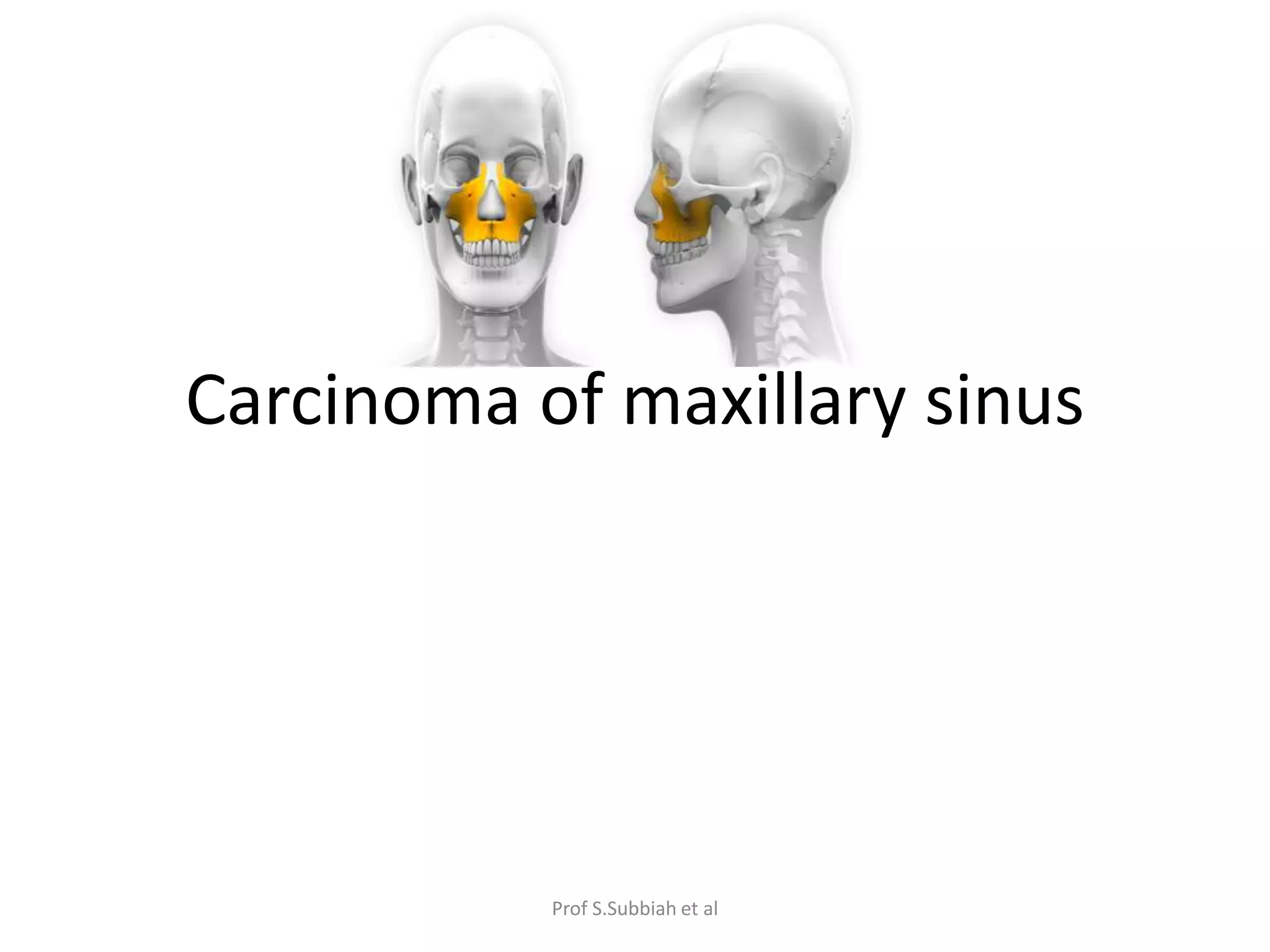
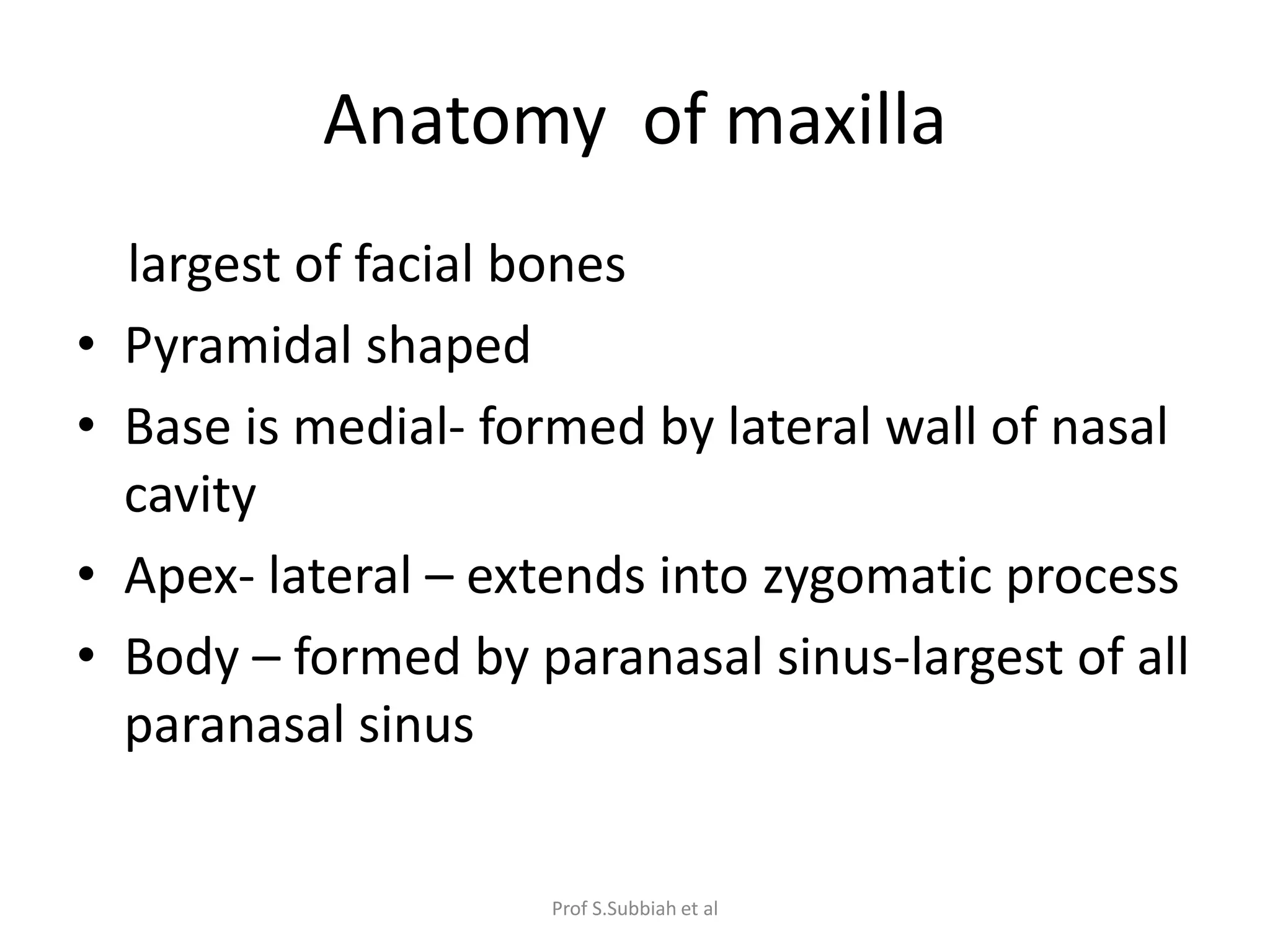
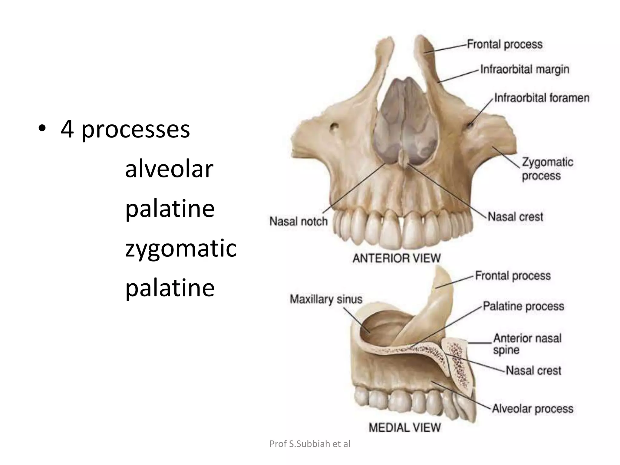
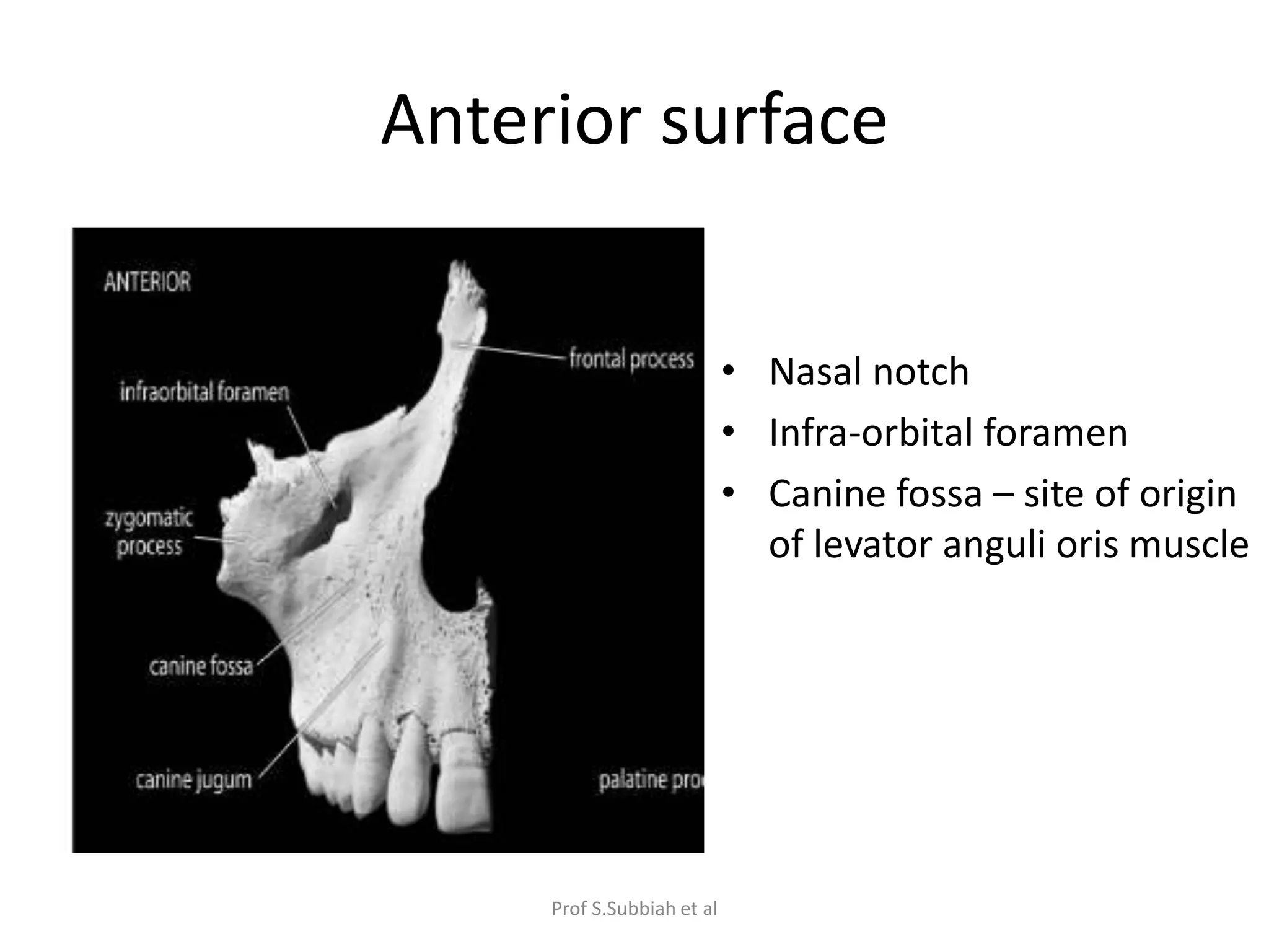
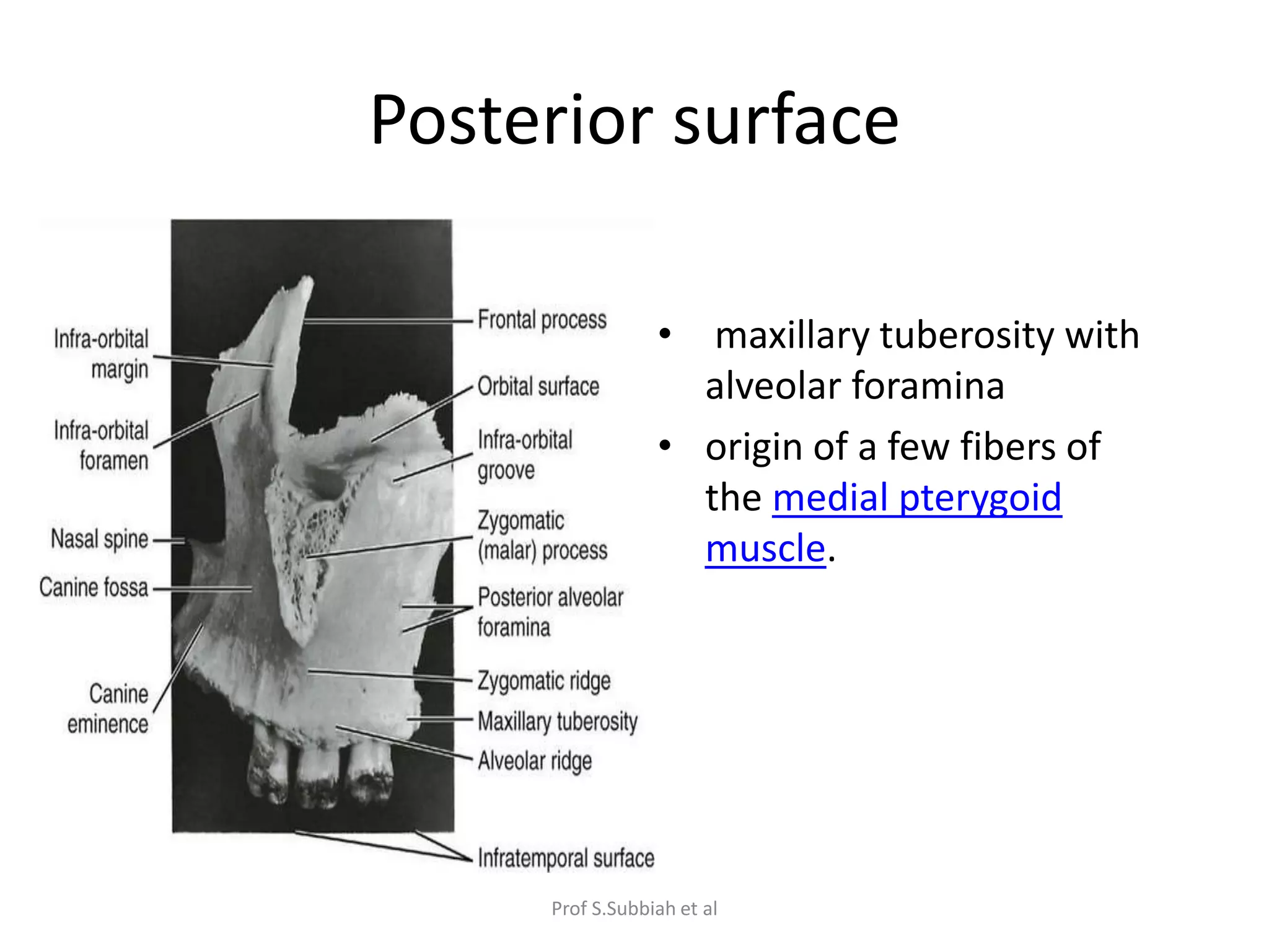
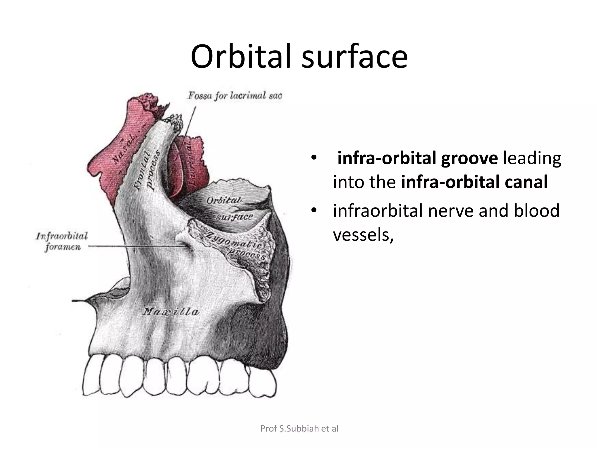
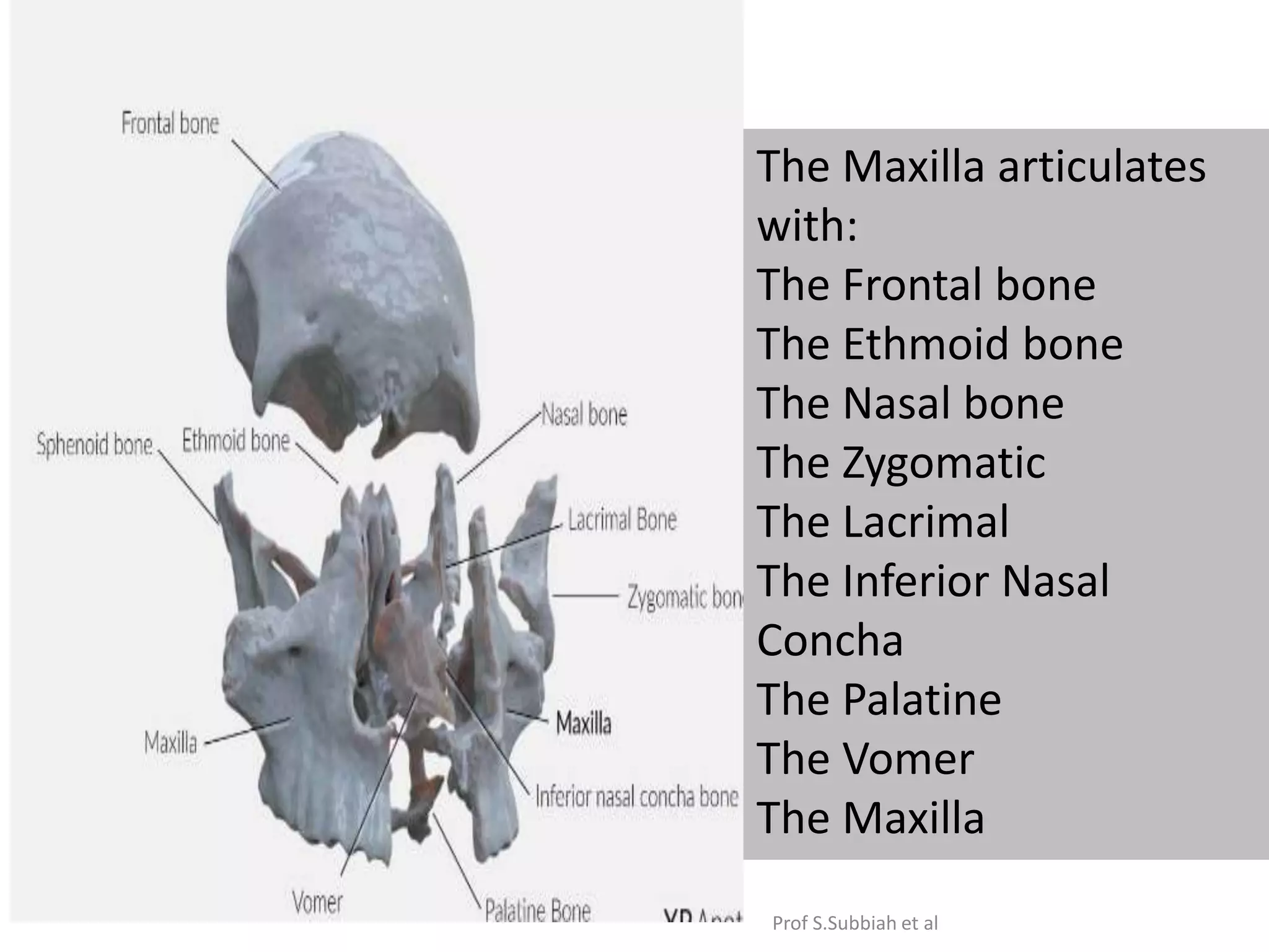
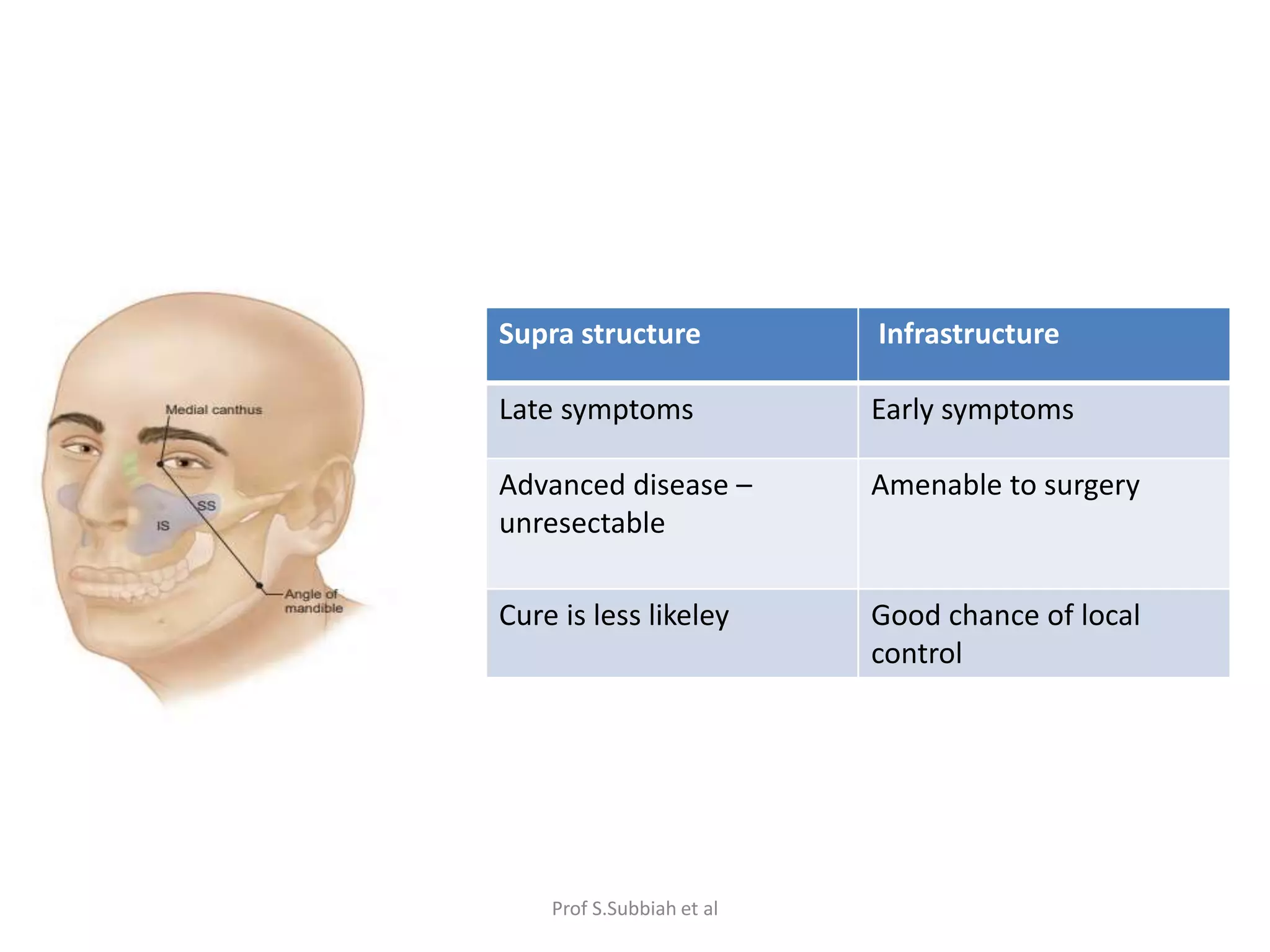
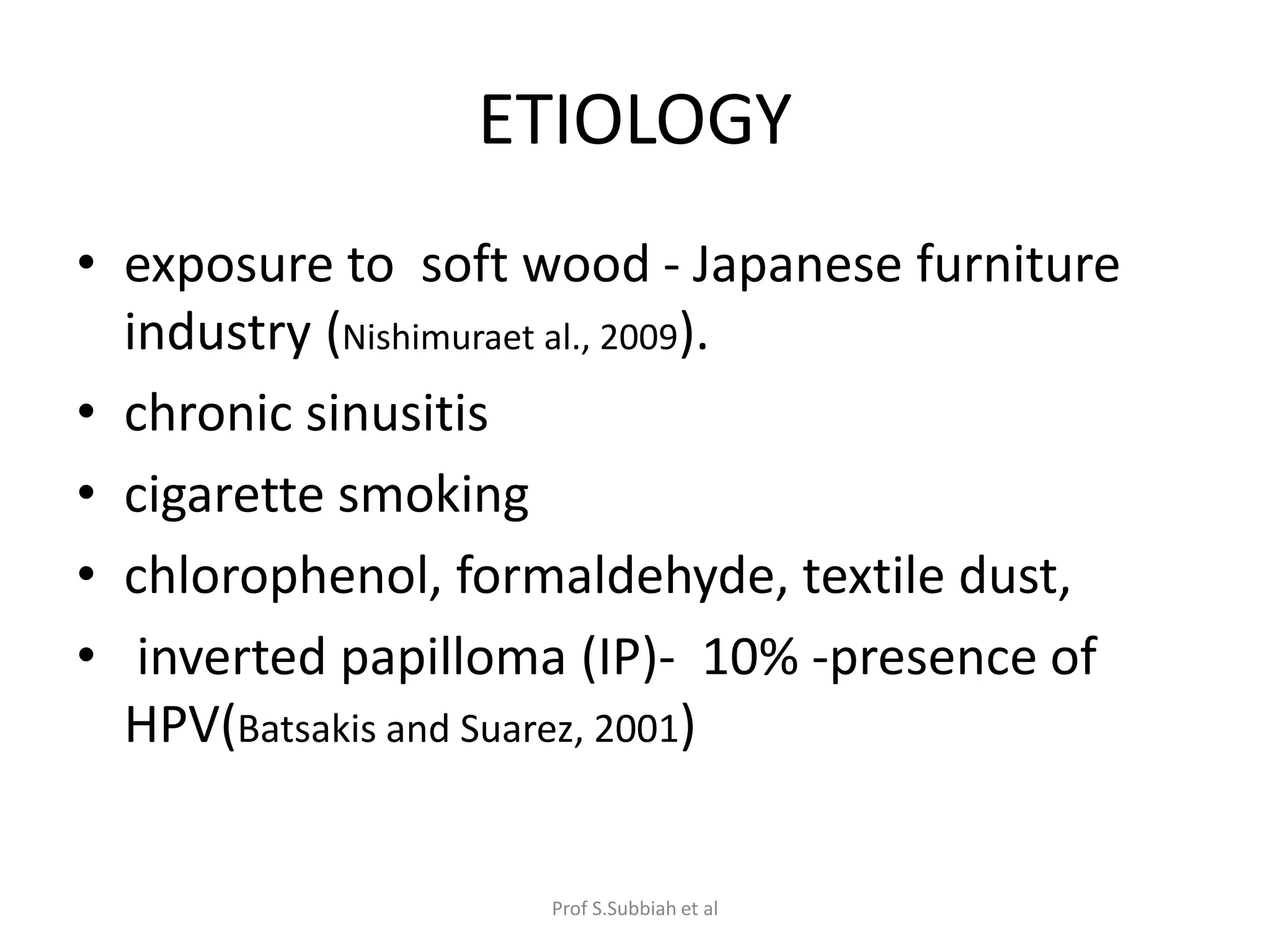
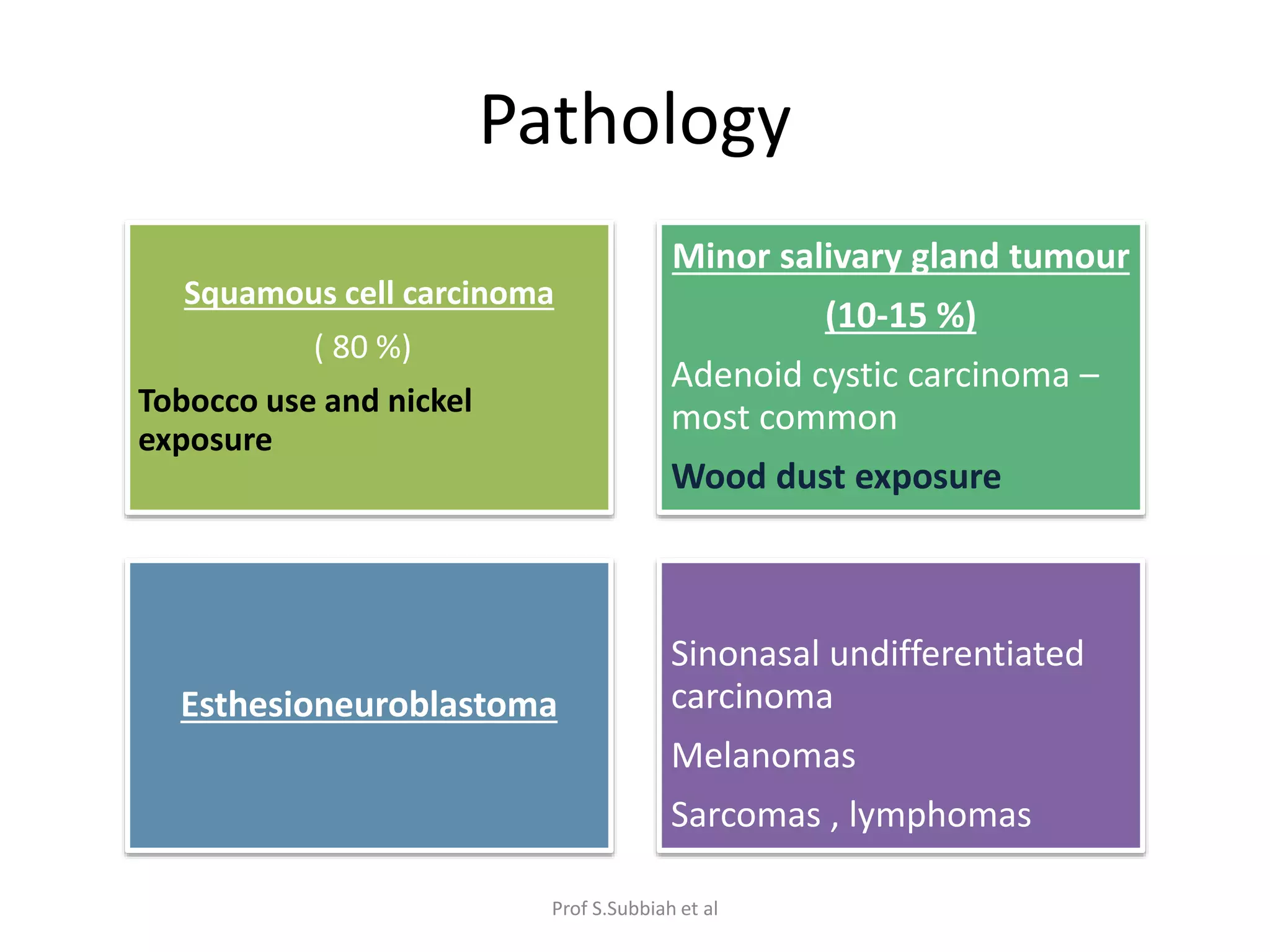
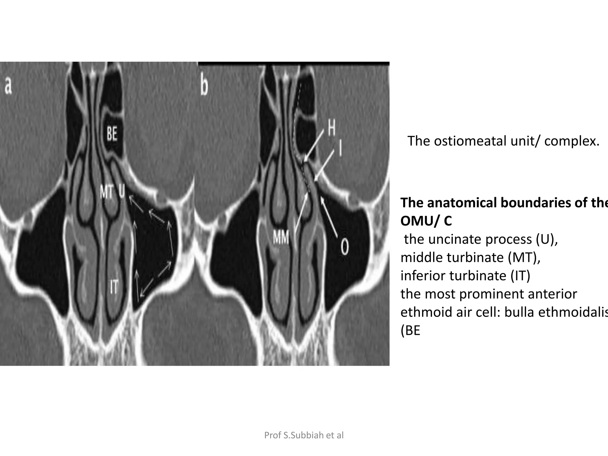
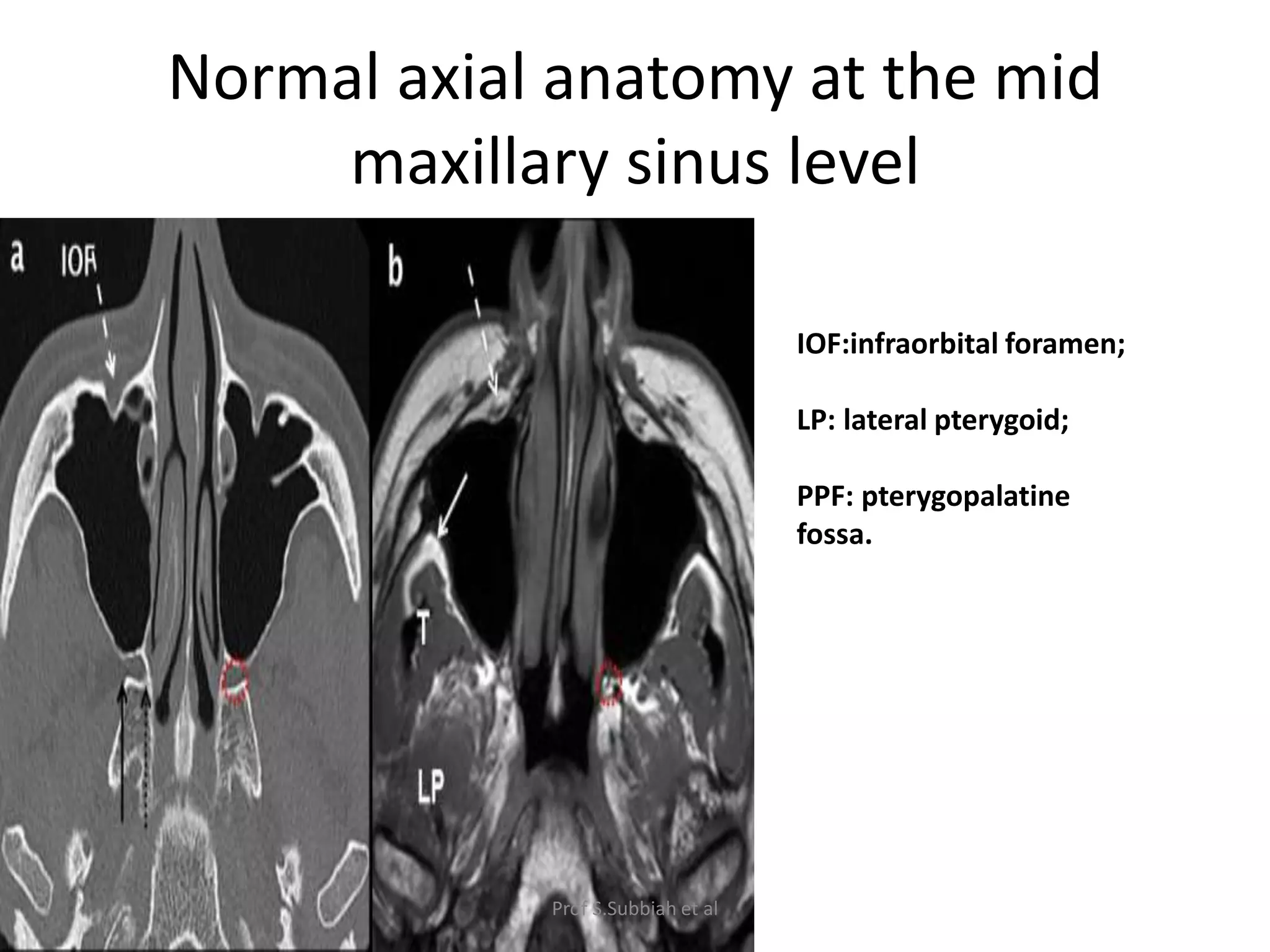
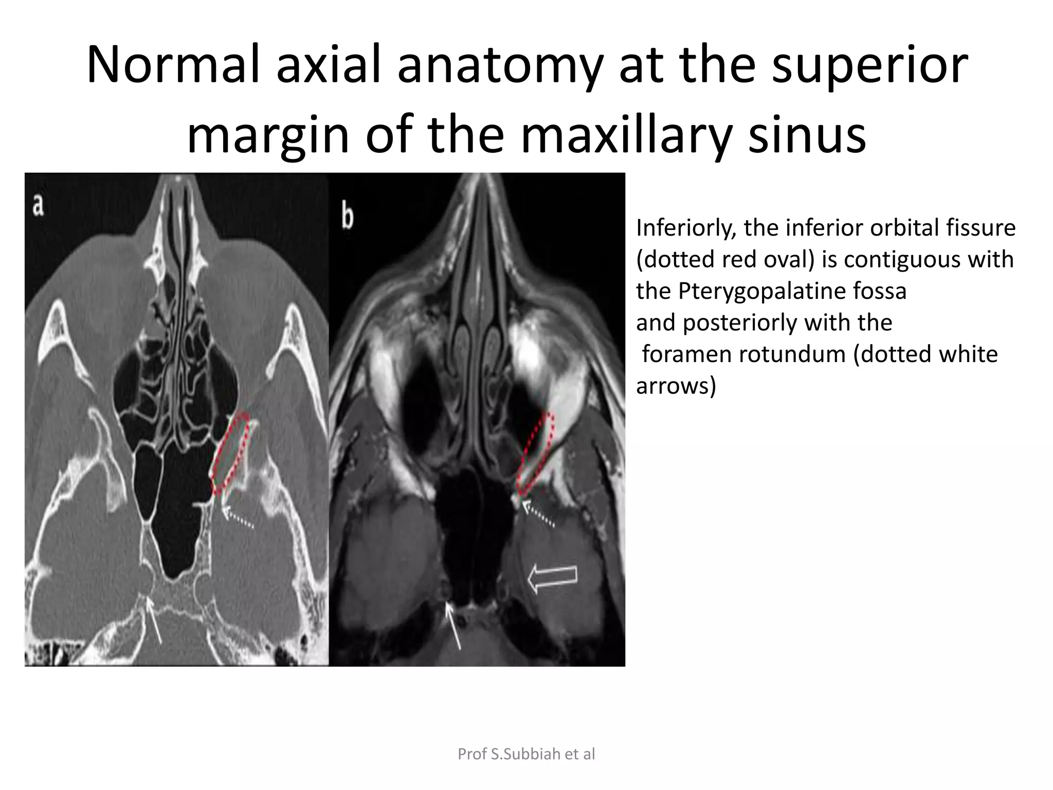
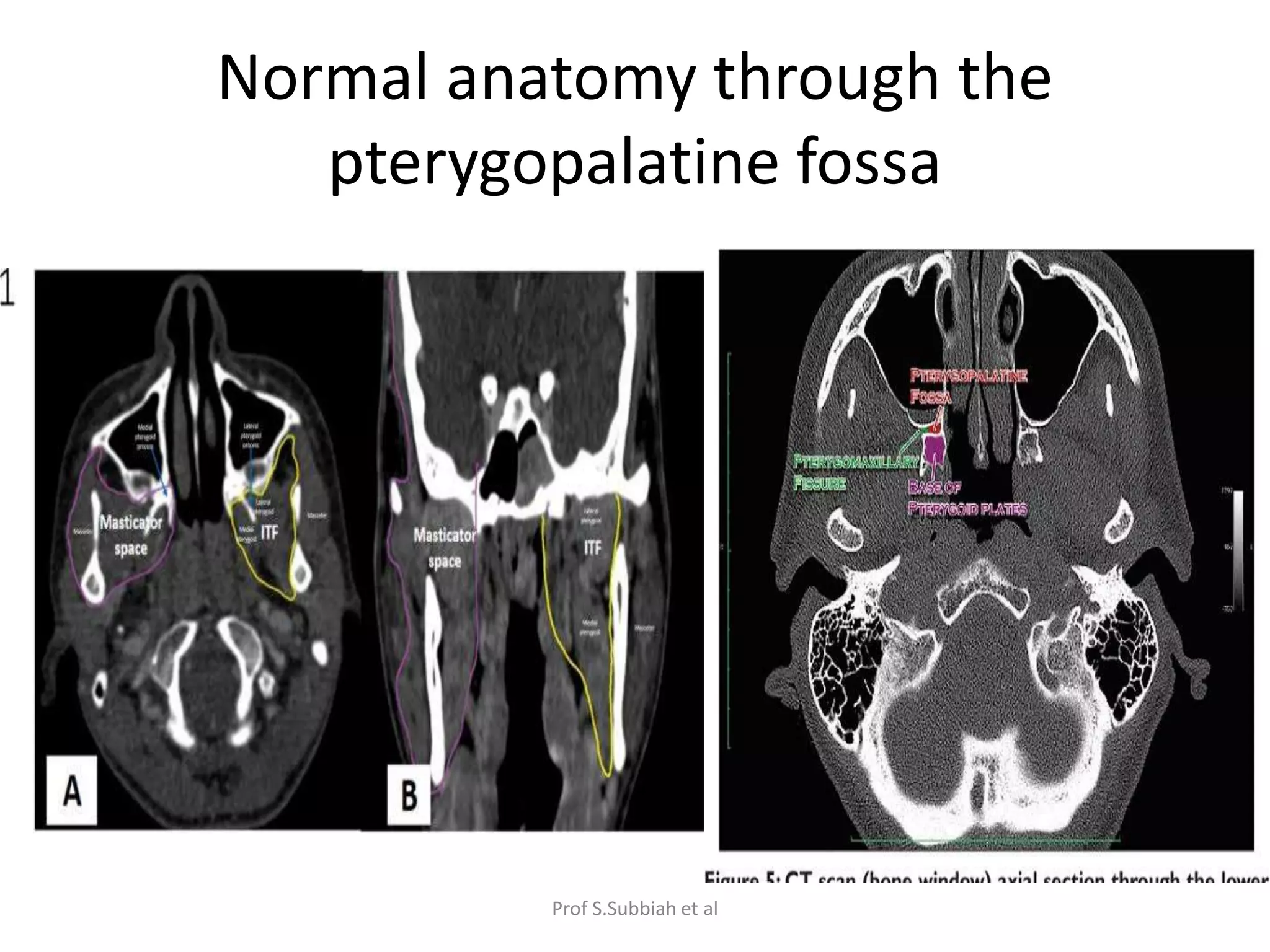
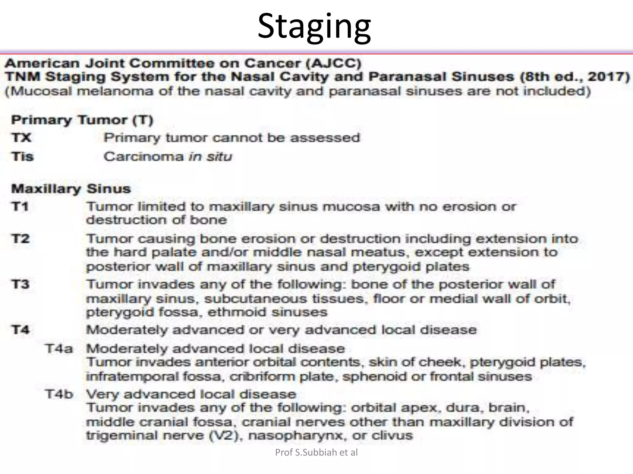
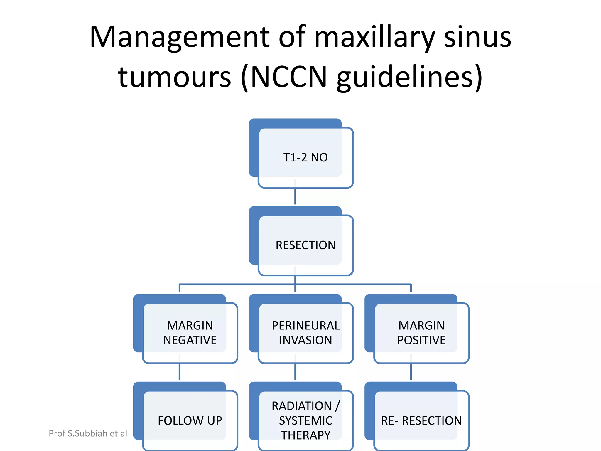
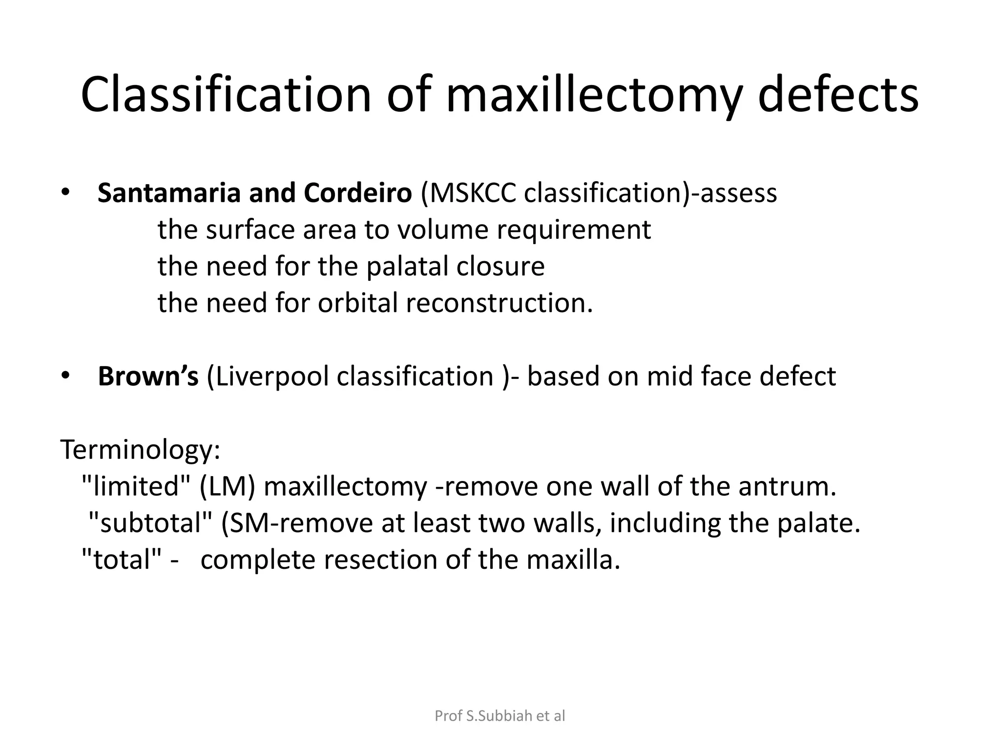
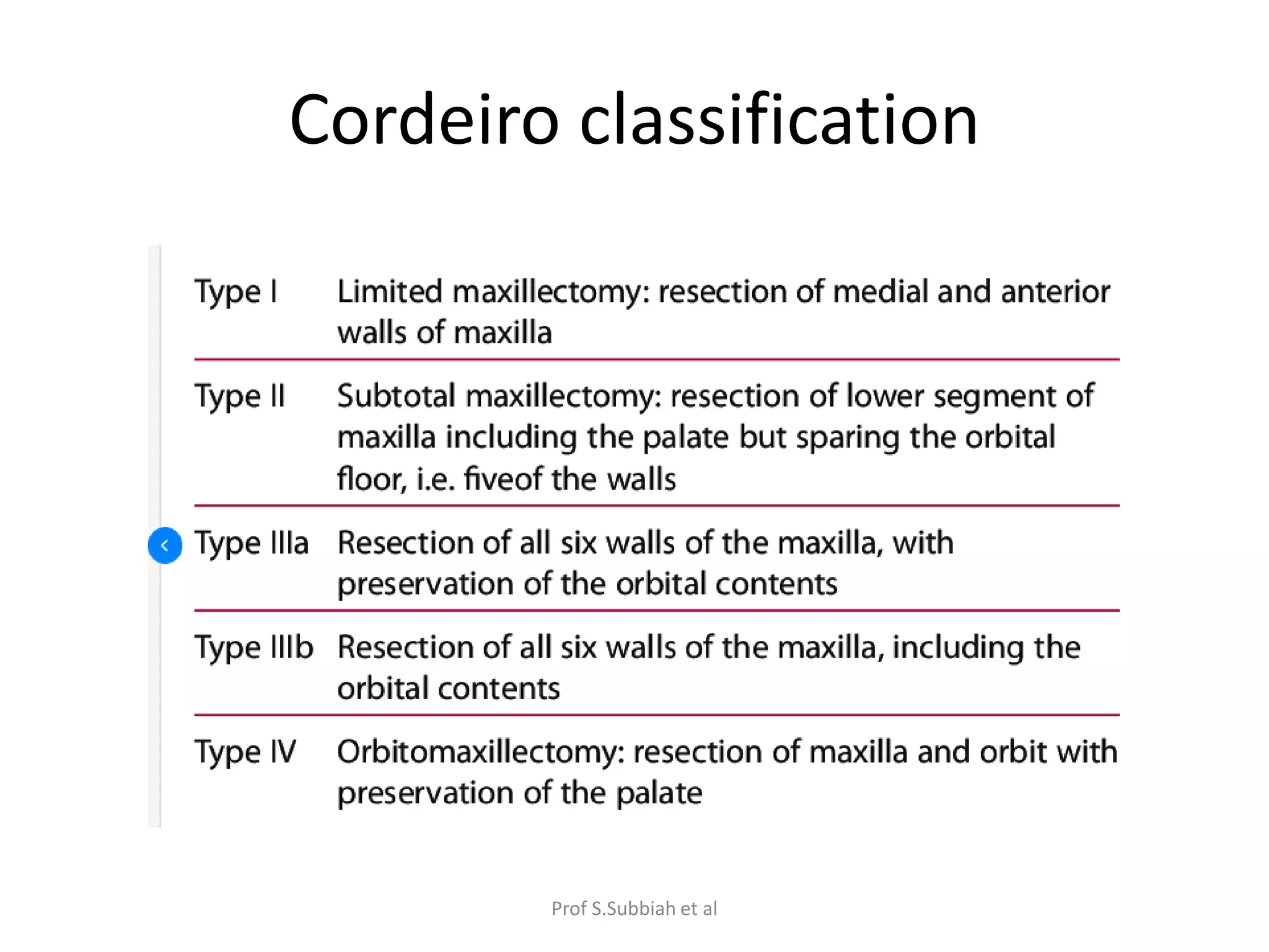
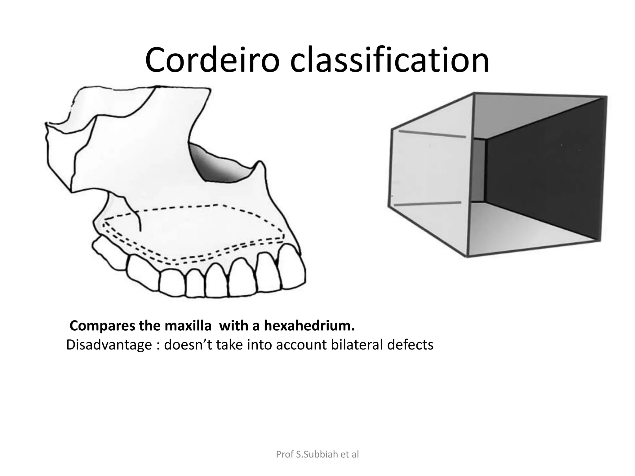
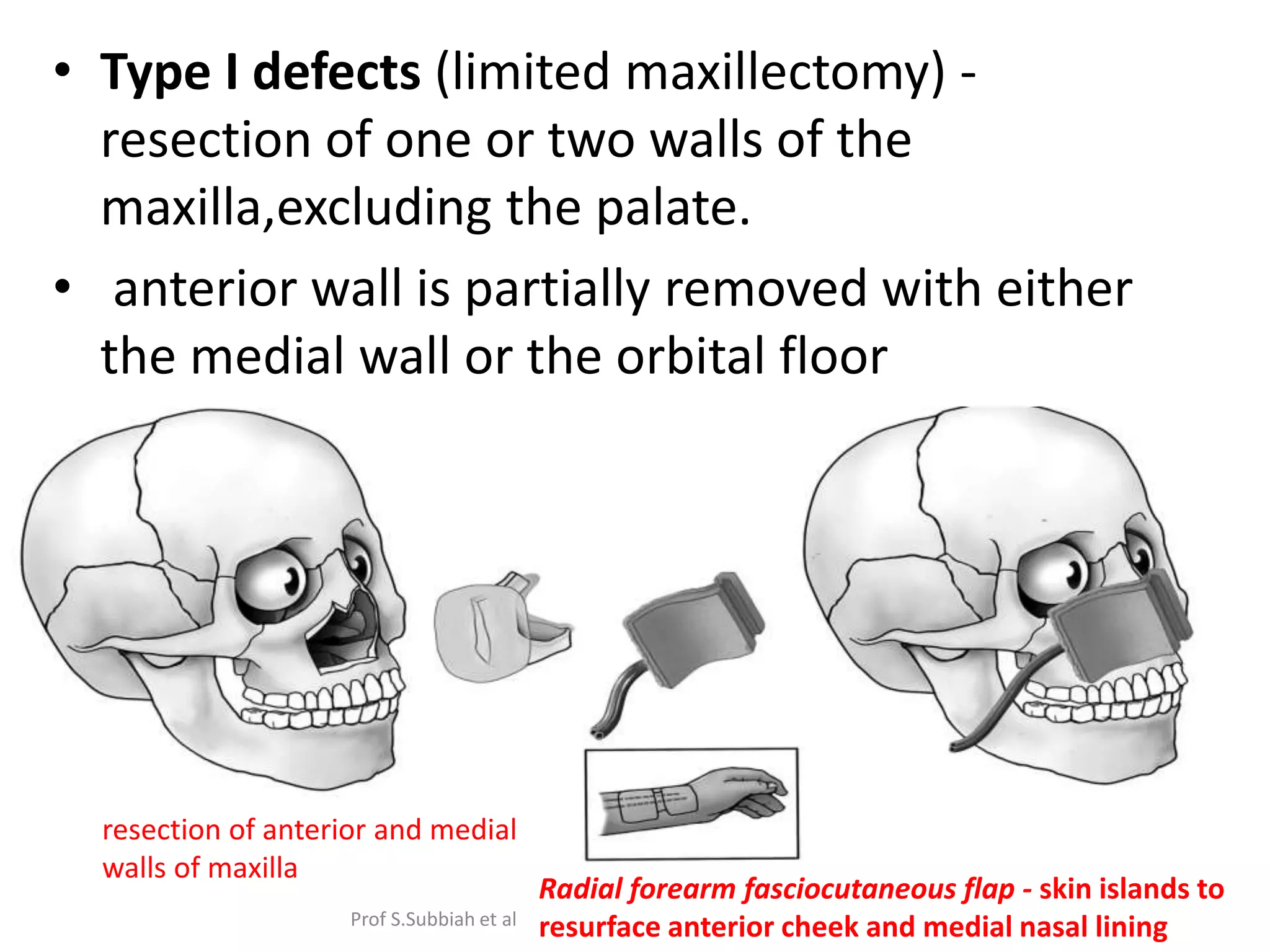
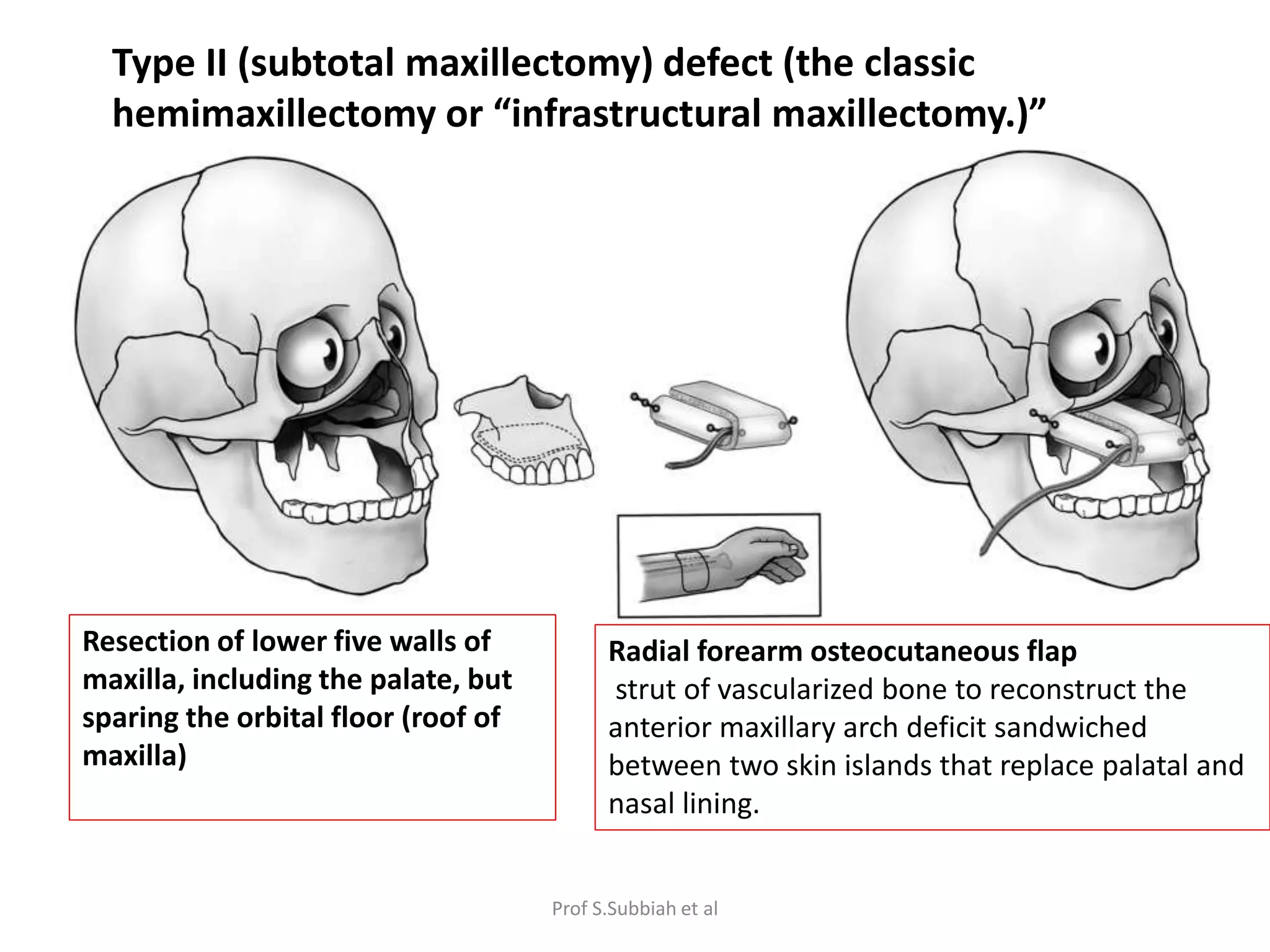
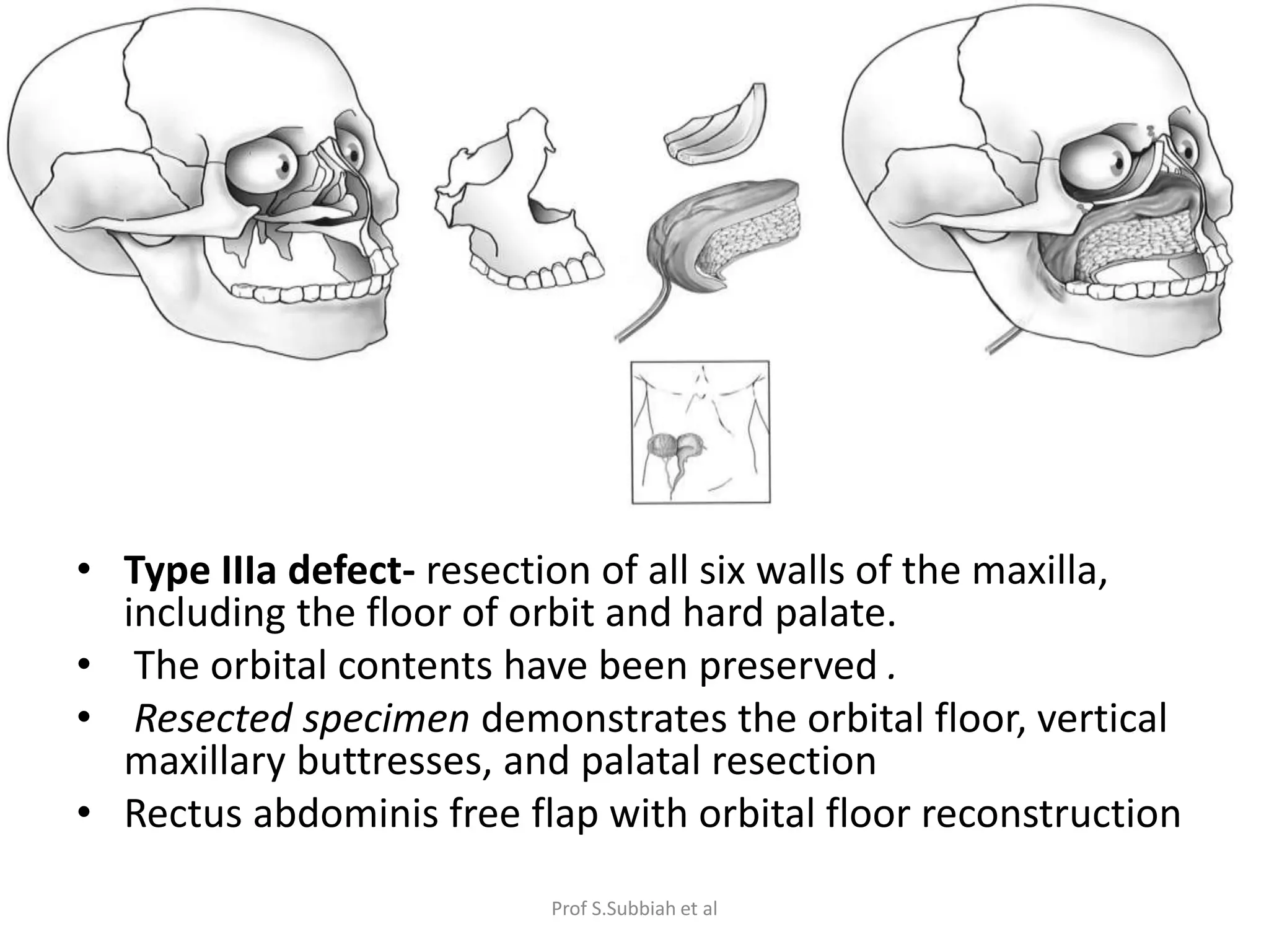
Carcinoma of maxillary sinus
Prof S.Subbiah et al
Introduction
• Carcinoma of the paranasal sinus are rare -
0.2–0.8% of all cancer
• Constitute 3–5% of the malignant tumors in
the head and neck region
• The maxillary sinus - most frequent site
Prof S.Subbiah et al
Anatomy of maxilla
largest of facial bones
• Pyramidal shaped
• Base is medial- formed by lateral wall of nasal
cavity
• Apex- lateral – extends into zygomatic process
• Body – formed by paranasal sinus-largest of all
paranasal sinus
Prof S.Subbiah et al
• 4 processes
alveolar
palatine
zygomatic
palatine
Prof S.Subbiah et al
• 4 surfaces
anterior
posterior (infratemporal)
orbital
nasal
Prof S.Subbiah et al
Anterior surface
• Nasal notch
• Infra-orbital foramen
• Canine fossa – site of origin
of levator anguli oris muscle
Prof S.Subbiah et al
Posterior surface
• maxillary tuberosity with
alveolar foramina
• origin of a few fibers of
the medial pterygoid
muscle.
Prof S.Subbiah et al
Orbital surface
• infra-orbital groove leading
into the infra-orbital canal
• infraorbital nerve and blood
vessels,
Prof S.Subbiah et al
The Maxilla articulates
with:
The Frontal bone
The Ethmoid bone
The Nasal bone
The Zygomatic
The Lacrimal
The Inferior Nasal
Concha
The Palatine
The Vomer
The Maxilla
Prof S.Subbiah et al
Boundaries of maxillary sinus
• Roof: inferior wall of orbit
• Medial wall: lateral wall of the nasal cavity
and part of the lacrimal system
• Floor: anterior portion of the hard palate and
alveolar ridge
• Buttresses: 2 horizontal and 3 vertical
buttresses provide projection & height
Prof S.Subbiah et al
Ohngren’s line
• .The superior wall- orbit
• Posterior wall - the pterygoid
plates, pterygoid space, and
infratemporal fossa.
• Ohngren (1933) - involvement
of these superior and
posterior areas signifies a poor
prognosis .
• imaginary line extending from
the medial canthus through
the angle of the mandible.
• Lesions posteriorsuperior –
suprastructure
• Lesions anteriorinferior to this
line-infrastructure
Prof S.Subbiah et al
Supra structure Infrastructure
Late symptoms Early symptoms
Advanced disease –
unresectable
Amenable to surgery
Cure is less likeley Good chance of local
control
Prof S.Subbiah et al
Routes of spread
Supra structure
Pterygomaxillary space , infratemporal fossa via
posterior wall
Middle cranial fossa via orbital roof
Anterior cranial fossa via ethmoidal air cells
Infrastructure
Floor of mouth to oral cavity
Nasal cavity via medial wall
Soft tissues of cheek via anterior wall
Regional lymph node spread < 10 %
Prof S.Subbiah et al
ETIOLOGY
• exposure to soft wood - Japanese furniture
industry (Nishimuraet al., 2009).
• chronic sinusitis
• cigarette smoking
• chlorophenol, formaldehyde, textile dust,
• inverted papilloma (IP)- 10% -presence of
HPV(Batsakis and Suarez, 2001)
Prof S.Subbiah et al
Pathology
Squamous cell carcinoma
( 80 %)
Tobocco use and nickel
exposure
Minor salivary gland tumour
(10-15 %)
Adenoid cystic carcinoma –
most common
Wood dust exposure
Esthesioneuroblastoma
Sinonasal undifferentiated
carcinoma
Melanomas
Sarcomas , lymphomas
Prof S.Subbiah et al
Natural history of disease
• locally advanced at diagnosis -symptoms are
nonspecific
• localized for a long time
• invade adjacent structures after sometime
• The most effective barrier against tumors
spread -periosteum that is particularly more
resistant in two critical areas: The skull base
and orbit
Prof S.Subbiah et al
SIGNS AND SYMPTOMS
oral
•pain involving the maxillary dentition,
• trismus,
•palatal and alveolar ridge fullness
• frank erosion into the oral cavity.
nasal
• persistent sinusitis,
• unilateral nasal obstruction,
• nasal discharge
• epistaxis
auditory
• hearing impairment secondary to serous otitis media due to
nasopharyngeal extension.
Facial
• include infraorbital nerve hyperaesthesia, pain and facial
asymmetry.
intracranial
• headache, personality changes(frontal lobe involvement)
Prof S.Subbiah et al
DIAGNOSIS
• Panoramic radiography-
Destruction of maxillary sinus walls, especially the
inferior antral wall, -
• CT -
soft tissue masses in the maxillary sinus cavity
bony destruction.
• MRI-
evaluation of the posterior cranial fossa,
orbit and perineural/perivascular dissemination
differentiation between retained secretions and
neoplastic tissue
Prof S.Subbiah et al
The ostiomeatal unit/ complex.
The anatomical boundaries of the
OMU/ C
the uncinate process (U),
middle turbinate (MT),
inferior turbinate (IT)
the most prominent anterior
ethmoid air cell: bulla ethmoidalis
(BE
Prof S.Subbiah et al
Normal axial anatomy at the mid
maxillary sinus level
IOF:infraorbital foramen;
LP: lateral pterygoid;
PPF: pterygopalatine
fossa.
Prof S.Subbiah et al
Normal axial anatomy at the superior
margin of the maxillary sinus
Inferiorly, the inferior orbital fissure
(dotted red oval) is contiguous with
the Pterygopalatine fossa
and posteriorly with the
foramen rotundum (dotted white
arrows)
Prof S.Subbiah et al
Normal anatomy through the
pterygopalatine fossa
Prof S.Subbiah et al
The infratemporal fossa lies peripheral to the lateral antral wall and is an
important deep fascial space which can be involved by spread of
infection or tumour
Prof S.Subbiah et al
Differential diagnosis include
• sinonasal undifferentiated carcinoma,
nasopharyngeal carcinoma, lymphoma,
esthesioneuroblastoma,
• primary sinonasal melanoma
• adenocarcinoma of minor salivary gland
• metastatic disease
Prof S.Subbiah et al
Staging
Prof S.Subbiah et al
Management of maxillary sinus
carcinoma
• Operable tumors – combined modality -
surgery + radiation
• Incidence of lymph node metastases -10%
• Recommend neck dissection or elective neck
radiation therapy - node positive
• Pre- op radiation therapy - stage II and stage III
tumors
Prof S.Subbiah et al
Management of maxillary sinus
tumours (NCCN guidelines)
T1-2 NO
RESECTION
MARGIN
NEGATIVE
FOLLOW UP
PERINEURAL
INVASION
RADIATION /
SYSTEMIC
THERAPY
MARGIN
POSITIVE
RE- RESECTION
Prof S.Subbiah et al
T1 – T4a, N+
Resection +
neck
dissection
Adverse
features
Yes
RT or consider
systemic
therapy
no
Only RT to
primary &
neck
T1 – T4a, N+
Resection +
neck
dissection
Adverse
features
Yes
RT or consider
systemic
therapy
no
Only RT to
primary &
neck
Prof S.Subbiah et al
T4b, N0-3
Clinical trial PS 0-1
Concurrent
Chemoradiation
PS 2
RT or concurrent
chemoradiation
PS 3
Palliative RT or
single agent CT or
best supportive
care
Prof S.Subbiah et al
Contraindications for surgery
• invasion of pterygoid muscles and soft tissues in
masticator space around temporomandibular
joint and pterygomaxillary fossa
• Invasion of skull base with destruction of
posterolateral wall and lateral walls of sphenoid
sinus
• Brain invasion
• Invasion of cavernous sinus
• Cranial nerve paralysis (CN ii, iii, iv, v,vi)
• Inavsion of carotid artery
Prof S.Subbiah et al
Classification of maxillectomy defects
• Santamaria and Cordeiro (MSKCC classification)-assess
the surface area to volume requirement
the need for the palatal closure
the need for orbital reconstruction.
• Brown’s (Liverpool classification )- based on mid face defect
Terminology:
"limited" (LM) maxillectomy -remove one wall of the antrum.
"subtotal" (SM-remove at least two walls, including the palate.
"total" - complete resection of the maxilla.
Prof S.Subbiah et al
Cordeiro classification
Prof S.Subbiah et al
Cordeiro classification
Compares the maxilla with a hexahedrium.
Disadvantage : doesn’t take into account bilateral defects
Prof S.Subbiah et al
• Type I defects (limited maxillectomy) -
resection of one or two walls of the
maxilla,excluding the palate.
• anterior wall is partially removed with either
the medial wall or the orbital floor
resection of anterior and medial
walls of maxilla
Radial forearm fasciocutaneous flap - skin islands to
resurface anterior cheek and medial nasal lining
Prof S.Subbiah et al
Resection of lower five walls of
maxilla, including the palate, but
sparing the orbital floor (roof of
maxilla)
Radial forearm osteocutaneous flap
strut of vascularized bone to reconstruct the
anterior maxillary arch deficit sandwiched
between two skin islands that replace palatal and
nasal lining.
Type II (subtotal maxillectomy) defect (the classic
hemimaxillectomy or “infrastructural maxillectomy.)”
Prof S.Subbiah et al
• Type IIIa defect- resection of all six walls of the maxilla,
including the floor of orbit and hard palate.
• The orbital contents have been preserved .
• Resected specimen demonstrates the orbital floor, vertical
maxillary buttresses, and palatal resection
• Rectus abdominis free flap with orbital floor reconstruction
Prof S.Subbiah et al
Patients who are not free-flap candidates - reconstruct with split calvarial
bone grafts, covered with the temporalis muscle, transposed anteriorly.
The zygomatic arch should be osteotomized and removed temporarily to
increase excursion of the temporalis muscle.
Prof S.Subbiah et al
TYPE IIIB MAXILLECTOMY
resection of all six walls of the maxilla,
including the floor of the orbit as well
as orbital contents .
resection of external eyelid, cheek skin,
and orbital contents, in combination
with entire maxilla and palate
A three-skin-island rectus abdominis myocutaneous flap
Orbital floor reconstruction –
alloplasts- titanium mesh
autologous tissues- free cartilage or bone grafts or
osteocutaneous free microvascular flap
Prof S.Subbiah et al
resection of upper five walls of maxilla,
including the orbital contents but sparing the
palate
Type IV (orbitomaxillectomy) defect
Prof S.Subbiah et al
Browns classification
Horizontal classification:
a Palatal defect only, not involving the dental alveolus.
b Palatal defect less than or equal to half unilateral.
c Palatal defect less than or equal to half bilateral or transverse
anterior.
d Palatal defect greater than half maxillectomy
Prof S.Subbiah et al
Vertical classification:
I Maxillectomy not causing an oronasal fistula.
II Maxillectomy not involving the orbit.
III Maxillectomy involving the orbital adnexae with orbital retention.
IV Maxillectomy with orbital enucleation or exenteration.
V Maxillectomy with orbitomaxillary defect.
VI Maxillectomy with nasomaxillary defect.
Loss of the vertical
component causes
more of an aesthetic
problem
Prof S.Subbiah et al
Orbital Involvement
• Carcinoma of the maxillary sinus commonest to invade
the orbit (Dukes Elder et.al)
• Three stages of orbital invasion (Iannetti17 et al)
Grade I- erosion or destruction of the medial orbital
wall
Grade II- extraconal invasion of the periorbital fat
Grade III- invasion of the medial rectus muscle, optic
nerve, ocular bulb, or the skin overlying the eyelid.
only grade III orbital invasion warrants orbital
clearance or exenteration.
Prof S.Subbiah et al
Approaches for maxillectomy
• lateral rhinotomy- extending anteriorly to nasal bone or
soft tissue or the lateral nasal wall
• Open Medial Maxillectomy- lateral wall of the nasal cavity
or the medial wall of the maxillary antrum
• A transoral partial maxillectomy- tumors of the
“infrastructure” of the maxilla, including tumors of the
upper gum, hard palate, or floor of the maxillary antrum
• Sublabial degloving approach-larger tumors of the
anteroinferior aspect of the nasal cavity and the
infrastructure of the maxilla
• open craniofacial approach -involve the skull base
Prof S.Subbiah et al
• Classic Weber –Ferguson-Diffenbach
1) excellent exposure
2)unacceptable scar
3)distortion of facial contour and expression
4) ectropion
5) alteration of shape of nasal vestibule
Prof S.Subbiah et al
• Starts from midline upper lip
• Philtrum
• Columella
• Around vestibule and nasal ala
• Nasolabial crease and medial canthus
Prof S.Subbiah et al
Modified Weber-Ferguson incision
• A-lynch extension;
• B- subciliary extension;
• C- infraorbital
extension.
D – Borle’s extension –
ALLOWS HAREVEST OF
TEMPORALIS FLAP
Prof S.Subbiah et al
Conceptual Approach to Reconstruction of
Midface/ Maxillary Defects
extent of resection of the maxillary bone- key
building block to the structure of the midface.
• Address the bony defect first- followed by
soft-tissue, skin, palate, and cheek lining
deficits.
• Finally, critical functional structures –
palate, oral commissure, nasal airway, and
eyelids.
Prof S.Subbiah et al
• The anterior (cheek), superior (orbital floor), and inferior (palatal) walls
most commonly need to be reconstructed for the following reasons.
• Bone replacement (floor of the orbit) –
1) maintain position of the ocular globe.
2)maxillary arch to provide anterior projection of the midface
3)obliterate any communication between the mouth and the nose
and the nasopharynx and anterior cranial base to prevent
ascending infections,
• Palatal reconstruction - to enhance articulation and deglutition,
• lacrimal apparatus
• The space between the restored anterior, superior, and inferior walls of
the maxilla can usually be filled with soft tissue (muscle/fat), and nasal
lining may or may not be necessarily restored.
Prof S.Subbiah et al
.
• If the cavity is not reconstructed (no free microvascular
flap)-
1) the inner aspect should be lined with a skin graft or
acellular dermis.
2) promote rapid healing
3) prevent contraction of
the surgical cavity –prevent subsequent collapse
of the mid face.
4)The scar band that develops at the interface of the skin
graft and the remaining buccal mucosa often aids to
secure the prosthesis.
Prof S.Subbiah et al
Prosthesis after maxillectomy
OBTURATOR MAXILLOFACIAL PROSTHESIS
CAD / CAM
PROSTHESIS
( computer aided design /
computer assisted
manufacture
Prof S.Subbiah et al
Postoperative rehabilitation
• Preoperatively designed
surgical obturator with clasps-
fitted after the bolster is
removed postoperative day
5th to 7th day.
• Holes drilled in the posterior
aspect - allow sutures to be
placed in the soft palate to the
splint- reduces oro-nasal
reflux and facilitates the early
return to a normal diet.
• final prosthesis - after the
incisions are healed and the
edema has resolved
(usually after the adjuvant
treatment) Prof S.Subbiah et al
Definitive obturator prosthesis- only 3
months to 1 year after the resection surgery.
Dimensional changes due to remodeling and
scarring of the defect's contours last
approximately 1 year
( soft tissue >bone tissue remodeling)
• Maxillary defect < a quarter of the hard
palate- the obturator gives excellent
functional results
Prof S.Subbiah et al
POST OPEARATIVE COMPLICATIONS
• Resorption of orbital fat and wound contracture I
enophthalmos
diplopia
restricted range of motion.
cataract -Postoperative radiation therapy
• Trismus-
Soft tissue fibrosis
• Lacrimal duct obstruction-
dacryocystorhinostomy prevents it
Prof S.Subbiah et al
Role of definitive chemoradiation
Prof S.Subbiah et al
• Eighteen patients with inoperable MSC with neck
lymph node metastasis
• 70 Gy in 35 fractions +tri-weekly systemic
administration of cisplatin at 100 mg per square meter
of body surface area
• the 2-year LC, PFS and OS rates for all patients were 34,
31 and 46%, respectively
Prof S.Subbiah et al
Recurrence
• After surgery - radiation therapy or
craniofacial resection with postoperative
radiation therapy.
• After radiation therapy -craniofacial resection
if indicated.
• Chemotherapy - does not respond
• Clinical trials
Prof S.Subbiah et al
Prognosis
• Advanced stage -dismal prognosis- 5-year
overall survival rates of 35–49%
• A meta-analysis revealed that disease specific
survival after five years is significantly higher
for patients treated with surgery plus adjuvant
radiotherapy (RT) (66%) than for patients
treated with RT alone (46%)
Prof S.Subbiah et al
PROGNOSIS
• Orbital involvement -significant reduction in
survival
• skull base and orbital involvement - only factors
significantly associated with disease-specific
survival (Gullaneet al., 1983)
5-year survival - 17% vs 49% when there was
no invasion .
• no survival benefit - by orbital clearance
5 year survival- 11% despite complete
extirpation of orbital contents (Carrilloet al.,
2003)
Prof S.Subbiah et al
Take home message
• Suprastructure lesions present late and have
poor prognosis
• Orbital involvement is T3 disease and has poor
prognosis and reduced survival
• Lymph nodal metastasis is rare and nodal
dissection is indicated only in node positive
cases
• Only grade 3 orbital invasion requires orbital
exenteration
Prof S.Subbiah et al
Thank you
Prof S.Subbiah et al

Carcinoma Maxillary sinus

  • 1.
    Carcinoma of maxillarysinus Prof S.Subbiah et al
  • 2.
    Introduction • Carcinoma ofthe paranasal sinus are rare - 0.2–0.8% of all cancer • Constitute 3–5% of the malignant tumors in the head and neck region • The maxillary sinus - most frequent site Prof S.Subbiah et al
  • 3.
    Anatomy of maxilla largestof facial bones • Pyramidal shaped • Base is medial- formed by lateral wall of nasal cavity • Apex- lateral – extends into zygomatic process • Body – formed by paranasal sinus-largest of all paranasal sinus Prof S.Subbiah et al
  • 4.
  • 5.
    • 4 surfaces anterior posterior(infratemporal) orbital nasal Prof S.Subbiah et al
  • 6.
    Anterior surface • Nasalnotch • Infra-orbital foramen • Canine fossa – site of origin of levator anguli oris muscle Prof S.Subbiah et al
  • 7.
    Posterior surface • maxillarytuberosity with alveolar foramina • origin of a few fibers of the medial pterygoid muscle. Prof S.Subbiah et al
  • 8.
    Orbital surface • infra-orbitalgroove leading into the infra-orbital canal • infraorbital nerve and blood vessels, Prof S.Subbiah et al
  • 9.
    The Maxilla articulates with: TheFrontal bone The Ethmoid bone The Nasal bone The Zygomatic The Lacrimal The Inferior Nasal Concha The Palatine The Vomer The Maxilla Prof S.Subbiah et al
  • 10.
    Boundaries of maxillarysinus • Roof: inferior wall of orbit • Medial wall: lateral wall of the nasal cavity and part of the lacrimal system • Floor: anterior portion of the hard palate and alveolar ridge • Buttresses: 2 horizontal and 3 vertical buttresses provide projection & height Prof S.Subbiah et al
  • 11.
    Ohngren’s line • .Thesuperior wall- orbit • Posterior wall - the pterygoid plates, pterygoid space, and infratemporal fossa. • Ohngren (1933) - involvement of these superior and posterior areas signifies a poor prognosis . • imaginary line extending from the medial canthus through the angle of the mandible. • Lesions posteriorsuperior – suprastructure • Lesions anteriorinferior to this line-infrastructure Prof S.Subbiah et al
  • 12.
    Supra structure Infrastructure Latesymptoms Early symptoms Advanced disease – unresectable Amenable to surgery Cure is less likeley Good chance of local control Prof S.Subbiah et al
  • 13.
    Routes of spread Suprastructure Pterygomaxillary space , infratemporal fossa via posterior wall Middle cranial fossa via orbital roof Anterior cranial fossa via ethmoidal air cells Infrastructure Floor of mouth to oral cavity Nasal cavity via medial wall Soft tissues of cheek via anterior wall Regional lymph node spread < 10 % Prof S.Subbiah et al
  • 14.
    ETIOLOGY • exposure tosoft wood - Japanese furniture industry (Nishimuraet al., 2009). • chronic sinusitis • cigarette smoking • chlorophenol, formaldehyde, textile dust, • inverted papilloma (IP)- 10% -presence of HPV(Batsakis and Suarez, 2001) Prof S.Subbiah et al
  • 15.
    Pathology Squamous cell carcinoma (80 %) Tobocco use and nickel exposure Minor salivary gland tumour (10-15 %) Adenoid cystic carcinoma – most common Wood dust exposure Esthesioneuroblastoma Sinonasal undifferentiated carcinoma Melanomas Sarcomas , lymphomas Prof S.Subbiah et al
  • 16.
    Natural history ofdisease • locally advanced at diagnosis -symptoms are nonspecific • localized for a long time • invade adjacent structures after sometime • The most effective barrier against tumors spread -periosteum that is particularly more resistant in two critical areas: The skull base and orbit Prof S.Subbiah et al
  • 17.
    SIGNS AND SYMPTOMS oral •paininvolving the maxillary dentition, • trismus, •palatal and alveolar ridge fullness • frank erosion into the oral cavity. nasal • persistent sinusitis, • unilateral nasal obstruction, • nasal discharge • epistaxis auditory • hearing impairment secondary to serous otitis media due to nasopharyngeal extension. Facial • include infraorbital nerve hyperaesthesia, pain and facial asymmetry. intracranial • headache, personality changes(frontal lobe involvement) Prof S.Subbiah et al
  • 18.
    DIAGNOSIS • Panoramic radiography- Destructionof maxillary sinus walls, especially the inferior antral wall, - • CT - soft tissue masses in the maxillary sinus cavity bony destruction. • MRI- evaluation of the posterior cranial fossa, orbit and perineural/perivascular dissemination differentiation between retained secretions and neoplastic tissue Prof S.Subbiah et al
  • 19.
    The ostiomeatal unit/complex. The anatomical boundaries of the OMU/ C the uncinate process (U), middle turbinate (MT), inferior turbinate (IT) the most prominent anterior ethmoid air cell: bulla ethmoidalis (BE Prof S.Subbiah et al
  • 20.
    Normal axial anatomyat the mid maxillary sinus level IOF:infraorbital foramen; LP: lateral pterygoid; PPF: pterygopalatine fossa. Prof S.Subbiah et al
  • 21.
    Normal axial anatomyat the superior margin of the maxillary sinus Inferiorly, the inferior orbital fissure (dotted red oval) is contiguous with the Pterygopalatine fossa and posteriorly with the foramen rotundum (dotted white arrows) Prof S.Subbiah et al
  • 22.
    Normal anatomy throughthe pterygopalatine fossa Prof S.Subbiah et al
  • 23.
    The infratemporal fossalies peripheral to the lateral antral wall and is an important deep fascial space which can be involved by spread of infection or tumour Prof S.Subbiah et al
  • 24.
    Differential diagnosis include •sinonasal undifferentiated carcinoma, nasopharyngeal carcinoma, lymphoma, esthesioneuroblastoma, • primary sinonasal melanoma • adenocarcinoma of minor salivary gland • metastatic disease Prof S.Subbiah et al
  • 25.
  • 26.
  • 27.
    Management of maxillarysinus carcinoma • Operable tumors – combined modality - surgery + radiation • Incidence of lymph node metastases -10% • Recommend neck dissection or elective neck radiation therapy - node positive • Pre- op radiation therapy - stage II and stage III tumors Prof S.Subbiah et al
  • 28.
    Management of maxillarysinus tumours (NCCN guidelines) T1-2 NO RESECTION MARGIN NEGATIVE FOLLOW UP PERINEURAL INVASION RADIATION / SYSTEMIC THERAPY MARGIN POSITIVE RE- RESECTION Prof S.Subbiah et al
  • 29.
    T1 – T4a,N+ Resection + neck dissection Adverse features Yes RT or consider systemic therapy no Only RT to primary & neck T1 – T4a, N+ Resection + neck dissection Adverse features Yes RT or consider systemic therapy no Only RT to primary & neck Prof S.Subbiah et al
  • 30.
    T4b, N0-3 Clinical trialPS 0-1 Concurrent Chemoradiation PS 2 RT or concurrent chemoradiation PS 3 Palliative RT or single agent CT or best supportive care Prof S.Subbiah et al
  • 31.
    Contraindications for surgery •invasion of pterygoid muscles and soft tissues in masticator space around temporomandibular joint and pterygomaxillary fossa • Invasion of skull base with destruction of posterolateral wall and lateral walls of sphenoid sinus • Brain invasion • Invasion of cavernous sinus • Cranial nerve paralysis (CN ii, iii, iv, v,vi) • Inavsion of carotid artery Prof S.Subbiah et al
  • 32.
    Classification of maxillectomydefects • Santamaria and Cordeiro (MSKCC classification)-assess the surface area to volume requirement the need for the palatal closure the need for orbital reconstruction. • Brown’s (Liverpool classification )- based on mid face defect Terminology: "limited" (LM) maxillectomy -remove one wall of the antrum. "subtotal" (SM-remove at least two walls, including the palate. "total" - complete resection of the maxilla. Prof S.Subbiah et al
  • 33.
  • 34.
    Cordeiro classification Compares themaxilla with a hexahedrium. Disadvantage : doesn’t take into account bilateral defects Prof S.Subbiah et al
  • 35.
    • Type Idefects (limited maxillectomy) - resection of one or two walls of the maxilla,excluding the palate. • anterior wall is partially removed with either the medial wall or the orbital floor resection of anterior and medial walls of maxilla Radial forearm fasciocutaneous flap - skin islands to resurface anterior cheek and medial nasal lining Prof S.Subbiah et al
  • 36.
    Resection of lowerfive walls of maxilla, including the palate, but sparing the orbital floor (roof of maxilla) Radial forearm osteocutaneous flap strut of vascularized bone to reconstruct the anterior maxillary arch deficit sandwiched between two skin islands that replace palatal and nasal lining. Type II (subtotal maxillectomy) defect (the classic hemimaxillectomy or “infrastructural maxillectomy.)” Prof S.Subbiah et al
  • 37.
    • Type IIIadefect- resection of all six walls of the maxilla, including the floor of orbit and hard palate. • The orbital contents have been preserved . • Resected specimen demonstrates the orbital floor, vertical maxillary buttresses, and palatal resection • Rectus abdominis free flap with orbital floor reconstruction Prof S.Subbiah et al
  • 38.
    Patients who arenot free-flap candidates - reconstruct with split calvarial bone grafts, covered with the temporalis muscle, transposed anteriorly. The zygomatic arch should be osteotomized and removed temporarily to increase excursion of the temporalis muscle. Prof S.Subbiah et al
  • 39.
    TYPE IIIB MAXILLECTOMY resectionof all six walls of the maxilla, including the floor of the orbit as well as orbital contents . resection of external eyelid, cheek skin, and orbital contents, in combination with entire maxilla and palate A three-skin-island rectus abdominis myocutaneous flap Orbital floor reconstruction – alloplasts- titanium mesh autologous tissues- free cartilage or bone grafts or osteocutaneous free microvascular flap Prof S.Subbiah et al
  • 40.
    resection of upperfive walls of maxilla, including the orbital contents but sparing the palate Type IV (orbitomaxillectomy) defect Prof S.Subbiah et al
  • 41.
    Browns classification Horizontal classification: aPalatal defect only, not involving the dental alveolus. b Palatal defect less than or equal to half unilateral. c Palatal defect less than or equal to half bilateral or transverse anterior. d Palatal defect greater than half maxillectomy Prof S.Subbiah et al
  • 42.
    Vertical classification: I Maxillectomynot causing an oronasal fistula. II Maxillectomy not involving the orbit. III Maxillectomy involving the orbital adnexae with orbital retention. IV Maxillectomy with orbital enucleation or exenteration. V Maxillectomy with orbitomaxillary defect. VI Maxillectomy with nasomaxillary defect. Loss of the vertical component causes more of an aesthetic problem Prof S.Subbiah et al
  • 43.
    Orbital Involvement • Carcinomaof the maxillary sinus commonest to invade the orbit (Dukes Elder et.al) • Three stages of orbital invasion (Iannetti17 et al) Grade I- erosion or destruction of the medial orbital wall Grade II- extraconal invasion of the periorbital fat Grade III- invasion of the medial rectus muscle, optic nerve, ocular bulb, or the skin overlying the eyelid. only grade III orbital invasion warrants orbital clearance or exenteration. Prof S.Subbiah et al
  • 44.
    Approaches for maxillectomy •lateral rhinotomy- extending anteriorly to nasal bone or soft tissue or the lateral nasal wall • Open Medial Maxillectomy- lateral wall of the nasal cavity or the medial wall of the maxillary antrum • A transoral partial maxillectomy- tumors of the “infrastructure” of the maxilla, including tumors of the upper gum, hard palate, or floor of the maxillary antrum • Sublabial degloving approach-larger tumors of the anteroinferior aspect of the nasal cavity and the infrastructure of the maxilla • open craniofacial approach -involve the skull base Prof S.Subbiah et al
  • 45.
    • Classic Weber–Ferguson-Diffenbach 1) excellent exposure 2)unacceptable scar 3)distortion of facial contour and expression 4) ectropion 5) alteration of shape of nasal vestibule Prof S.Subbiah et al
  • 46.
    • Starts frommidline upper lip • Philtrum • Columella • Around vestibule and nasal ala • Nasolabial crease and medial canthus Prof S.Subbiah et al
  • 47.
    Modified Weber-Ferguson incision •A-lynch extension; • B- subciliary extension; • C- infraorbital extension. D – Borle’s extension – ALLOWS HAREVEST OF TEMPORALIS FLAP Prof S.Subbiah et al
  • 48.
    Conceptual Approach toReconstruction of Midface/ Maxillary Defects extent of resection of the maxillary bone- key building block to the structure of the midface. • Address the bony defect first- followed by soft-tissue, skin, palate, and cheek lining deficits. • Finally, critical functional structures – palate, oral commissure, nasal airway, and eyelids. Prof S.Subbiah et al
  • 49.
    • The anterior(cheek), superior (orbital floor), and inferior (palatal) walls most commonly need to be reconstructed for the following reasons. • Bone replacement (floor of the orbit) – 1) maintain position of the ocular globe. 2)maxillary arch to provide anterior projection of the midface 3)obliterate any communication between the mouth and the nose and the nasopharynx and anterior cranial base to prevent ascending infections, • Palatal reconstruction - to enhance articulation and deglutition, • lacrimal apparatus • The space between the restored anterior, superior, and inferior walls of the maxilla can usually be filled with soft tissue (muscle/fat), and nasal lining may or may not be necessarily restored. Prof S.Subbiah et al
  • 50.
    . • If thecavity is not reconstructed (no free microvascular flap)- 1) the inner aspect should be lined with a skin graft or acellular dermis. 2) promote rapid healing 3) prevent contraction of the surgical cavity –prevent subsequent collapse of the mid face. 4)The scar band that develops at the interface of the skin graft and the remaining buccal mucosa often aids to secure the prosthesis. Prof S.Subbiah et al
  • 51.
  • 52.
    Prosthesis after maxillectomy OBTURATORMAXILLOFACIAL PROSTHESIS CAD / CAM PROSTHESIS ( computer aided design / computer assisted manufacture Prof S.Subbiah et al
  • 53.
    Postoperative rehabilitation • Preoperativelydesigned surgical obturator with clasps- fitted after the bolster is removed postoperative day 5th to 7th day. • Holes drilled in the posterior aspect - allow sutures to be placed in the soft palate to the splint- reduces oro-nasal reflux and facilitates the early return to a normal diet. • final prosthesis - after the incisions are healed and the edema has resolved (usually after the adjuvant treatment) Prof S.Subbiah et al
  • 54.
    Definitive obturator prosthesis-only 3 months to 1 year after the resection surgery. Dimensional changes due to remodeling and scarring of the defect's contours last approximately 1 year ( soft tissue >bone tissue remodeling) • Maxillary defect < a quarter of the hard palate- the obturator gives excellent functional results Prof S.Subbiah et al
  • 55.
    POST OPEARATIVE COMPLICATIONS •Resorption of orbital fat and wound contracture I enophthalmos diplopia restricted range of motion. cataract -Postoperative radiation therapy • Trismus- Soft tissue fibrosis • Lacrimal duct obstruction- dacryocystorhinostomy prevents it Prof S.Subbiah et al
  • 56.
    Role of definitivechemoradiation Prof S.Subbiah et al
  • 57.
    • Eighteen patientswith inoperable MSC with neck lymph node metastasis • 70 Gy in 35 fractions +tri-weekly systemic administration of cisplatin at 100 mg per square meter of body surface area • the 2-year LC, PFS and OS rates for all patients were 34, 31 and 46%, respectively Prof S.Subbiah et al
  • 58.
    Recurrence • After surgery- radiation therapy or craniofacial resection with postoperative radiation therapy. • After radiation therapy -craniofacial resection if indicated. • Chemotherapy - does not respond • Clinical trials Prof S.Subbiah et al
  • 59.
    Prognosis • Advanced stage-dismal prognosis- 5-year overall survival rates of 35–49% • A meta-analysis revealed that disease specific survival after five years is significantly higher for patients treated with surgery plus adjuvant radiotherapy (RT) (66%) than for patients treated with RT alone (46%) Prof S.Subbiah et al
  • 60.
    PROGNOSIS • Orbital involvement-significant reduction in survival • skull base and orbital involvement - only factors significantly associated with disease-specific survival (Gullaneet al., 1983) 5-year survival - 17% vs 49% when there was no invasion . • no survival benefit - by orbital clearance 5 year survival- 11% despite complete extirpation of orbital contents (Carrilloet al., 2003) Prof S.Subbiah et al
  • 61.
    Take home message •Suprastructure lesions present late and have poor prognosis • Orbital involvement is T3 disease and has poor prognosis and reduced survival • Lymph nodal metastasis is rare and nodal dissection is indicated only in node positive cases • Only grade 3 orbital invasion requires orbital exenteration Prof S.Subbiah et al
  • 62.