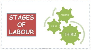
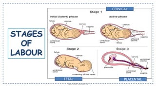
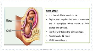
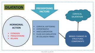
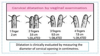
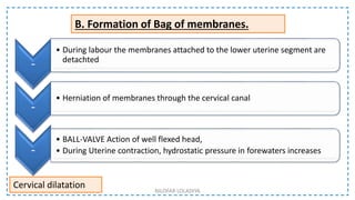
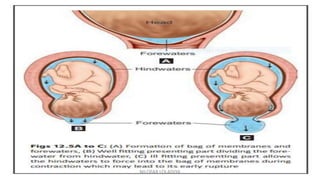
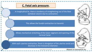
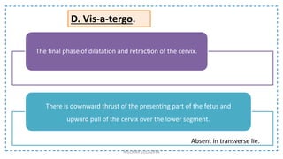
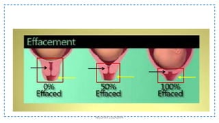
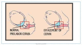
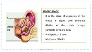
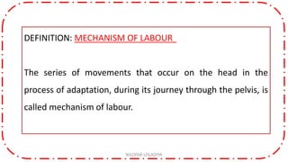
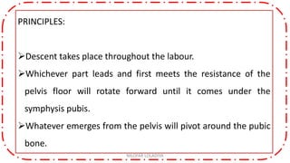
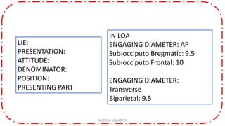
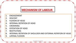
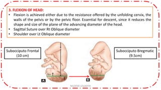
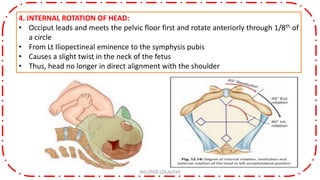
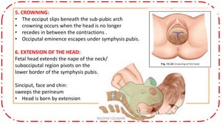
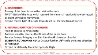
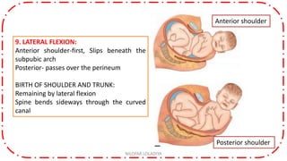
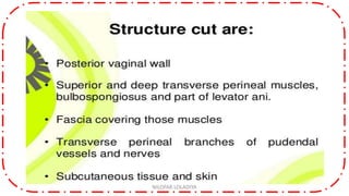
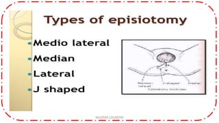
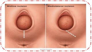
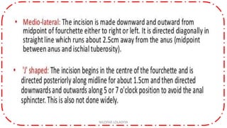
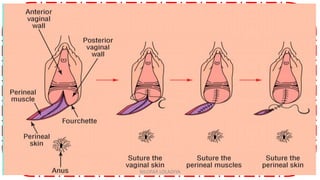
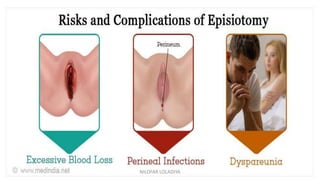
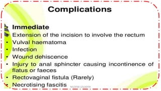
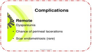
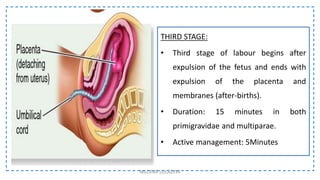
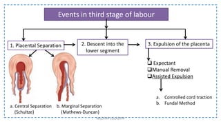
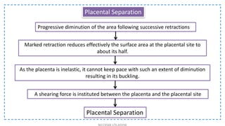
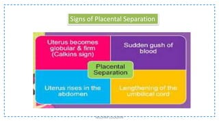
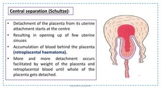
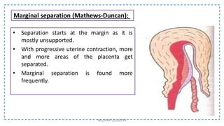
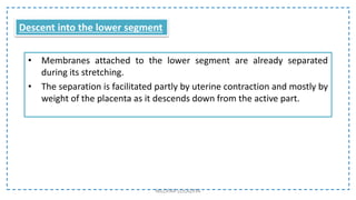
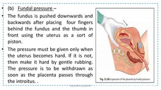
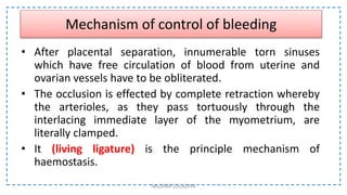
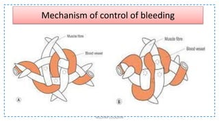
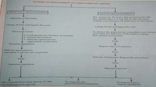
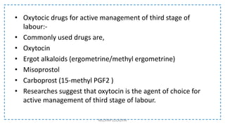
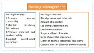
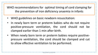
NILOFAR LOLADIYA
MSN: OBGY
Labor consists of a series of rhythmic, involuntary or medically induced contractions of the uterus that result in effacement (thinning and shortening) and dilation of the uterine cervix. The World Health Organization (WHO) defines normal birth as follows:
The birth is spontaneous in onset and low risk at the start of labor and remains so throughout labor and delivery.
The infant is born spontaneously in the vertex position between 37 and 42 weeks of pregnancy.
After birth, mother and infant are in good condition (1).
The stimulus for labor is unknown, but digitally manipulating or mechanically stretching the cervix during examination enhances uterine contractile activity, most likely by stimulating release of oxytocin by the posterior pituitary gland.
Normal labor usually begins within 2 weeks (before or after) the estimated delivery date. In a first pregnancy, labor usually lasts 12 to 18 hours on average; subsequent labors are often shorter, averaging 6 to 8 hours.
Labor begins with irregular uterine contractions of varying intensity; they apparently soften (ripen) the cervix, which begins to efface and dilate. As labor progresses, contractions increase in duration, intensity, and frequency.
Stages of labor
There are 3 stages of labor.
The 1st stage—from onset of labor to full dilation of the cervix (about 10 cm)—has 2 phases, latent and active.
During the latent phase, irregular contractions become progressively coordinated, discomfort is minimal, and the cervix effaces and dilates to 4 cm. The latent phase is difficult to time precisely, and duration varies, averaging 8 hours in nulliparas and 5 hours in multiparas; duration is considered abnormal if it lasts > 20 hours in nulliparas or > 12 hours in multiparas.
During the active phase, the cervix becomes fully dilated, and the presenting part descends well into the midpelvis. On average, the active phase lasts 5 to 7 hours in nulliparas and 2 to 4 hours in multiparas. Traditionally, the cervix was expected to dilate about 1.2 cm/hour in nulliparas and 1.5 cm/hour in multiparas. However, recent data suggest that slower progression of cervical dilation from 4 to 6 cm may be normal (1). Pelvic examinations are done every 2 to 3 hours to evaluate labor progress. Lack of progress in dilation and descent of the presenting part may indicate dystocia (fetopelvic disproportion).
Standing and walking shorten the first stage of labor by > 1 hour and reduce the rate of cesarean delivery
The placenta is said to be retained when it is not expelled from the uterus even 30 minutes after the delivery of the baby
Manual placenta removal is a procedure to remove a retained placenta from the uterus after childbirth.
The placenta is said to be retained when it is not expelled from the uterus even 30 minutes after the delivery of the baby
Manual placenta removal is a procedure to remove a retained placenta from the uterus after childbirth.
Definition-
The destructive operations are designed to diminish the bulk of the fetus so as to facilitate easy delivery through the birth canal
types
Craniotomy
Eviceration
Decapitation
Cleidotomy
CRANIOTOMY
Definition
It is an operation to make a perforation on the fetal head to evacuate the contents followed by extraction of the fetus
DECAPITATION
Definition
It is a destructive operation whereby the fetal head is severed from the trunk and the delivery is completed with the extraction of the trunk and that of the decapitated head per vaginam
CLEIDOTOMY
Definition
The operation consist of reduction in the bulk of the shoulder girdle by division of one or both the clavicles
Indications
Dead fetus with shoulder dystocia
Procedure
The clavicles are divided by the embryotomy scissors or long straight scissors introduced under the guidance of left two fingers placed inside the vagina
Fourth stage of labor: The hour or two after delivery when the tone of the uterus is reestablished as the uterus contracts again, expelling any remaining contents. These contractions are hastened by breastfeeding, which stimulates production of the hormone oxytocin.
What is the normal placenta
what is the Placental Abnormalities and
Hemorrhagic Complications during pregnancy
What is APH
How to manage The Hemorrhage
Physiological changes in second stage of laborDR MUKESH SAH
There is an interplay of physiological processes occurring during the second stage of labour. Second stage is said to have two phases, latent and active. It is during the latent phase that the presenting part passes through the fully dilated cervix to the birth canal.
The second stage of labor begins when the cervix is completely dilated (open), and ends with the birth of your baby. Contractions push the baby down the birth canal, and you may feel intense pressure, similar to an urge to have a bowel movement. Your health care provider may ask you to push with each contraction.
This topic contains definition, incidence, types, causes, diagnosis, mechanism, management of occipito posterior position and deep transverse arrest and manual rotation of occipito posterior position
Mrs. Nilofar Loladiya
Asst Professor
OBGY Dept
Dive into the world of nursing research with our comprehensive presentation on various types of research designs. This informative PPT delves into the intricacies of quantitative, qualitative, and mixed-methods research, highlighting their unique designs, applications, and implications in the nursing field. Understand the strengths and weaknesses of each approach and gain insights into how they contribute to evidence-based nursing practices. Whether you're a seasoned nurse or a nursing student, this presentation will provide valuable insights to enhance your research knowledge.
Comprehensive exploration of the diverse landscape of research designs within the nursing field. This presentation serves as a valuable resource for nurses, healthcare professionals, and students keen on understanding the nuances of various research methodologies and their impact on evidence-based nursing practices. With a focus on types of research designs, their advantages, and disadvantages, this presentation offers an in-depth journey into the world of nursing research.
In the dynamic realm of healthcare, research plays a pivotal role in shaping clinical practices and enhancing patient outcomes. This presentation begins by elucidating the fundamental categories of research designs: quantitative, qualitative, and mixed-methods. Each design is dissected to provide a clear understanding of its core principles, methodologies, and applications in nursing contexts.
Quantitative research, known for its structured and numerical approach, is explored with a spotlight on its ability to yield statistically significant results. The presentation delves into the process of hypothesis formulation, data collection through surveys or experiments, and the subsequent analysis methods. However, the presentation also acknowledges the challenges of quantitative research, such as potential bias and limited exploration of complex phenomena.
In contrast, qualitative research is presented as a qualitative exploration of experiences, beliefs, and motivations. The presentation explains the various techniques for data collection, including interviews, focus groups, and observations. It underscores the value of capturing rich, contextual insights that quantitative methods might overlook. Yet, qualitative research is not without its limitations, including potential subjectivity and difficulty in generalizing findings.
To bridge the gap between these two approaches, the presentation introduces mixed-methods research as a harmonious fusion of quantitative and qualitative methodologies. This section outlines the benefits of triangulating data, providing a more comprehensive understanding of research questions. However, it also acknowledges the challenges of managing the intricacies of both approaches within a single study.
Nilofar Loladiya
Assistant professor
nilofarlsalim@gmail.com
The Ethics are the moral principles that govern a person’s behaviour. Research ethics may be referred to as doing what is mostly and legally right in research.
Slides on the famous medical experiments, exploitation in research and development of ethical codes in research.
Major agencies and principles given by them on ethical standards in conducting research.
Details about ANA and INC ethical principles in nursing research.
How to write informed consent? What is ethical committee?
What is risk benefit ratio?
Nilofar Loladiya
MSN OBG
Simulation has been used widely in the clinical training of health-care students and
professionals. It is a valuable strategy for teaching, learning and evaluating clinical skills
at different levels of nursing and midwifery education: undergraduate, postgraduate and
lifelong education (Park et al., 2016; Martins, 2017).
Simulation has a positive impact on students, educators, and the individuals, groups
and communities they care for, as well as on education and health organizations. The
principal aims of simulation as a teaching method are to improve quality of care and
ensure patient safety.
The WHO document Transforming and scaling up health professionals’ education and training (WHO,
2013) strongly recommends the use of simulation. Recommendation 5 states:
Health professionals’ education and training institutions should use simulation methods
(high fidelity methods in settings with appropriate resources and lower fidelity methods in
resource limited settings) of contextually appropriate fidelity levels in the education of health
professionals.
A large proportion of nursing and midwifery education curricula worldwide is dedicated
to the acquisition of clinical skills. At the beginning of the learning period in clinical
settings, students should be able to develop safe and timely evidence-based interventions
without being interrupted by supervisors due to technical errors that may jeopardize
patients’ and students’ safety. In clinical practice with actual patients, students should
be self-confident and feel that others trust them; they should feel capable of performing
tasks without errors and be confident that the supervisor and other team members
believe in their abilities.
From an ethical perspective, invasive procedures should not be taught or practised on
real people; instead, trainees should be able to train in simulated, controlled and safe
environments, allowing them to make errors and learn from them with no harmful
consequences to any person. This ensures absolute respect for human rights by protecting
patients’ dignity and guarantees the quality of nursing care, even during health
professionals’ learning processes.
NILOFAR LOLADIYA
MSN: OBGY
The common thread uniting different types of nurses who work in varied areas is the nursing process—the essential core of practice for the registered nurse to deliver holistic, patient-focused care
One of the most important tools a nurse can use in practice is the nursing process. Although nursing schools teach first-year students about the nursing process, some nurses fail to grasp the impact its proper use can have on patient care. In this article, I will share information about the nursing process, its history, its purpose, its main characteristics, and the 5 steps involved in carrying out the nursing process.
• Establishes plans to meet patient needs
• Guides nurses in the delivery of high-quality evidence-based care
• Protects nurses against potential legal problems
• Promotes a systematic approach to patient care that all members of the nursing team can follow
The nursing process consists of five steps which encompass the care provided. The five nursing process steps are:
1. Assessment
2. Diagnosis
3. Planning
4. Implementation
5. Evaluation
NILOFAR LOLADIYA
MSN: OBGY
Pressure ulcers (also known as pressure sores or bedsores) are injuries to the skin and underlying tissue, primarily caused by prolonged pressure on the skin. They can happen to anyone, but usually affect people confined to bed or who sit in a chair or wheelchair for long periods of time.
t is widely believed that other factors can influence the tolerance of skin for pressure and shear, thereby increasing the risk of pressure ulcer development. These factors are protein-calorie malnutrition, microclimate (skin wetness caused by sweating or incontinence), diseases that reduce blood flow to the skin, such as arteriosclerosis, or diseases that reduce the sensation in the skin, such as paralysis or neuropathy. The healing of pressure ulcers may be slowed by the age of the person, medical conditions (such as arteriosclerosis, diabetes or infection), smoking or medications such as anti-inflammatory drugs.
Nilofar Loladiya
MSN: OBGY
Group Dynamics is a system of behaviors and psychological processes occurring within a social group or between social groups.
It is one of the important topics in nursing service and management.
263778731218 Abortion Clinic /Pills In Harare ,sisternakatoto
263778731218 Abortion Clinic /Pills In Harare ,ABORTION WOMEN’S CLINIC +27730423979 IN women clinic we believe that every woman should be able to make choices in her pregnancy. Our job is to provide compassionate care, safety,affordable and confidential services. That’s why we have won the trust from all generations of women all over the world. we use non surgical method(Abortion pills) to terminate…Dr.LISA +27730423979women Clinic is committed to providing the highest quality of obstetrical and gynecological care to women of all ages. Our dedicated staff aim to treat each patient and her health concerns with compassion and respect.Our dedicated group ABORTION WOMEN’S CLINIC +27730423979 IN women clinic we believe that every woman should be able to make choices in her pregnancy. Our job is to provide compassionate care, safety,affordable and confidential services. That’s why we have won the trust from all generations of women all over the world. we use non surgical method(Abortion pills) to terminate…Dr.LISA +27730423979women Clinic is committed to providing the highest quality of obstetrical and gynecological care to women of all ages. Our dedicated staff aim to treat each patient and her health concerns with compassion and respect.Our dedicated group of receptionists, nurses, and physicians have worked together as a teamof receptionists, nurses, and physicians have worked together as a team wwww.lisywomensclinic.co.za/
Title: Sense of Taste
Presenter: Dr. Faiza, Assistant Professor of Physiology
Qualifications:
MBBS (Best Graduate, AIMC Lahore)
FCPS Physiology
ICMT, CHPE, DHPE (STMU)
MPH (GC University, Faisalabad)
MBA (Virtual University of Pakistan)
Learning Objectives:
Describe the structure and function of taste buds.
Describe the relationship between the taste threshold and taste index of common substances.
Explain the chemical basis and signal transduction of taste perception for each type of primary taste sensation.
Recognize different abnormalities of taste perception and their causes.
Key Topics:
Significance of Taste Sensation:
Differentiation between pleasant and harmful food
Influence on behavior
Selection of food based on metabolic needs
Receptors of Taste:
Taste buds on the tongue
Influence of sense of smell, texture of food, and pain stimulation (e.g., by pepper)
Primary and Secondary Taste Sensations:
Primary taste sensations: Sweet, Sour, Salty, Bitter, Umami
Chemical basis and signal transduction mechanisms for each taste
Taste Threshold and Index:
Taste threshold values for Sweet (sucrose), Salty (NaCl), Sour (HCl), and Bitter (Quinine)
Taste index relationship: Inversely proportional to taste threshold
Taste Blindness:
Inability to taste certain substances, particularly thiourea compounds
Example: Phenylthiocarbamide
Structure and Function of Taste Buds:
Composition: Epithelial cells, Sustentacular/Supporting cells, Taste cells, Basal cells
Features: Taste pores, Taste hairs/microvilli, and Taste nerve fibers
Location of Taste Buds:
Found in papillae of the tongue (Fungiform, Circumvallate, Foliate)
Also present on the palate, tonsillar pillars, epiglottis, and proximal esophagus
Mechanism of Taste Stimulation:
Interaction of taste substances with receptors on microvilli
Signal transduction pathways for Umami, Sweet, Bitter, Sour, and Salty tastes
Taste Sensitivity and Adaptation:
Decrease in sensitivity with age
Rapid adaptation of taste sensation
Role of Saliva in Taste:
Dissolution of tastants to reach receptors
Washing away the stimulus
Taste Preferences and Aversions:
Mechanisms behind taste preference and aversion
Influence of receptors and neural pathways
Impact of Sensory Nerve Damage:
Degeneration of taste buds if the sensory nerve fiber is cut
Abnormalities of Taste Detection:
Conditions: Ageusia, Hypogeusia, Dysgeusia (parageusia)
Causes: Nerve damage, neurological disorders, infections, poor oral hygiene, adverse drug effects, deficiencies, aging, tobacco use, altered neurotransmitter levels
Neurotransmitters and Taste Threshold:
Effects of serotonin (5-HT) and norepinephrine (NE) on taste sensitivity
Supertasters:
25% of the population with heightened sensitivity to taste, especially bitterness
Increased number of fungiform papillae
Knee anatomy and clinical tests 2024.pdfvimalpl1234
This includes all relevant anatomy and clinical tests compiled from standard textbooks, Campbell,netter etc..It is comprehensive and best suited for orthopaedicians and orthopaedic residents.
Rasamanikya is a excellent preparation in the field of Rasashastra, it is used in various Kushtha Roga, Shwasa, Vicharchika, Bhagandara, Vatarakta, and Phiranga Roga. In this article Preparation& Comparative analytical profile for both Formulationon i.e Rasamanikya prepared by Kushmanda swarasa & Churnodhaka Shodita Haratala. The study aims to provide insights into the comparative efficacy and analytical aspects of these formulations for enhanced therapeutic outcomes.
These lecture slides, by Dr Sidra Arshad, offer a quick overview of the physiological basis of a normal electrocardiogram.
Learning objectives:
1. Define an electrocardiogram (ECG) and electrocardiography
2. Describe how dipoles generated by the heart produce the waveforms of the ECG
3. Describe the components of a normal electrocardiogram of a typical bipolar lead (limb II)
4. Differentiate between intervals and segments
5. Enlist some common indications for obtaining an ECG
6. Describe the flow of current around the heart during the cardiac cycle
7. Discuss the placement and polarity of the leads of electrocardiograph
8. Describe the normal electrocardiograms recorded from the limb leads and explain the physiological basis of the different records that are obtained
9. Define mean electrical vector (axis) of the heart and give the normal range
10. Define the mean QRS vector
11. Describe the axes of leads (hexagonal reference system)
12. Comprehend the vectorial analysis of the normal ECG
13. Determine the mean electrical axis of the ventricular QRS and appreciate the mean axis deviation
14. Explain the concepts of current of injury, J point, and their significance
Study Resources:
1. Chapter 11, Guyton and Hall Textbook of Medical Physiology, 14th edition
2. Chapter 9, Human Physiology - From Cells to Systems, Lauralee Sherwood, 9th edition
3. Chapter 29, Ganong’s Review of Medical Physiology, 26th edition
4. Electrocardiogram, StatPearls - https://www.ncbi.nlm.nih.gov/books/NBK549803/
5. ECG in Medical Practice by ABM Abdullah, 4th edition
6. Chapter 3, Cardiology Explained, https://www.ncbi.nlm.nih.gov/books/NBK2214/
7. ECG Basics, http://www.nataliescasebook.com/tag/e-c-g-basics
These simplified slides by Dr. Sidra Arshad present an overview of the non-respiratory functions of the respiratory tract.
Learning objectives:
1. Enlist the non-respiratory functions of the respiratory tract
2. Briefly explain how these functions are carried out
3. Discuss the significance of dead space
4. Differentiate between minute ventilation and alveolar ventilation
5. Describe the cough and sneeze reflexes
Study Resources:
1. Chapter 39, Guyton and Hall Textbook of Medical Physiology, 14th edition
2. Chapter 34, Ganong’s Review of Medical Physiology, 26th edition
3. Chapter 17, Human Physiology by Lauralee Sherwood, 9th edition
4. Non-respiratory functions of the lungs https://academic.oup.com/bjaed/article/13/3/98/278874
Muktapishti is a traditional Ayurvedic preparation made from Shoditha Mukta (Purified Pearl), is believed to help regulate thyroid function and reduce symptoms of hyperthyroidism due to its cooling and balancing properties. Clinical evidence on its efficacy remains limited, necessitating further research to validate its therapeutic benefits.
2. Clausen et all, 1973
Labour is rhythmic contraction and relaxation of the
uterine muscles with progressive effacement i.e thinning
and dilatation i.e. opening of the cervix, leading to
expulsion of the products of conception.
DEFINITION
NILOFAR LOLADIYA
3. D.C DUTTA
A series of events that take place in the genital organs
in an effort to expel the viable products of conception
out of the womb into vagina into the outer world is
called labour.
DEFINITION
NILOFAR LOLADIYA
7. FIRST STAGE:
• It is that of dilatation of cervix.
• Begins with regular rhythmic contraction
and is complete when cervix is fully
dilated and effaced.
• In other words it is the cervical stage.
• Primigravida- 12 hours
• Multipara- 6 hours
NILOFAR LOLADIYA
12. Too much Fibrosis as in chronic cervicitis, prolapse or
organic lesion in the cervix as in carcinoma results in
deficiency of these factors. As a result cervix may fail to
dilate.
NILOFAR LOLADIYA
13. A. Uterine Contraction and retraction.
B. Formation of Bag of membranes.
C. Fetal axis pressure.
D. Vis-a-tergo.
1. DILATATION
2. EFFACEMENT
3.FORMATION OF LOWER UTERINE SEGMENT
EVENTS IN FIRST STAGE OF LABOUR
NILOFAR LOLADIYA
14. • The longitudinal muscle fibres of the upper segment are attached with
circular muscle fibres of the lower segment and upper part of the cervix in
the bucket holding fashion.
• Thus, with each uterine contraction not only the canal is opened up from
above down but it also becomes shortened and retracted.
• Upper segment contract, retracts and pushes the foetus,
• The lower segment and cervix dilate in response to the forces of contraction
of upper segment.
A. Uterine Contraction and retraction
NILOFAR LOLADIYA
16. B. Formation of Bag of membranes.
-
• During labour the membranes attached to the lower uterine segment are
detachted
-
• Herniation of membranes through the cervical canal
-
• BALL-VALVE Action of well flexed head,
• During Uterine contraction, hydrostatic pressure in forewaters increases
Cervical dilatation NILOFAR LOLADIYA
18. C. Fetal axis pressure.
In longitudinal lie, there is tendency of straightening out of the fetal
vertebral column due to contraction.
This allows the fundal contraction to transmit.
Allows mechanical stretching of the lower segment and opening of the
cervical Canal.
With each uterine contraction, there is elongation of the uterine ovoid and
decrease in the transverse diameter.
Absent in transverse lie.
NILOFAR LOLADIYA
19. D. Vis-a-tergo.
The final phase of dilatation and retraction of the cervix.
There is downward thrust of the presenting part of the fetus and
upward pull of the cervix over the lower segment.
Absent in transverse lie.
NILOFAR LOLADIYA
20. Effacement is the process by which the muscular fibres of the
cervix are pulled upwards and merges with the fibres of the lower
uterine segment.
Shortening of cervical Canal from its usual length of 2 to 3
centimetres to one in which the cervical Canal is obliterated
leaving only the external os as a circular orifice with thin edges.
NILOFAR LOLADIYA
2. EFFACEMENT or ‘taking up’ of the Cervix.
23. Expulsion of mucus plug is caused by effacement.
The cervix becomes thin during first stage of labour or even
before that in primigravida.
In primigravida effacement proceed dilatation of the cervix,
where as in multipara both occur simultaneously.
NILOFAR LOLADIYA
25. 3.FORMATION OF LOWER UTERINE SEGMENT
Before the onset of labour there is no complete Anatomical or functional
division of the uterus.
During labour, the demarcation of an active upper segment and a relatively
passive lower segment..
The wall of the upper segment becomes progressively thickened with
progressive thinning of the lower segment.
More pronounced in late first stage, especially after rupture of membranes.
NILOFAR LOLADIYA
28. ANATOMICAL FEATURES:
Developed from Isthmus.
Above: Physiological ring.
Below: Fibromuscular junction
of cervix and uterus.
7.5-10 cm when fully formed.
Gradually becomes thin.
NILOFAR LOLADIYA
29. CLINICAL SIGNIFICANCE:
Enables expulsion of fetus.
Caesarean section is performed at this segment.
Poor decidual reaction facilitates morbid adherent placenta.
In obstructed labour, it gets stretched and thinned and ultimately
ruptures.
Passive segment, Poor retractile property
Chances of PPH if placenta implanted here.
NILOFAR LOLADIYA
31. SECOND STAGE:
• It is the stage of expansion of the
foetus it begins with complete
dilation of the cervix through
complete birth of a baby.
• Primigravida- 2 hours
• Multipara- 30 mins
NILOFAR LOLADIYA
32. The second stage begins with the complete dilatation of the cervix
and ends with the expulsion of the fetus.
It is concerned with the descent and delivery of the fetus through
the birth canal.
A. Propulsive Phase:
Full Dilatation-head touches the pelvic floor
B. Expulsive Phase:
Irresistible desire to ‘bear down’ and push until the baby is
delivered.
NILOFAR LOLADIYA
33. Expulsive uterine contractions
Rupture of the forewaters
Dilatation and gaping of the anus
Appearance of the presenting part
Show
Congestion of the vulva
SIGNS
NILOFAR LOLADIYA
34. DEFINITION: MECHANISM OF LABOUR
The series of movements that occur on the head in the
process of adaptation, during its journey through the pelvis, is
called mechanism of labour.
NILOFAR LOLADIYA
35. PRINCIPLES:
Descent takes place throughout the labour.
Whichever part leads and first meets the resistance of the
pelvis floor will rotate forward until it comes under the
symphysis pubis.
Whatever emerges from the pelvis will pivot around the pubic
bone.
NILOFAR LOLADIYA
37. 1. ENGAGEMENT
2. DESCENT
3. FLEXION OF HEAD
4. INTERNAL ROTATION OF HEAD
5. CROWNING
6. EXTENTION OF HEAD
7. RESTITUTION:
8. INTERNAL ROTATION OF SHOULDER AND EXTERNAL ROTATION OF HEAD
9. LATERAL FLEXION
NILOFAR LOLADIYA
MECHANISM OF LABOUR
38. 1. ENGAGEMENT:
The head enters the brim of the pelvis and gets fixed.
2. DESCENT:
Slow progressive throughout the labour
Factors responsible are-
• Uterine contractions and retractions
• Bearing down efforts
• Straightening of fetal ovoid specially after rupture of membranes
NILOFAR LOLADIYA
39. 3. FLEXION OF HEAD:
• Flexion is achieved either due to the resistance offered by the unfolding cervix, the
walls of the pelvis or by the pelvic floor. Essential for descent, since it reduces the
shape and size of the plane of the advancing diameter of the head.
• Sagittal Suture over Rt Oblique diameter
• Shoulder over Lt Oblique diameter
NILOFAR LOLADIYA
Subocciputo Frontal
(10 cm)
Subocciputo Bregmatic
(9.5cm)
40. 4. INTERNAL ROTATION OF HEAD:
• Occiput leads and meets the pelvic floor first and rotate anteriorly through 1/8th of
a circle
• From Lt Iliopectineal eminence to the symphysis pubis
• Causes a slight twist in the neck of the fetus
• Thus, head no longer in direct alignment with the shoulder
NILOFAR LOLADIYA
41. 5. CROWNING:
• The occiput slips beneath the sub-pubic arch
• crowning occurs when the head is no longer
• recedes in between the contractions .
• Occiputal eminence escapes under symphysis pubis.
6. EXTENSION OF THE HEAD:
Fetal head extends the nape of the neck/
subocciputal region pivots on the
lower border of the symphysis pubis.
Sinciput, face and chin:
sweeps the perineum
• Head is born by extension
NILOFAR LOLADIYA
42. 7. RESTITUTION:
Turning of the head to undo the twist in the neck
TWIST- Neck of the fetus which resulted from internal rotation is now corrected
by slight untwisting movement.
Occiput moves 1/8th of a circle towards left i.e. the side from it started
8. INTERNAL ROTATION OF SHOULDER
From Lt oblique to AP diameter
Anterior shoulder reaches the Rt side of the pelvic floor
Rotates forward bringing shoulder into the AP diameter of outlet
EXTERNAL ROTATION OF HEAD: Occiput-further 1/8th circle the same direction
as restitution
Occiput lies laterally, faces the opposite thigh
NILOFAR LOLADIYA
43. 9. LATERAL FLEXION:
Anterior shoulder-first, Slips beneath the
subpubic arch
Posterior- passes over the perineum
BIRTH OF SHOULDER AND TRUNK:
Remaining by lateral flexion
Spine bends sideways through the curved
canal
NILOFAR LOLADIYA
Anterior shoulder
Posterior shoulder
44. MANAGEMENT OF THE SECOND STAGE
The transition from the first stage to the second stage is
evidenced by the following features:
Increasing intensity of uterine contractions.
Appearance of bearing down efforts.
Urge to defecate with descent of the presenting part.
Complete dilatation of the cervix as evidenced on vaginal
examination
NILOFAR LOLADIYA
45. PRINCIPLES:
(1)To assist in the natural expulsion of the fetus slowly and steadily,
(2)To prevent perineal injuries.
GENERAL MEASURES:
— The patient should be in bed.
— Constant supervision is mandatory and the FHR is recorded at every 5 minutes.
— To administer inhalation analgesics, if available, in the form of Gas N2O and O2
to relieve pain during contractions.
— Vaginal examination is done at the beginning of the second stage not only to
confirm its onset but to detect any accidental cord prolapse.
The position and the station of the head are once more to be reviewed and the
progressive descent of the head is ensured
NILOFAR LOLADIYA
46. PREPARATION FOR DELIVERY
— Position: Positions of the woman during delivery may be lateral
or partial sitting. Dorsal position with 15° left lateral tilt is
commonly favored as it avoids aortocaval compression and
facilitates pushing effort.
—Scrubs up and puts on sterile gown, mask and gloves and stands
on the right side of the table.
— Toileting the external genitalia and inner side of the thighs is
done with cotton swabs soaked in Savlon or Dettol solution.
NILOFAR LOLADIYA
47. One sterile sheet is placed beneath the buttocks of the
patient and one over the abdomen. Sterilized leggings
are to be used.
Essential aseptic procedures are remembered as 3 ‘C’s:
(a)Clean hands,
(b)Clean surface and
(c)Clean cutting and ligaturing of the cord.
— To catheterize the bladder, if it is full.
NILOFAR LOLADIYA
49. Perineal lacerations:
1st = involving skin or vaginal mucosa BUT NOT into muscle
2nd = extending from skin & vaginal mucosa into m
muscles of perineum
3rd = extending from skin, vaginal muscosa into the anal
sphincter
4th = extending through the rectal mucosa into the lumen of
the rectum NILOFAR LOLADIYA
72. THIRD STAGE:
• Third stage of labour begins after
expulsion of the fetus and ends with
expulsion of the placenta and
membranes (after-births).
• Duration: 15 minutes in both
primigravidae and multiparae.
• Active management: 5Minutes
NILOFAR LOLADIYA
73. Events in third stage of labour
NILOFAR LOLADIYA
1. Placental Separation
a. Central Separation
(Schultze)
2. Descent into the
lower segment
3. Expulsion of the placenta
Expectant
Manual Removal
Assisted Expulsion
b. Marginal Separation
(Mathews-Duncan)
a. Controlled cord traction
b. Fundal Method
74. Placental Separation
Progressive diminution of the area following successive retractions
Marked retraction reduces effectively the surface area at the placental site to
about its half.
As the placenta is inelastic, it cannot keep pace with such an extent of diminution
resulting in its buckling.
A shearing force is instituted between the placenta and the placental site
Placental Separation
76. • Detachment of the placenta from its uterine
attachment starts at the centre
• Resulting in opening up of few uterine
sinuses
• Accumulation of blood behind the placenta
(retroplacental haematoma).
• More and more detachment occurs
facilitated by weight of the placenta and
retroplacental blood until whole of the
placenta gets detached.
NILOFAR LOLADIYA
Central separation (Schultze):
77. • Separation starts at the margin as it is
mostly unsupported.
• With progressive uterine contraction, more
and more areas of the placenta get
separated.
• Marginal separation is found more
frequently.
NILOFAR LOLADIYA
Marginal separation (Mathews-Duncan):
78. • Membranes attached to the lower segment are already separated
during its stretching.
• The separation is facilitated partly by uterine contraction and mostly by
weight of the placenta as it descends down from the active part.
NILOFAR LOLADIYA
Descent into the lower segment
79. Expulsion of placenta
• After complete separation of the placenta, it is forced
down into the flabby lower uterine segment or upper
part of the vagina by effective contraction and
retraction of the uterus.
• Thereafter, it is expelled out by either voluntary
contraction of abdominal muscles (bearing down
efforts) or by manual procedure.
NILOFAR LOLADIYA
80. • Expulsion of placenta and membranes:-
• The expulsion is achieved either by voluntary bearing
down efforts or more commonly aided by manipulative
procedure.
• The “after-birth” delivery is soon followed by slight to
moderate bleeding amounting to 100-250 ml.
• Maternal signs:- There may be chills and occasional
shivering. Slight transient hypotension is not unusual.
NILOFAR LOLADIYA
81. Assisted expulsion:
• (A) Controlled cord traction
(modified Brandt – Andrews
method) –
• The palmar surface of the
fingers of the left hand is
placed (above the symphysis
pubis) approximately at the
junction of upper and lower
uterine segment.
NILOFAR LOLADIYA
82. Assisted expulsion:
(A) Controlled cord traction
The body of the uterus is pushed
upwards and backwards, towards
the umbilicus while by the right
hand steady tension is given in
downward and backward direction
holding the clamp until the placenta
comes outside the introitus.
NILOFAR LOLADIYA
83. Assisted expulsion:
(A) Controlled cord traction
• It is thus more an uterine
elevation which facilitates
expulsion of the placenta.
The procedure is to be
adopted only when the
uterus is hard and
contracted.
NILOFAR LOLADIYA
84. • (b) Fundal pressure –
• The fundus is pushed downwards and
backwards after placing four fingers
behind the fundus and the thumb in
front using the uterus as a sort of
piston.
• The pressure must be given only when
the uterus becomes hard. If it is not,
then make it hard by gentle rubbing.
The pressure is to be withdrawn as
soon as the placenta passes through
the introitus. .
NILOFAR LOLADIYA
85. • (b) Fundal pressure –
• If the baby is macerated or premature, this method is
preferable to cord traction as the tensile strength of the
cord is much reduced in both instances.
• The cord may be accidentally torn which is not likely to
cause any problem. The sterile gloved hand should be
introduced and the placenta is to be grasped and
extracted.
NILOFAR LOLADIYA
86. Mechanism of control of bleeding
• After placental separation, innumerable torn sinuses
which have free circulation of blood from uterine and
ovarian vessels have to be obliterated.
• The occlusion is effected by complete retraction whereby
the arterioles, as they pass tortuously through the
interlacing immediate layer of the myometrium, are
literally clamped.
• It (living ligature) is the principle mechanism of
haemostasis.
NILOFAR LOLADIYA
89. • Oxytocic drugs for active management of third stage of
labour:-
• Commonly used drugs are,
• Oxytocin
• Ergot alkaloids (ergometrine/methyl ergometrine)
• Misoprostol
• Carboprost (15-methyl PGF2 )
• Researches suggest that oxytocin is the agent of choice for
active management of third stage of labour.
NILOFAR LOLADIYA
90. Nursing Management
Nursing Priorities:-
1.Promote uterine
contractility
2.Maintain circulating
fluid volume
3.Promote maternal and
newborn safety
4.Support parent-infant
interaction
NILOFAR LOLADIYA
Nursing assessment:
Blood pressure and pulse rate
Amount of blood loss
Leg cramps/body tremors
Uterine contractions
Shape and level of fundus
Signs of placental separation
Extent of perineal laceration/episiotomy
Completeness of placenta and membranes
91. WHO recommendations for optimal timing of cord clamping for
the prevention of iron deficiency anaemia in infants
• WHO guidelines on basic newborn resuscitation:
• In newly born term or preterm babies who do not require
positive-pressure ventilation, the cord should not be
clamped earlier than 1 min after birth.
• When newly born term or preterm babies require positive-
pressure ventilation, the cord should be clamped and cut
to allow effective ventilation to be performed.
NILOFAR LOLADIYA
92. • Newly born babies who do not breathe spontaneously after
thorough drying should be stimulated by rubbing the back 2–3
times before clamping the cord and initiating positive-pressure
ventilation.
• WHO recommendations for the prevention and treatment of
postpartum haemorrhage
• Late cord clamping (performed approximately 1–3 min after
birth) is recommended for all births, while initiating
simultaneous essential neonatal care.
• Early umbilical cord clamping (less than 1 min after birth) is not
recommended unless the neonate is asphyxiated and needs to
be moved immediately for resuscitation.
NILOFAR LOLADIYA