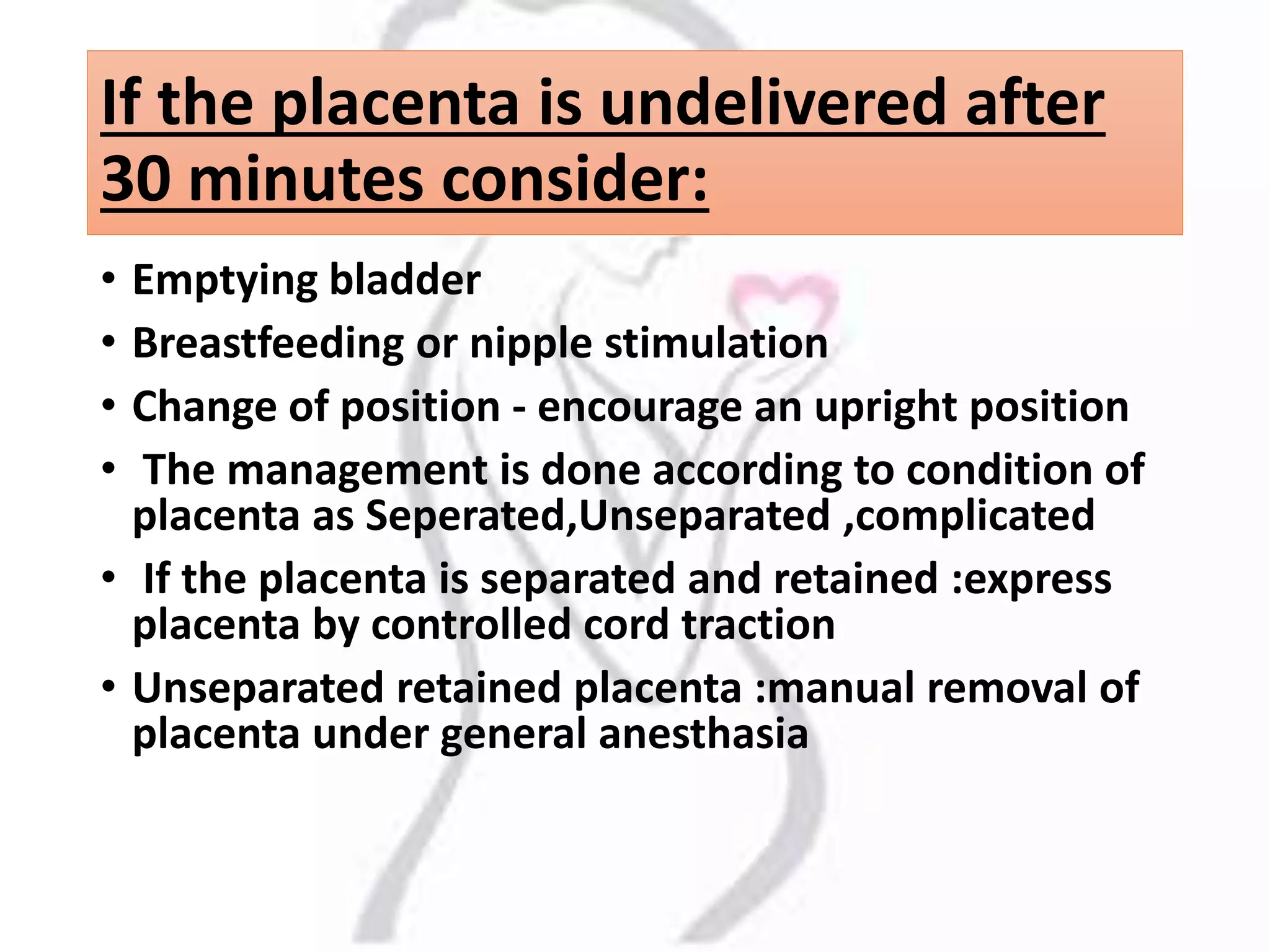
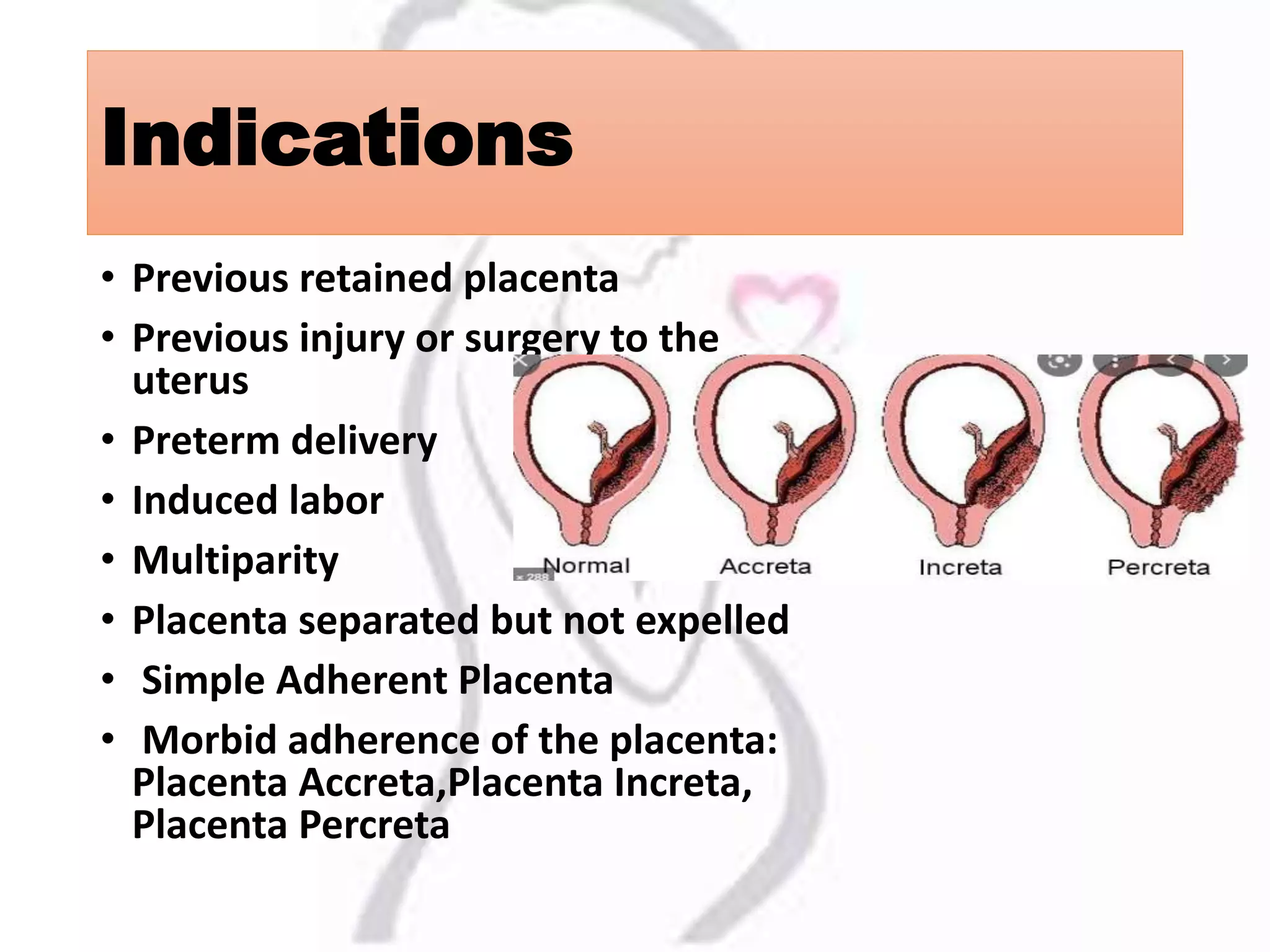
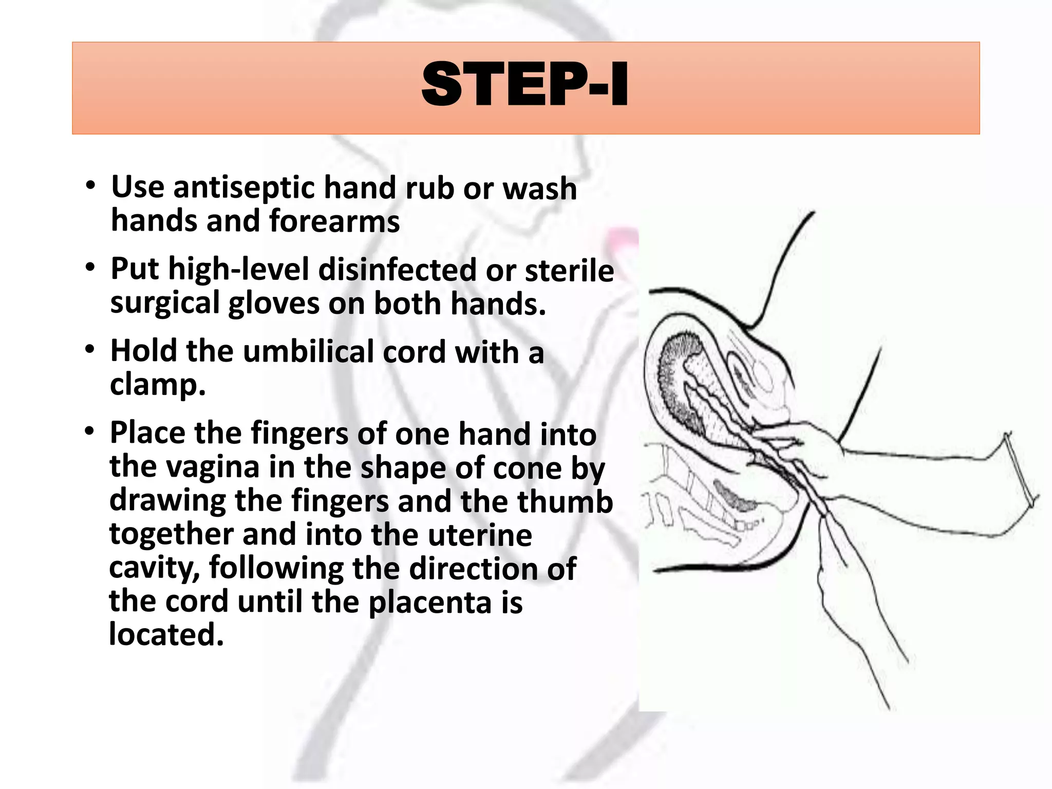
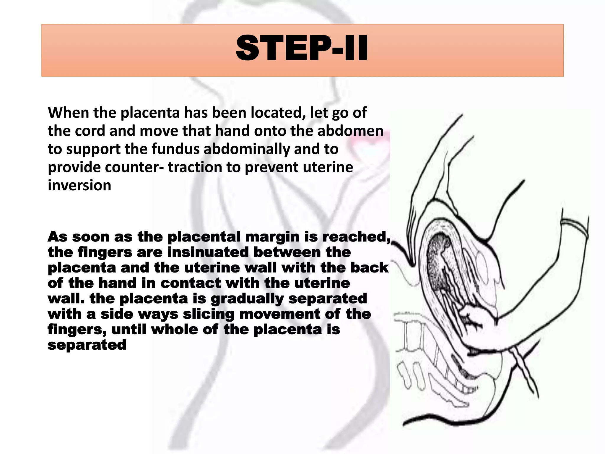
Manual removal of placenta
Ms. khushboo singh
Nursing tutor
Gold medalist
DEFINITION OF MANUAL
REMOVAL OF PLACENTA
• The placenta is said to be retained when it is not
expelled from the uterus even 30 minutes after
the delivery of the baby
• Manual placenta removal is a procedure to
remove a retained placenta from the uterus after
childbirth.
If the placenta is undelivered after
30 minutes consider:
• Emptying bladder
• Breastfeeding or nipple stimulation
• Change of position - encourage an upright position
• The management is done according to condition of
placenta as Seperated,Unseparated ,complicated
• If the placenta is separated and retained :express
placenta by controlled cord traction
• Unseparated retained placenta :manual removal of
placenta under general anesthasia
Indications
• Previous retained placenta
• Previous injury or surgery to the
uterus
• Preterm delivery
• Induced labor
• Multiparity
• Placenta separated but not expelled
• Simple Adherent Placenta
• Morbid adherence of the placenta:
Placenta Accreta,Placenta Increta,
Placenta Percreta
Prepare the necessary
equipment
• Antiseptic solution
•Sterile gloves
•Blood and subtitutes
• Anasthesia and analgesics
•Ergometrine and oxytocin
•Antibiotics
•
Give anesthetic and
analgesic as per need
• Give anesthesia (IV pethidine (25-50mg) and
diazepam (10 mg), or ketamine
• Give a single dose of prophylactic antibiotics:
Ampicillin 2 g IV PLUS metronidazole 500 mg IV,
OR Cefazolin 1 g IV PLUS metronidazole 500 mg IV
• Put on personal protective equipment.
STEP-I
STEP-II
When the placenta has been located, let go of
the cord and move that hand onto the abdomen
to support the fundus abdominally and to
provide counter- traction to prevent uterine
inversion
As soon as the placental margin is reached,
the fingers are insinuated between the
placenta and the uterine wall with the back
of the hand in contact with the uterine
wall. the placenta is gradually separated
with a side ways slicing movement of the
fingers, until whole of the placenta is
separated
STEP-III
When the placenta is completely
separated
Palpate the inside of the uterine
cavity to ensure that all placental
tissue has been removed.
Slowly withdraw the hand from
the uterus bringing the placenta
with it.
Continue to provide counter-
traction to the fundus by pushing
it in the opposite direction of the
hand that is being withdrawn
STEP IV
• Give oxytocin 20 units in 1 L IV fluid (normal saline or
Ringer’s lactate) at 60 drops/minute.
• Massage the fundus to encourage atonic uterine
contraction. If there is continued heavy bleeding.
• Give ergometrine 0.2 mg IM or give prostaglandins
Intravenous methergine 0.2 mg isgiven.
• Examine the uterine surface of the placenta to ensure
that it is complete.
• Examine the woman carefully and repair any tears to the
cervix or vagina, or repair episiotomy.
• Check for contracted uterus
POST-PROCEDURE CARE
• Observe the woman closely until the effect of IV
sedation has worn off.
• Monitor the vital signs (pulse, blood pressure,
respiration) every 30 minutes for the next 6 hours or
until stable.
• Palpate the uterine fundus to ensure that the uterus
remains contracted.
• Check for excessive lochia.
• Continue infusion of IV fluids.
• Transfuse as necessary.
MANUAL REMOVAL OF PLACENTA.

MANUAL REMOVAL OF PLACENTA.

  • 1.
    Manual removal ofplacenta Ms. khushboo singh Nursing tutor Gold medalist
  • 2.
    DEFINITION OF MANUAL REMOVALOF PLACENTA • The placenta is said to be retained when it is not expelled from the uterus even 30 minutes after the delivery of the baby • Manual placenta removal is a procedure to remove a retained placenta from the uterus after childbirth.
  • 3.
    If the placentais undelivered after 30 minutes consider: • Emptying bladder • Breastfeeding or nipple stimulation • Change of position - encourage an upright position • The management is done according to condition of placenta as Seperated,Unseparated ,complicated • If the placenta is separated and retained :express placenta by controlled cord traction • Unseparated retained placenta :manual removal of placenta under general anesthasia
  • 4.
    Indications • Previous retainedplacenta • Previous injury or surgery to the uterus • Preterm delivery • Induced labor • Multiparity • Placenta separated but not expelled • Simple Adherent Placenta • Morbid adherence of the placenta: Placenta Accreta,Placenta Increta, Placenta Percreta
  • 5.
    Prepare the necessary equipment •Antiseptic solution •Sterile gloves •Blood and subtitutes • Anasthesia and analgesics •Ergometrine and oxytocin •Antibiotics •
  • 6.
    Give anesthetic and analgesicas per need • Give anesthesia (IV pethidine (25-50mg) and diazepam (10 mg), or ketamine • Give a single dose of prophylactic antibiotics: Ampicillin 2 g IV PLUS metronidazole 500 mg IV, OR Cefazolin 1 g IV PLUS metronidazole 500 mg IV • Put on personal protective equipment.
  • 7.
  • 8.
    STEP-II When the placentahas been located, let go of the cord and move that hand onto the abdomen to support the fundus abdominally and to provide counter- traction to prevent uterine inversion As soon as the placental margin is reached, the fingers are insinuated between the placenta and the uterine wall with the back of the hand in contact with the uterine wall. the placenta is gradually separated with a side ways slicing movement of the fingers, until whole of the placenta is separated
  • 9.
    STEP-III When the placentais completely separated Palpate the inside of the uterine cavity to ensure that all placental tissue has been removed. Slowly withdraw the hand from the uterus bringing the placenta with it. Continue to provide counter- traction to the fundus by pushing it in the opposite direction of the hand that is being withdrawn
  • 10.
    STEP IV • Giveoxytocin 20 units in 1 L IV fluid (normal saline or Ringer’s lactate) at 60 drops/minute. • Massage the fundus to encourage atonic uterine contraction. If there is continued heavy bleeding. • Give ergometrine 0.2 mg IM or give prostaglandins Intravenous methergine 0.2 mg isgiven. • Examine the uterine surface of the placenta to ensure that it is complete. • Examine the woman carefully and repair any tears to the cervix or vagina, or repair episiotomy. • Check for contracted uterus
  • 12.
    POST-PROCEDURE CARE • Observethe woman closely until the effect of IV sedation has worn off. • Monitor the vital signs (pulse, blood pressure, respiration) every 30 minutes for the next 6 hours or until stable. • Palpate the uterine fundus to ensure that the uterus remains contracted. • Check for excessive lochia. • Continue infusion of IV fluids. • Transfuse as necessary.