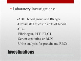
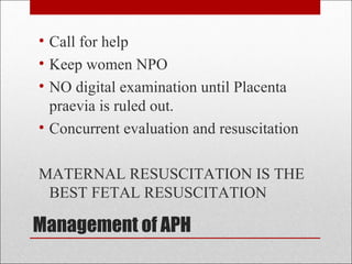
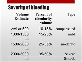
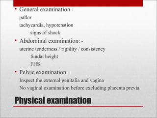
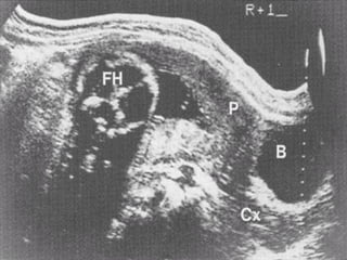
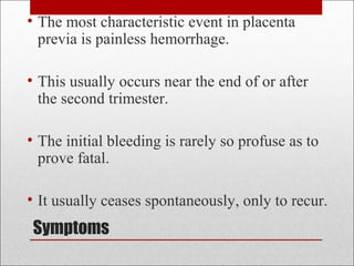
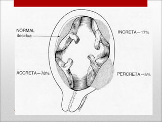
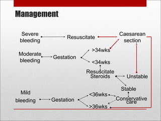
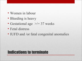
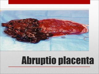
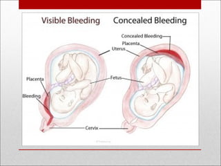
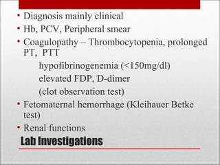
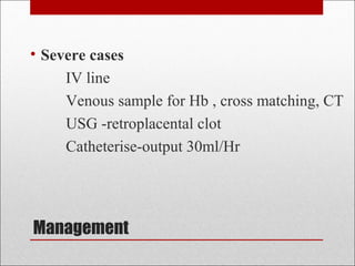
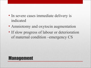
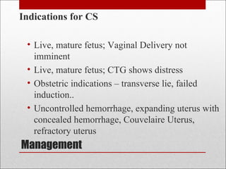
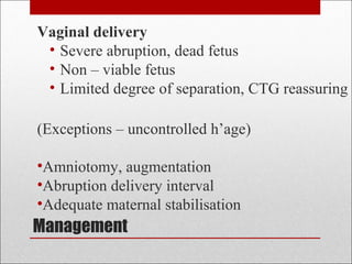
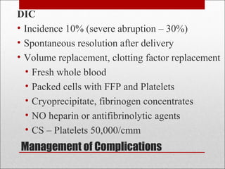
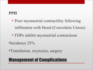
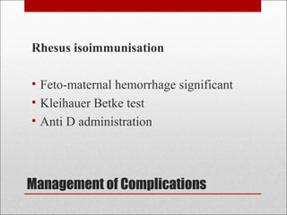
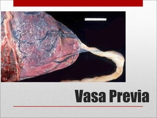
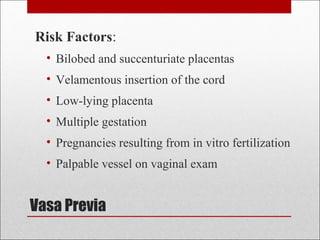
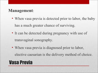
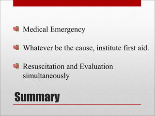
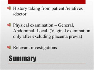
3. ⢠Bleeding per vaginum after 20 weeksâ
gestation.
⢠Complicates close to 4% of all pregnancies
and is a MEDICAL EMERGENCY!
⢠One of 3 leading causes of Maternal
mortality
⢠Major contributor to Perinatal loss
4. What are the most common causes of
Antepartum Hemorrhage ?
7. First aid management
⢠Insert 2 wide bore cannulae
⢠Blood sample
⢠Immediately start IV crystalloid solutions
⢠Provide 100% oxygen via mask
⢠Warm the women
⢠Insert Foley catheter
⢠Monitor blood pressure and pulse/ 5 min
⢠Monitor urine output /hour
8. Investigations
⢠Laboratory investigations:
-ABO blood group and Rh type
-Crossmatch atleast 2 units of blood
-CBC
-Fibrinogen, PTT, PT,CT
-Serum creatinine or BUN
-Urine analysis for protein and RBCs
9. Management of APH
⢠Call for help
⢠Keep women NPO
⢠NO digital examination until Placenta
praevia is ruled out.
⢠Concurrent evaluation and resuscitation
MATERNAL RESUSCITATION IS THE
BEST FETAL RESUSCITATION
10. Management of APH
MATERNAL EVALUATION
⢠History-
Trauma , Intercourse,
Pain? fetal movements?
Previous ANC , USG
Other complications like HT
Past Obstetric history
11. Management of APH
⢠Maternal Haemodynamic assessment
⢠Clinical signs â may be misleading
assess urine output
CVP catheter
⢠Treat Shock
IV line
Colloids / Blood & Blood Products
OXYGEN
Correct coagulopathy
13. Physical examination
⢠General examination:-
pallor
tachycardia, hypotenstion
signs of shock
⢠Abdominal examination: -
uterine tenderness / rigidity / consistency
fundal height
FHS
⢠Pelvic examination:
Inspect the external genitalia and vagina
No vaginal examination before excluding placenta previa
14. Ultrasound
⢠Confirm the fetal viability
⢠Localize the site of placenta
⢠Estimating the gestational age
⢠Retroplacental hematoma
⢠In case of severe bleeding, first aid management and
then quickly start active management .
⢠Even if the amount of bleeding is mild, NEVER
perform PV examination until placenta previa has
been excluded by US
15. Diagnosis â Placenta Previa
⢠Painless vaginal bleeding
⢠Symptoms and signs
-shock
-bleeding may be precipitated by intercourse
-relaxed uterus
-normal fetal condition
-abnormal presentations and lie
17. ⢠Mild vaginal bleeding after 24 w(mild)
⢠Symptoms and signs
-clinically stable
-fetal assessment showed fetal distress
that cannot be explained by the mild bleeding.
Diagnosis -Vasa previa
19. Placenta Previa
Defined as a placenta implanted in the
lower segment of the uterus, presenting
ahead of the leading pole of the fetus.
20. Placenta Previa
⢠Incidence about 1 in 300
⢠Perinatal morbidity and mortality are
primarily related to the complications of
prematurity, because the hemorrhage is
maternal.
25. Symptoms
⢠The most characteristic event in placenta
previa is painless hemorrhage.
⢠This usually occurs near the end of or after
the second trimester.
⢠The initial bleeding is rarely so profuse as to
prove fatal.
⢠It usually ceases spontaneously, only to recur.
26. Signs
⢠Tachycardia or hypotension
depending on blood loss
⢠Anemia
⢠Uterus relaxed
⢠Abnormal lie or presentation
⢠Fetal heart present
27. Complications
- shock
- postpartum hemorrhage
- high risk for PPH and
- placenta accreta/ increta/ percreta
a common finding is at the site of a previous
cesarean section
28.
29. Diagnosis
The diagnosis of placenta previa can seldom
be established firmly by clinical examination.
Vaginal examination is never permissible
because even the gentlest examination of this
sort can cause torrential hemorrhage.
30. Diagnosis
⢠The simplest and safest method of placental
localization is provided by transabdominal
sonography.
⢠Transvaginal ultrasonography has substantively
improved diagnostic accuracy of placenta previa.
⢠MRI
⢠At 18 weeks, 5-10% of placentas are low lying.
31. Transvaginal ultrasound is safe in the presence
of placenta praevia and is more accurate than
transabdominal ultrasound in locating the
placenta.
Diagnosis
33. Indications to terminate
⢠Women in labour
⢠Bleeding is heavy
⢠Gestational age >/= 37 weeks
⢠Fetal distress
⢠IUFD and /or fatal congenital anomalies
35. Management
⢠Delivery is by Caesarean section in
almost all cases.
⢠Occasionally Caesarean hysterectomy
necessary.
36. Management
⢠In cases of previous CS with anterior
placenta previa, high chance of adherent
placenta
⢠USG Doppler to confirm
⢠Elective classical caesarean hysterectomy in
such cases
39. Abruptio Placentae
⢠Incidence: 0.5 â 1.3%
severe abruption : 1 in 400 deliveries
⢠PNM rate â up to 25%
accounts for 12-14 % of all stillbirths
adverse sequelae in survivors
⢠Maternal morbidity and mortality
40. Abruptio placenta
⢠Bleeding with placental abruption is almost
always maternal.
⢠Significant fetal bleeding is more likely to be
seen with traumatic abruption.
⢠In this circumstance, fetal bleeding results from
a tear or fracture in the placenta rather than from
the placental separation itself.
41.
42.
43. Aetiology
⢠Increased age and parity
⢠Preeclampsia
⢠Chronic hypertension
⢠Preterm ruptured
membranes
⢠Multifetal gestation
⢠Hydramnios
⢠Cigarette smoking
⢠Thrombophilias
⢠Cocaine use
⢠Prior abruption(10-25%)
⢠Uterine leiomyoma
⢠External trauma
The primary cause is unknown, but there
are several associated conditions.
44. Symptoms & Signs
⢠The hallmark symptom of placental abruption is
pain which can vary from mild cramping to severe
pain.
⢠A firm, tender uterus
⢠The amount of external bleeding may not accurately
reflect the amount of blood loss.
⢠Importantly, negative findings with ultrasound
examination do not exclude placental abruption.
Ultrasound only shows 25% of abruptions.
45. Differential diagnosis
⢠Severe cases â diagnosis is obvious
⢠Milder cases â
Placenta previa
Other causes
Preterm labour
Role of USG
Sensitivity poor (<40%)
Rules out Placenta previa
52. Management
⢠Mild cases
If bleeding stops and fetus in good
condition ,pregnancy allowed to continue till
36-37weeks
Requires careful observation
53. Management
⢠Severe cases
IV line
Venous sample for Hb , cross matching, CT
USG -retroplacental clot
Catheterise-output 30ml/Hr
54. Management
⢠In severe cases immediate delivery is
indicated
⢠Amniotomy and oxytocin augmentation
⢠If slow progress of labour or deterioration
of maternal condition âemergency CS
55. Complications
⢠Maternal shock
⢠Fetal death
⢠Uterine atony leading to PPH
⢠Amniotic fluid embolism
⢠Consumptive Coagulopathy( 30%)
⢠Renal failure
The principal cause of maternal death is renal
failure due to prolonged hypotension .
56. Fetal, neonatal outcome
⢠Perinatal mortality -20-25%
⢠20% dead at admission
⢠20 â 40% die later
⢠Prematurity, intranatal hypoxia
⢠Neonatal coagulopathy
⢠SGA
⢠Congenital anomalies
58. Management
⢠Depends on â severity of abruption
- condition of mother and baby
- presence of complications
⢠3 factors influence management
Maternal haemodynamic status
Fetal condition
Fetal gestational age
62. Management
Vaginal delivery
⢠Severe abruption, dead fetus
⢠Non â viable fetus
⢠Limited degree of separation, CTG reassuring
(Exceptions â uncontrolled hâage)
â˘Amniotomy, augmentation
â˘Abruption delivery interval
â˘Adequate maternal stabilisation
63. Management
Early amniotomy
⢠Hastens delivery
⢠Reduces extravasation into myometrium
⢠Reduces entry of thromboplastin into circulation
⢠Encourages contractions â reduces bleeding
Augmentation
⢠If no rhythmic contractions
⢠Standard doses
70. Vasa Previa
⢠Rarely reported condition in which the fetal
vessels from the placenta cross the entrance to
the birth canal.
⢠Incidence varies, but most resources note
occurrence in 1:3000 pregnancies.
⢠Associated with a high fetal mortality rate (50-
95%) which can be attributed to rapid fetal
exsanguination resulting from the vessels tearing
during labor
71. Vasa Previa
There are three causes typically noted for vasa
previa:
1. Bi-lobed placenta
2. Velamentous insertion of the umbilical cord
3. Succenturiate (Accessory) lobe
74. Vasa Previa
Risk Factors:
⢠Bilobed and succenturiate placentas
⢠Velamentous insertion of the cord
⢠Low-lying placenta
⢠Multiple gestation
⢠Pregnancies resulting from in vitro fertilization
⢠Palpable vessel on vaginal exam
75. Vasa Previa
Management:
⢠When vasa previa is detected prior to labor, the baby
has a much greater chance of surviving.
⢠It can be detected during pregnancy with use of
transvaginal sonography.
⢠When vasa previa is diagnosed prior to labor,
elective caesarian is the delivery method of choice.
77. Summary
History taking from patient /relatives
/doctor
Physical examination â General,
Abdominal, Local, (Vaginal examination
only after excluding placenta previa)
Relevant investigations
78. Summary
Once diagnosis is confirmed, appropriate
management.
Team work would be of great help in
bringing down the Maternal & Perinatal
morbidity/mortality.
Editor's Notes
Normotensive â may have been hypertensive initially
Normal pulse â on hydration may show tachycardia
Momnitor urine out put , at least 30 ml / hr ensures good renal perfusion, and ARF is a common cause of death
Monitor CVP â hydrate accordingly
Maintain Hct of at least 30 %
Placenta previa or abruption should always be suspected in women with uterine bleeding during the latter half of pregnancy. The possibility of placenta previa should not be dismissed until appropriate evaluation, including sonography, has clearly proved its absence.
Peripheral smear - platelets reduced, schistocytes present indicating intravaxscular coagulation
Clot observation test â every hour, a venous sample is drawn and observed: failure to clot in 5 â 10 mins and lysis of a fromed clot when the tube is gently shaken indicates clotting def, mailnly reduced fibrinogen and platelets.
ATN â recovery is ususally complete
Cortical necrosis â prognosis depends on extent of necrosis
ARF in pregnant women â more than half are asso with abruption