Downloaded 54 times


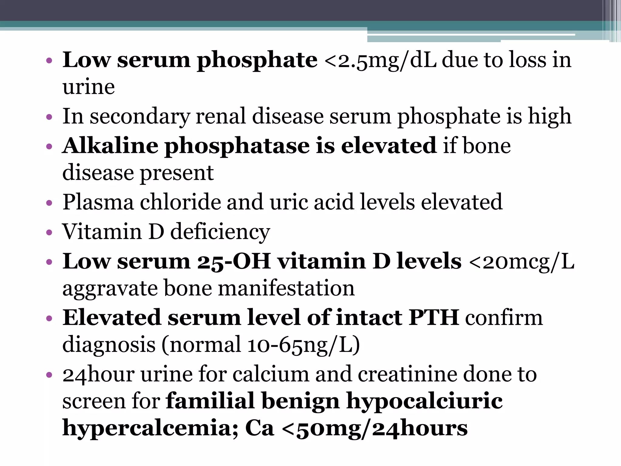
![Laboratory findings
• Hypercalcemia with serum adjusted total
calcium ≥10.5 mg/dL
Adjusted total calcium=measured serum
calcium in mg/dL +[0.8x(4-patient’s serum
albumin in g/dL)]
• Serum ionized calcium in hyperproteinemic
states and hyperparathyroidism is >5.4mg/dL](https://image.slidesharecdn.com/laboratoryandradiologicalfindingsinhyperparathyroidism-170427145539/75/laboratory-and-radiological-findings-in-hyperparathyroidism-13-2048.jpg)

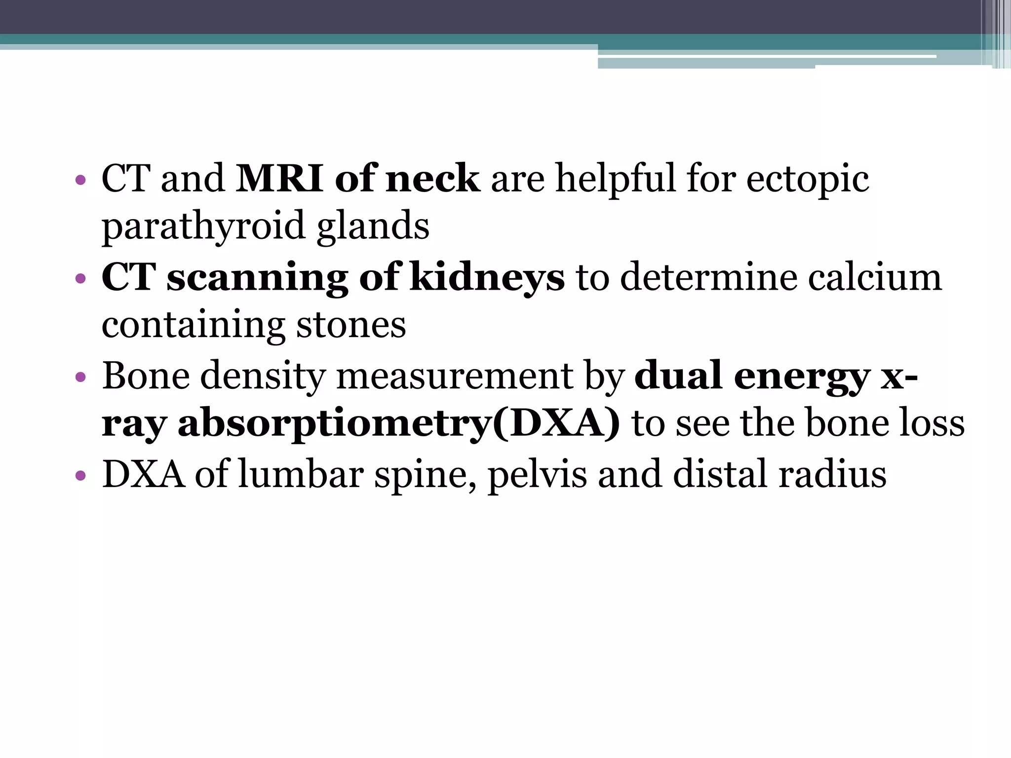
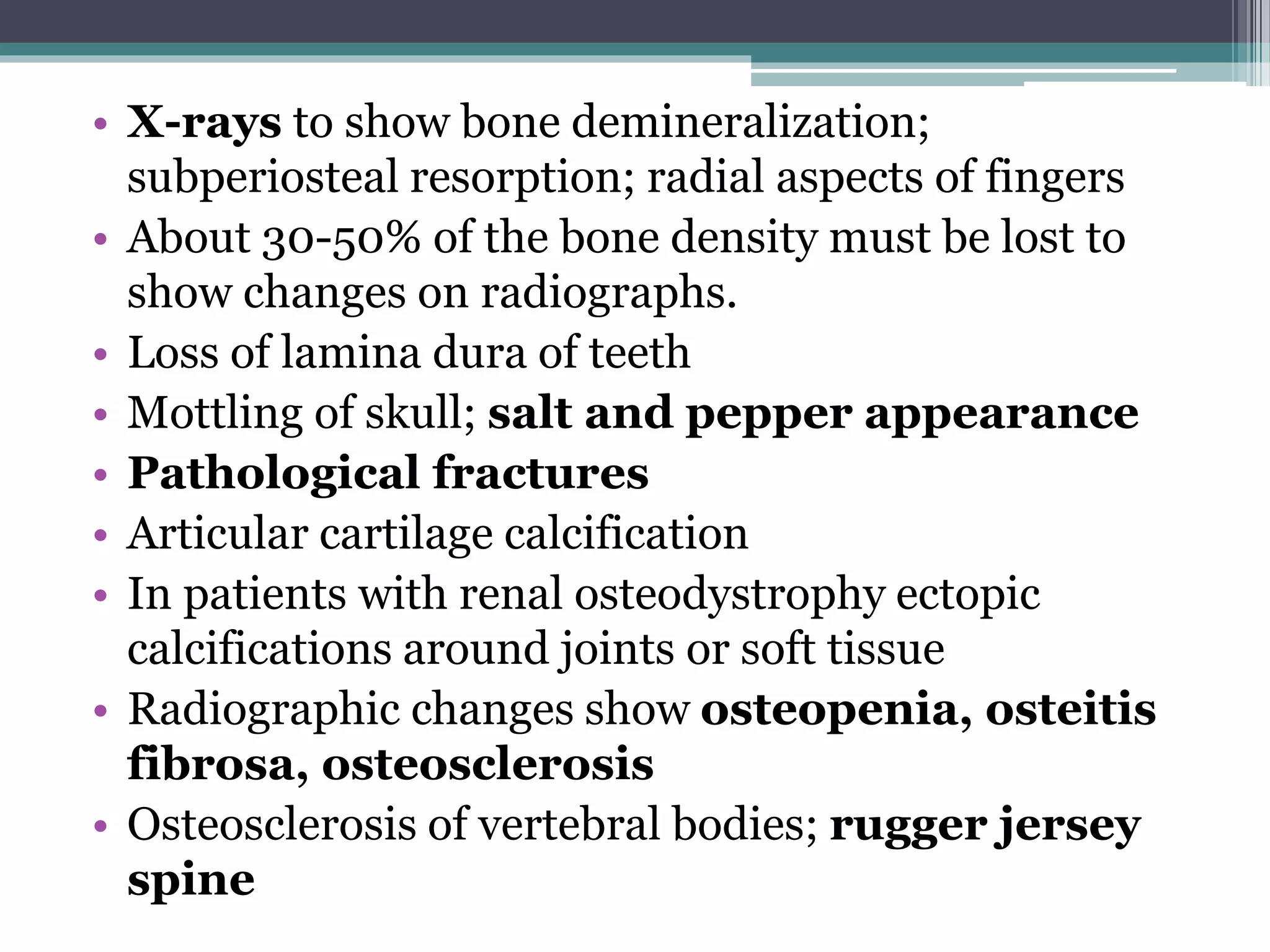


This document summarizes laboratory and radiological findings in hyperparathyroidism. It notes that hyperparathyroidism is usually caused by a single parathyroid adenoma and results in excessive calcium excretion by the kidneys due to overproduction of PTH. Common clinical findings include bone loss, kidney stones, abdominal pain, and fatigue. Laboratory tests show elevated serum calcium, PTH, and alkaline phosphatase levels. Imaging like sestamibi scans and ultrasound can detect parathyroid adenomas, while DXA scans measure bone mineral density loss and X-rays show bone demineralization patterns. Management involves IV fluids, bisphosphonates, vitamin D, and calcimimetic drugs.