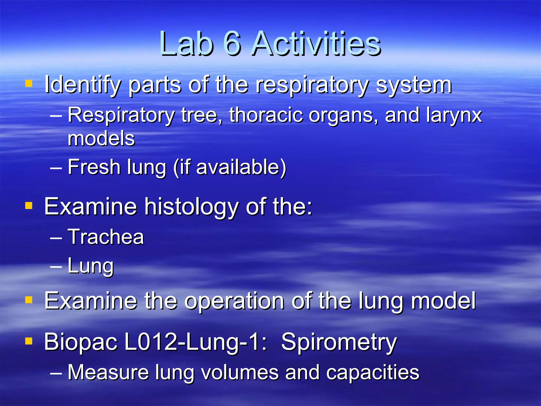
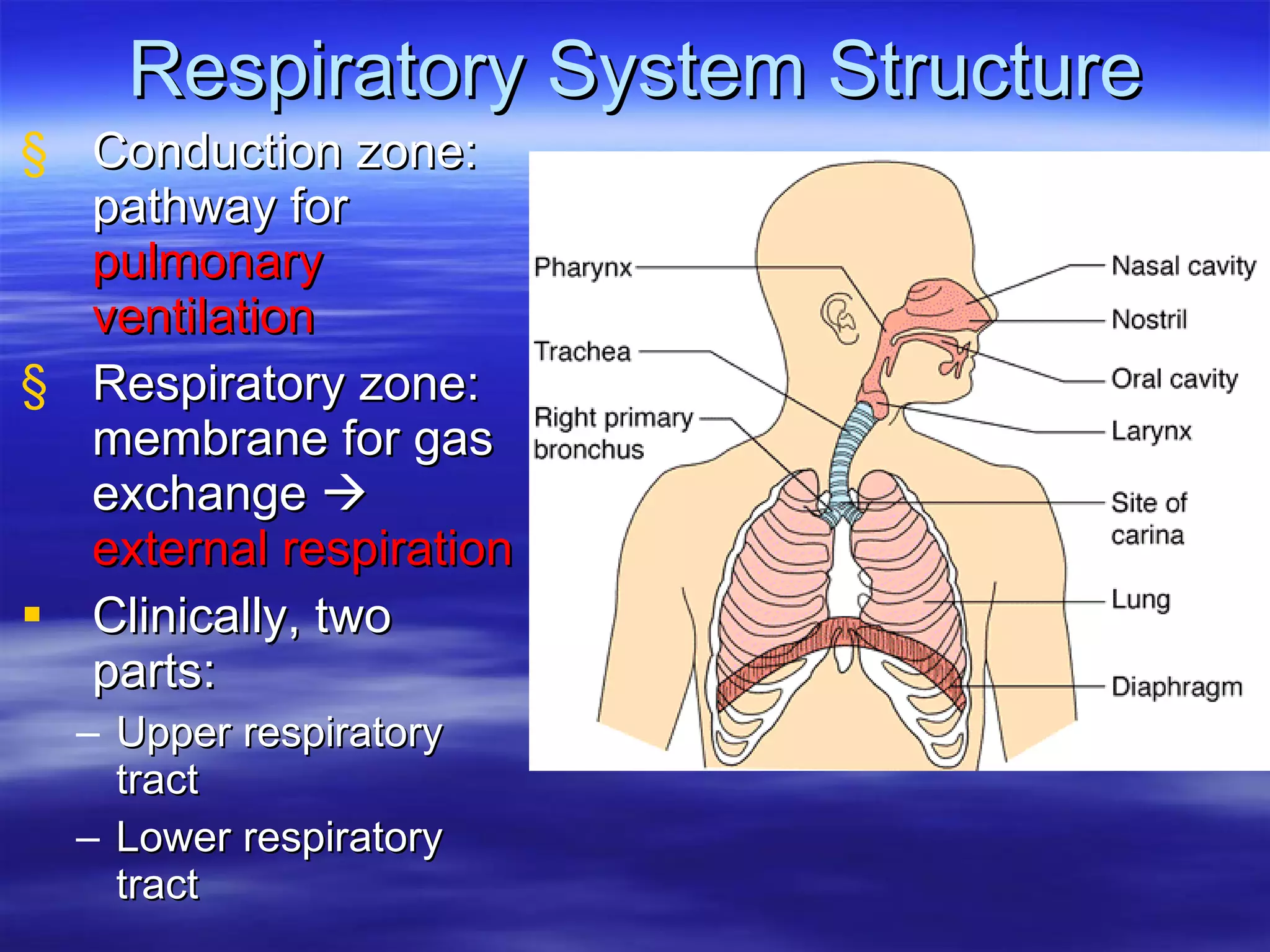
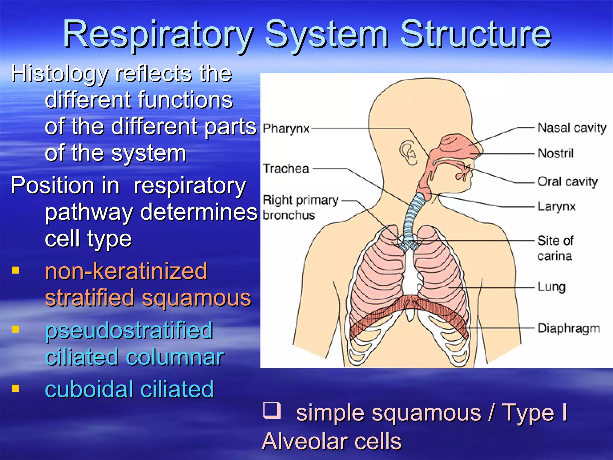
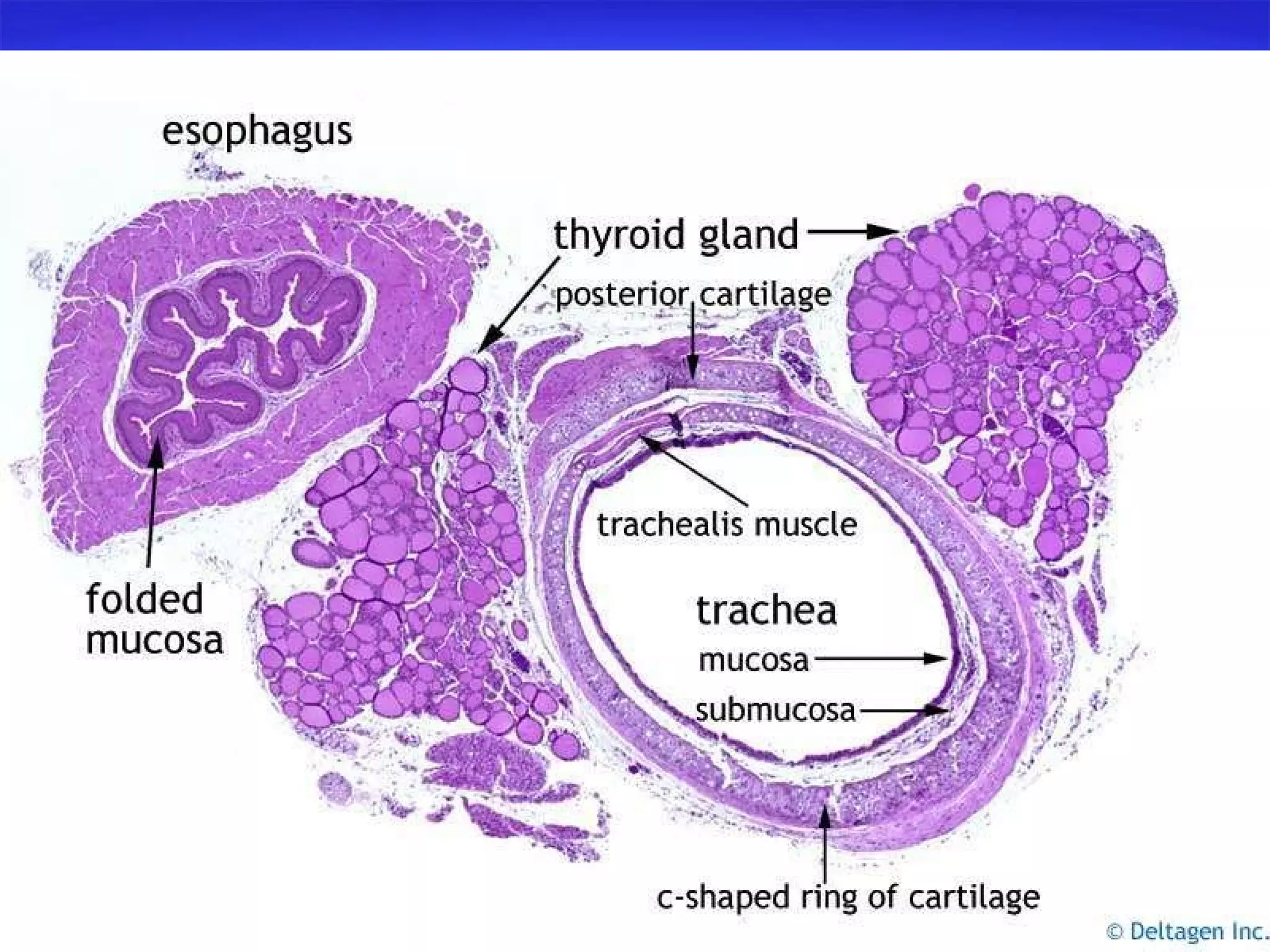
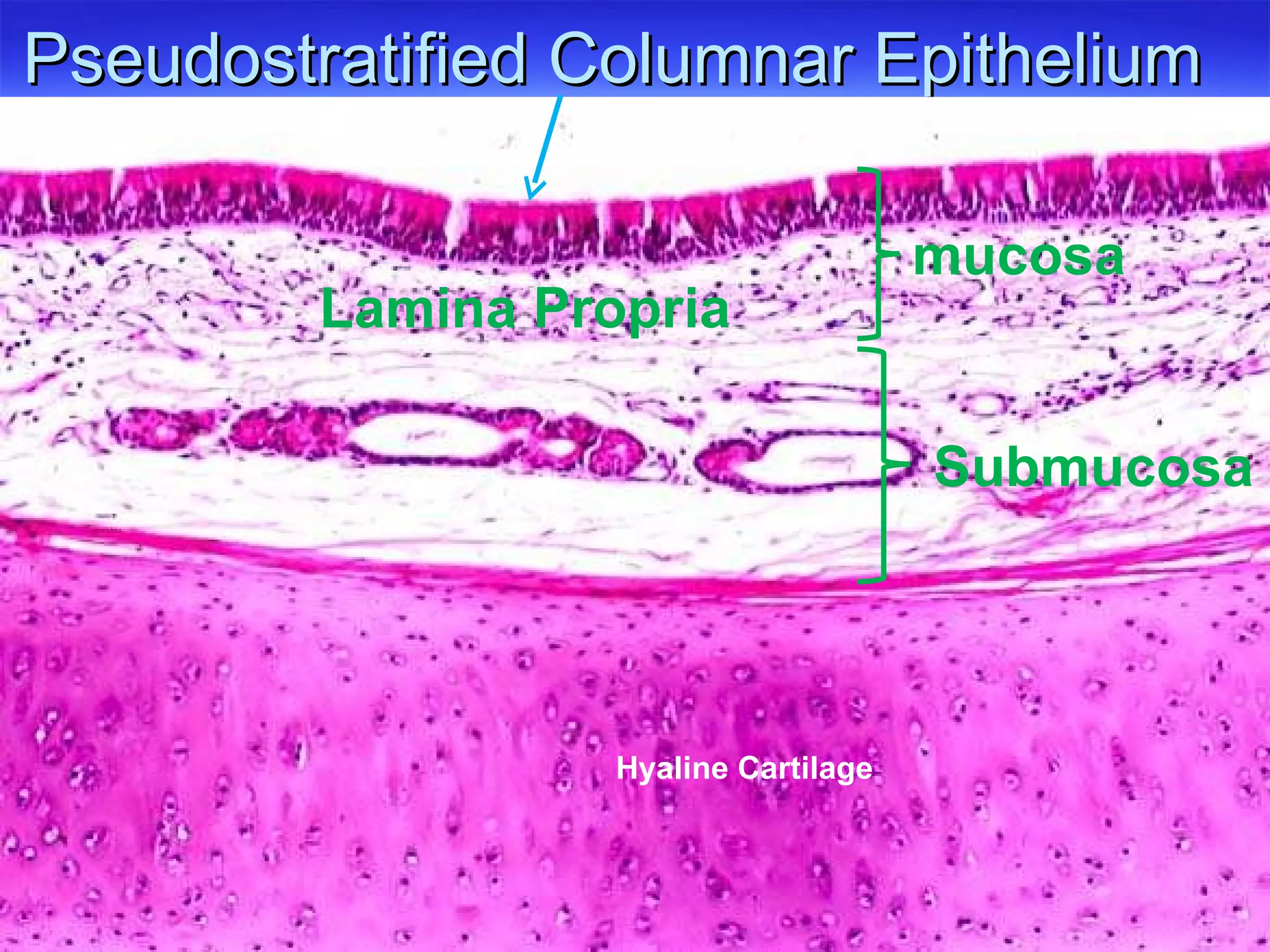
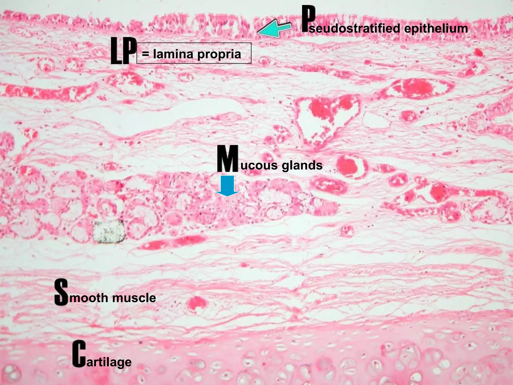
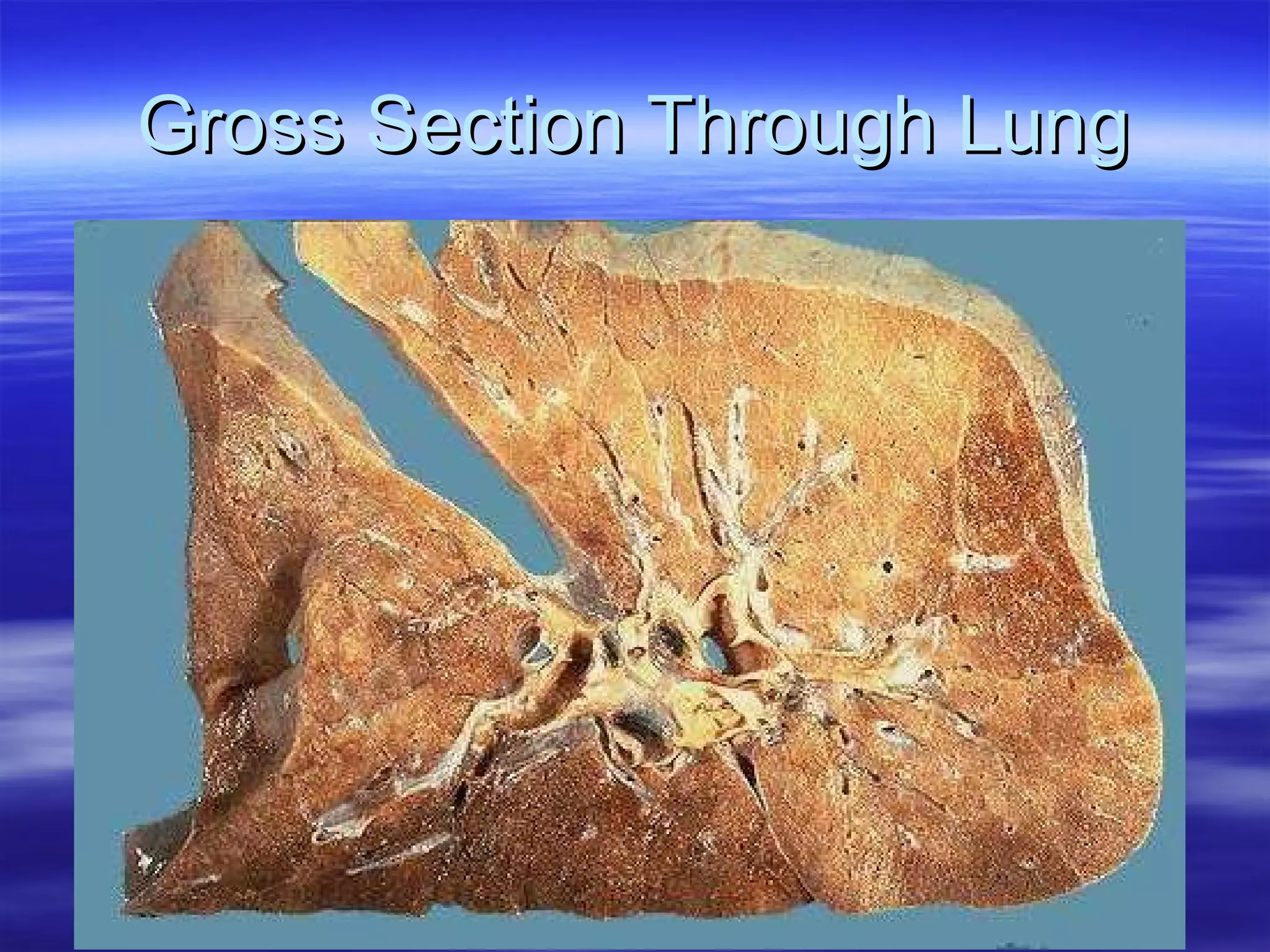
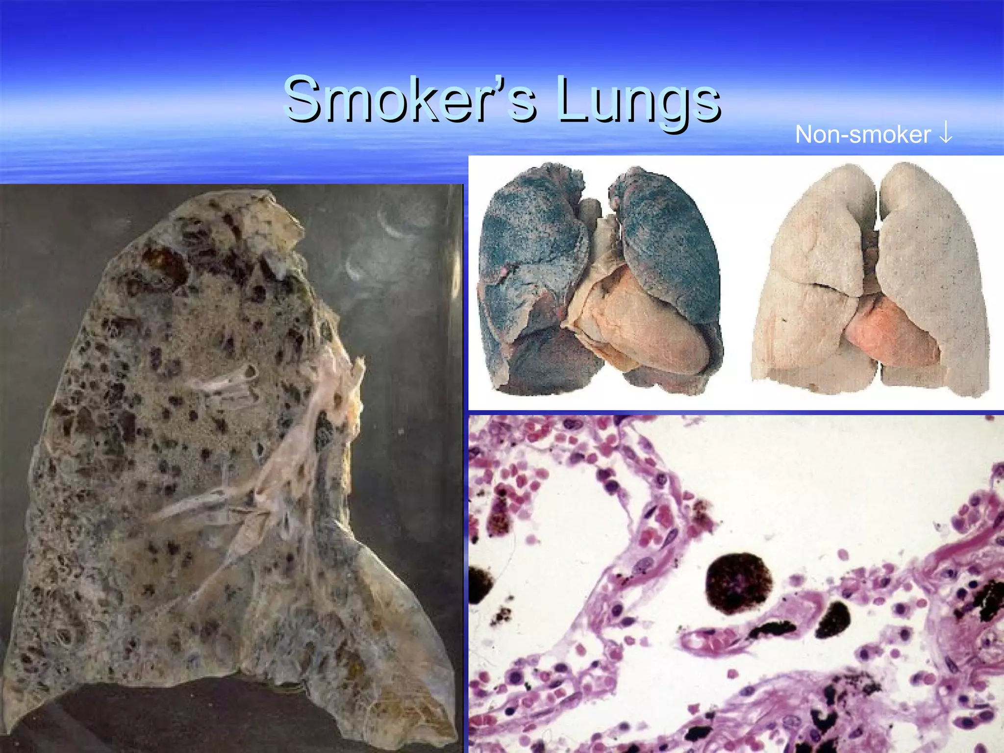
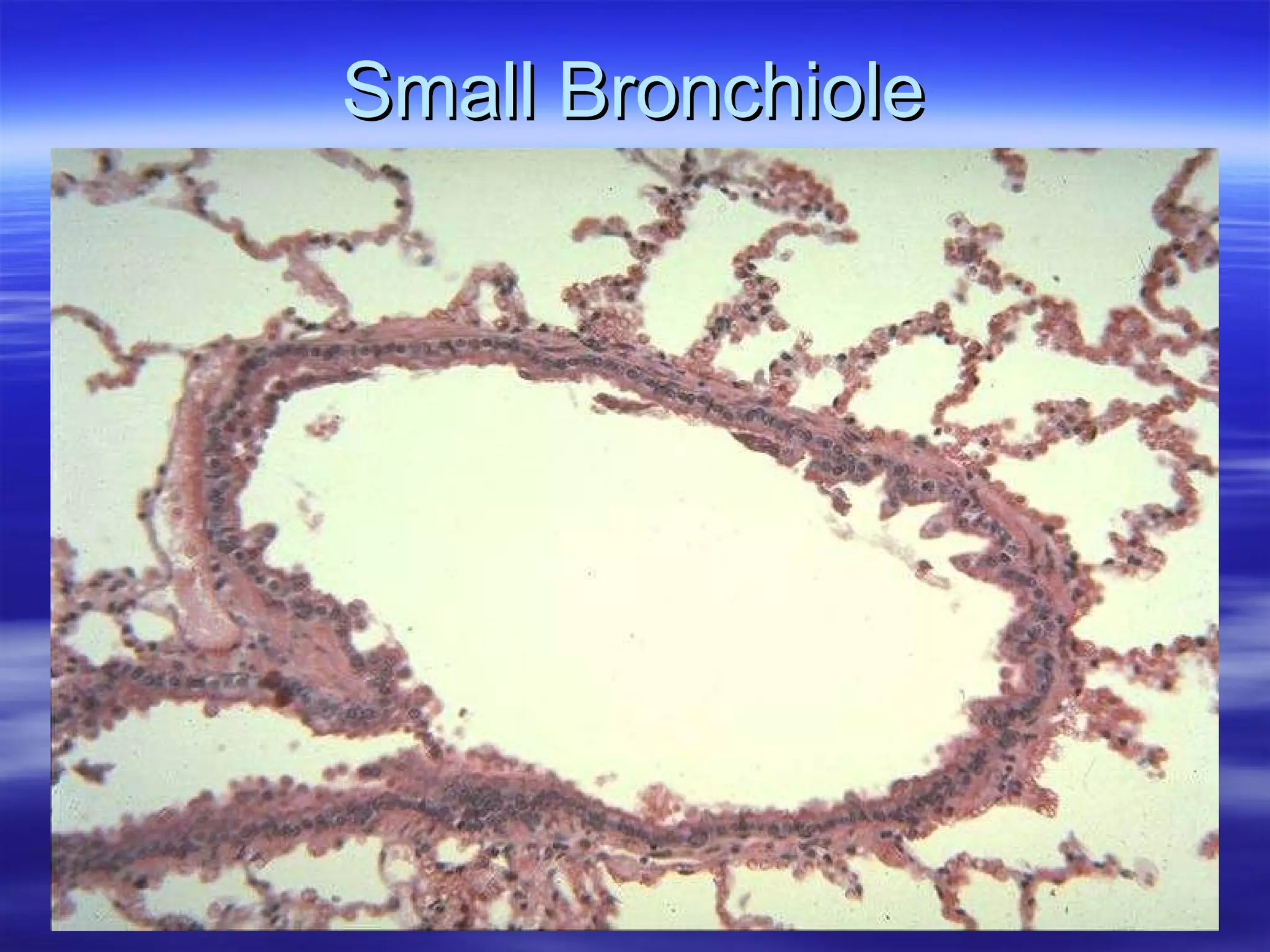
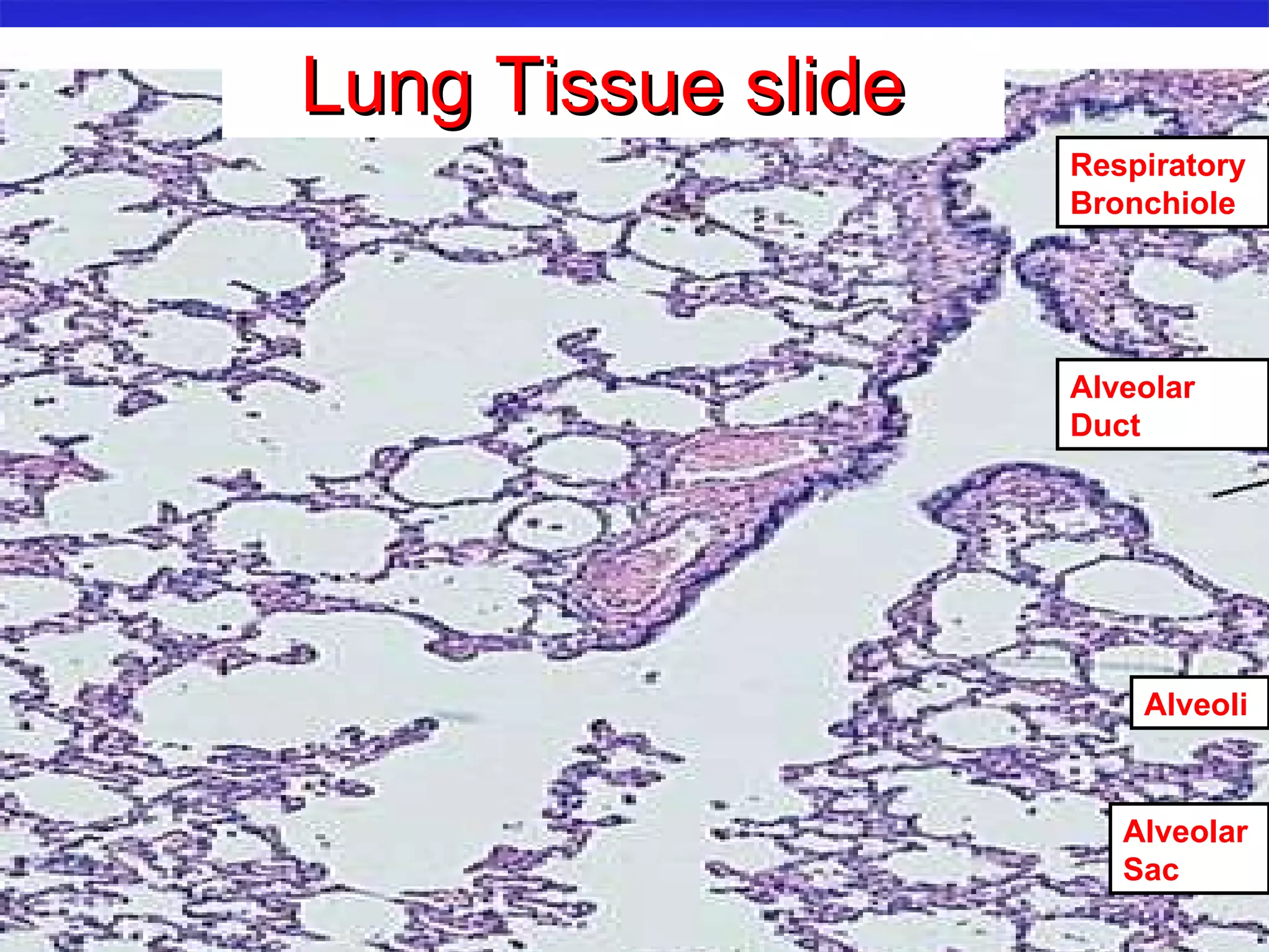
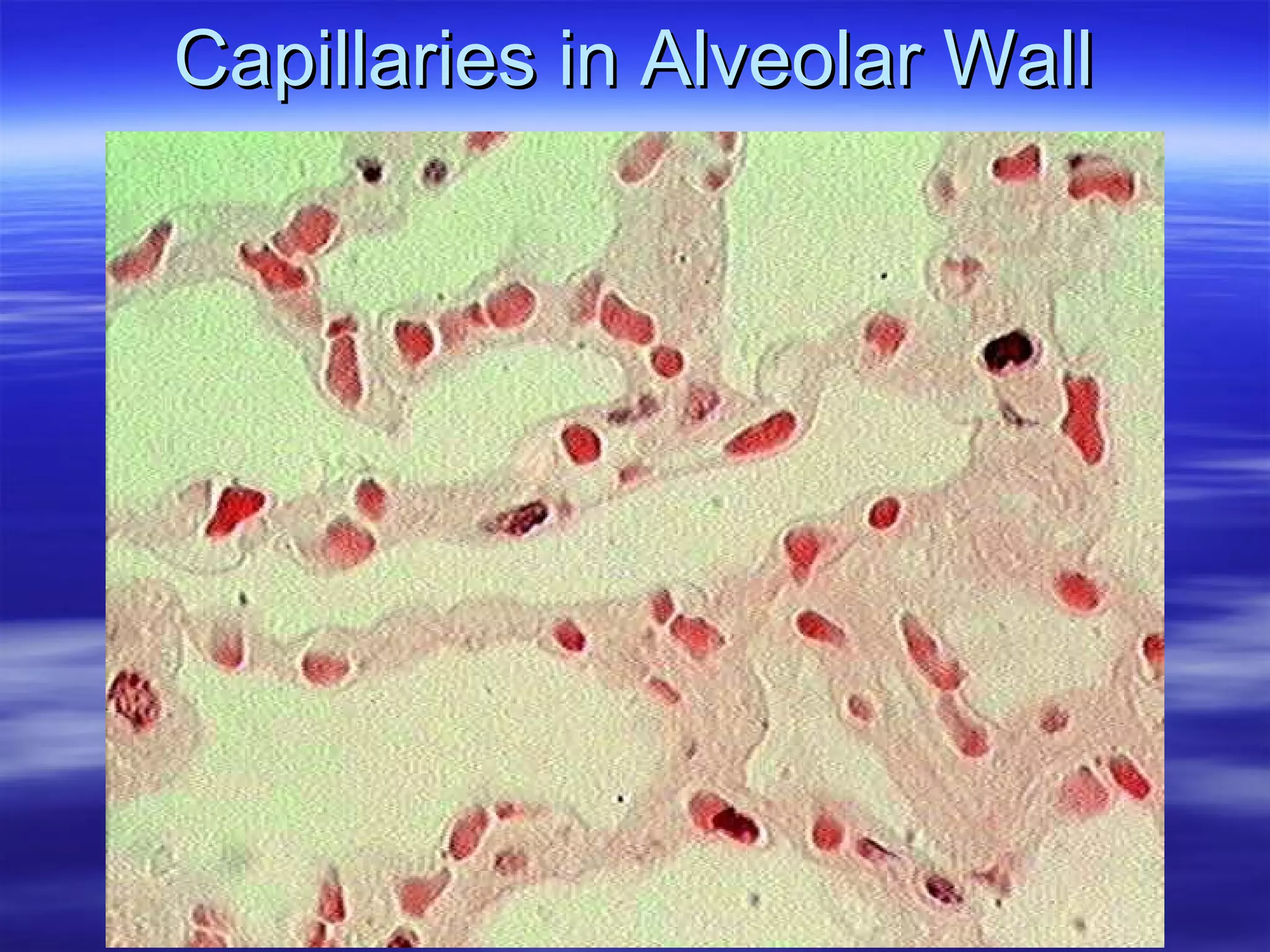
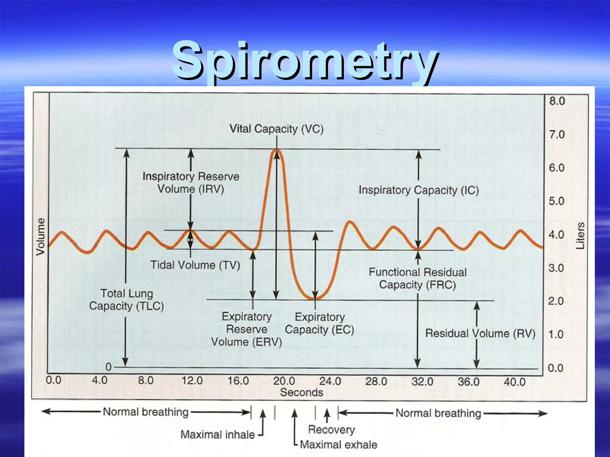
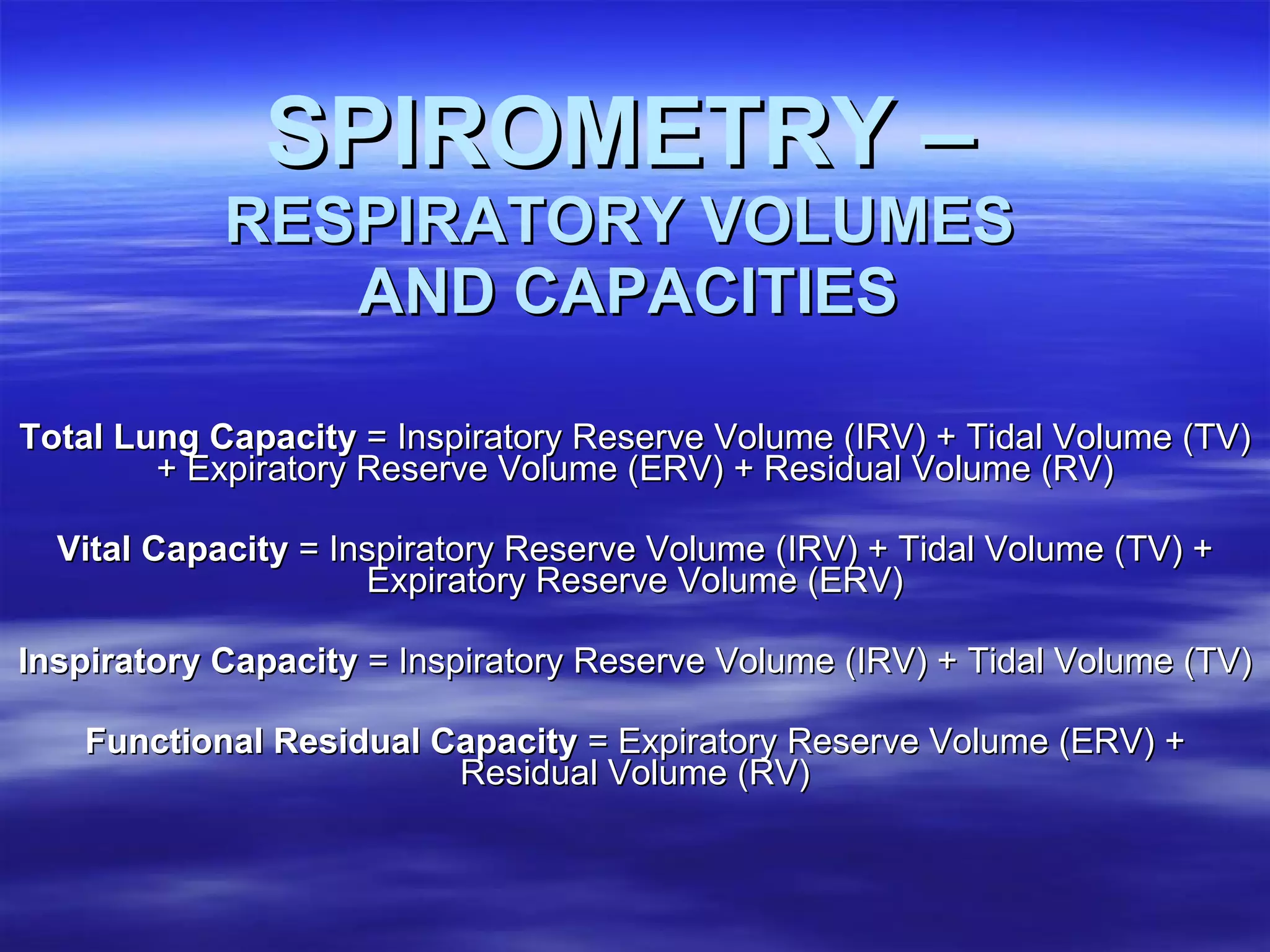
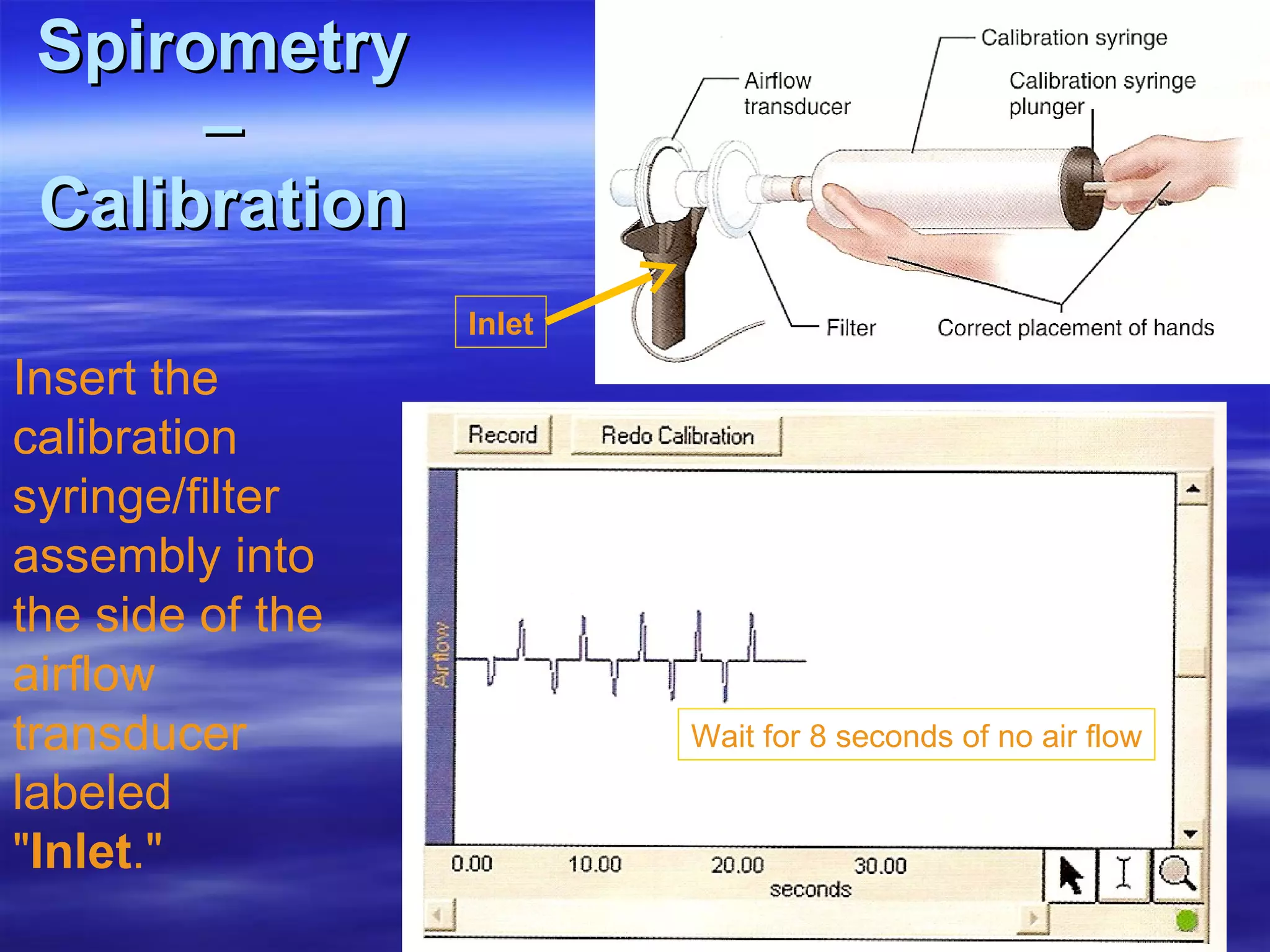
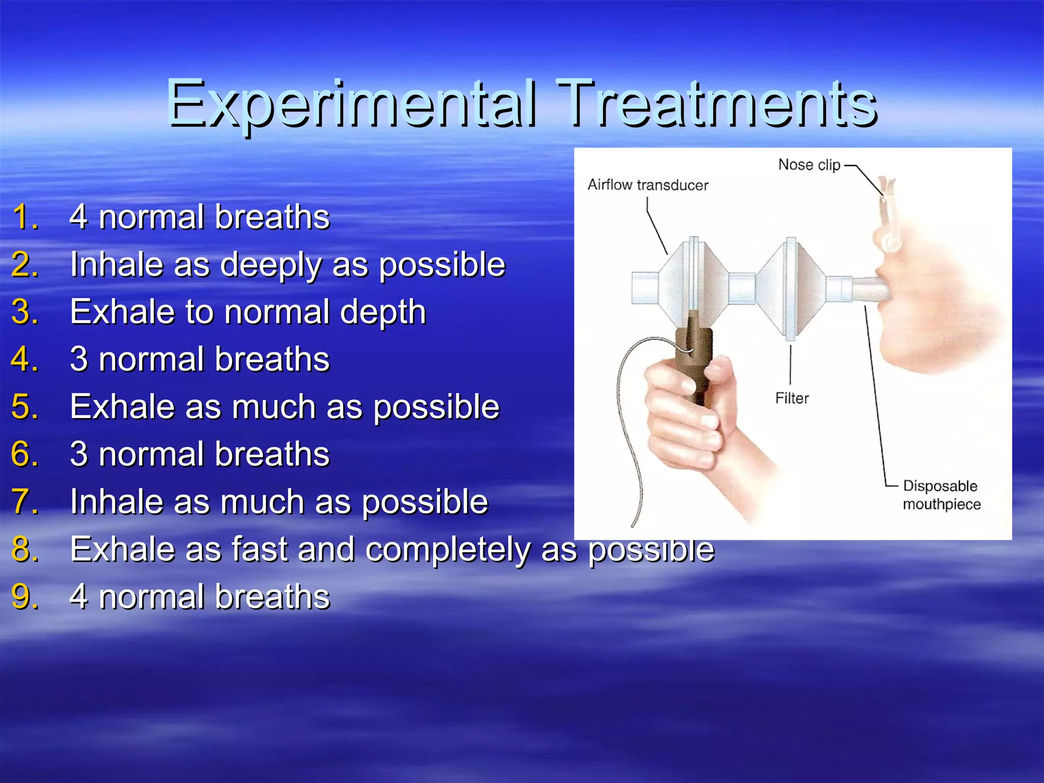
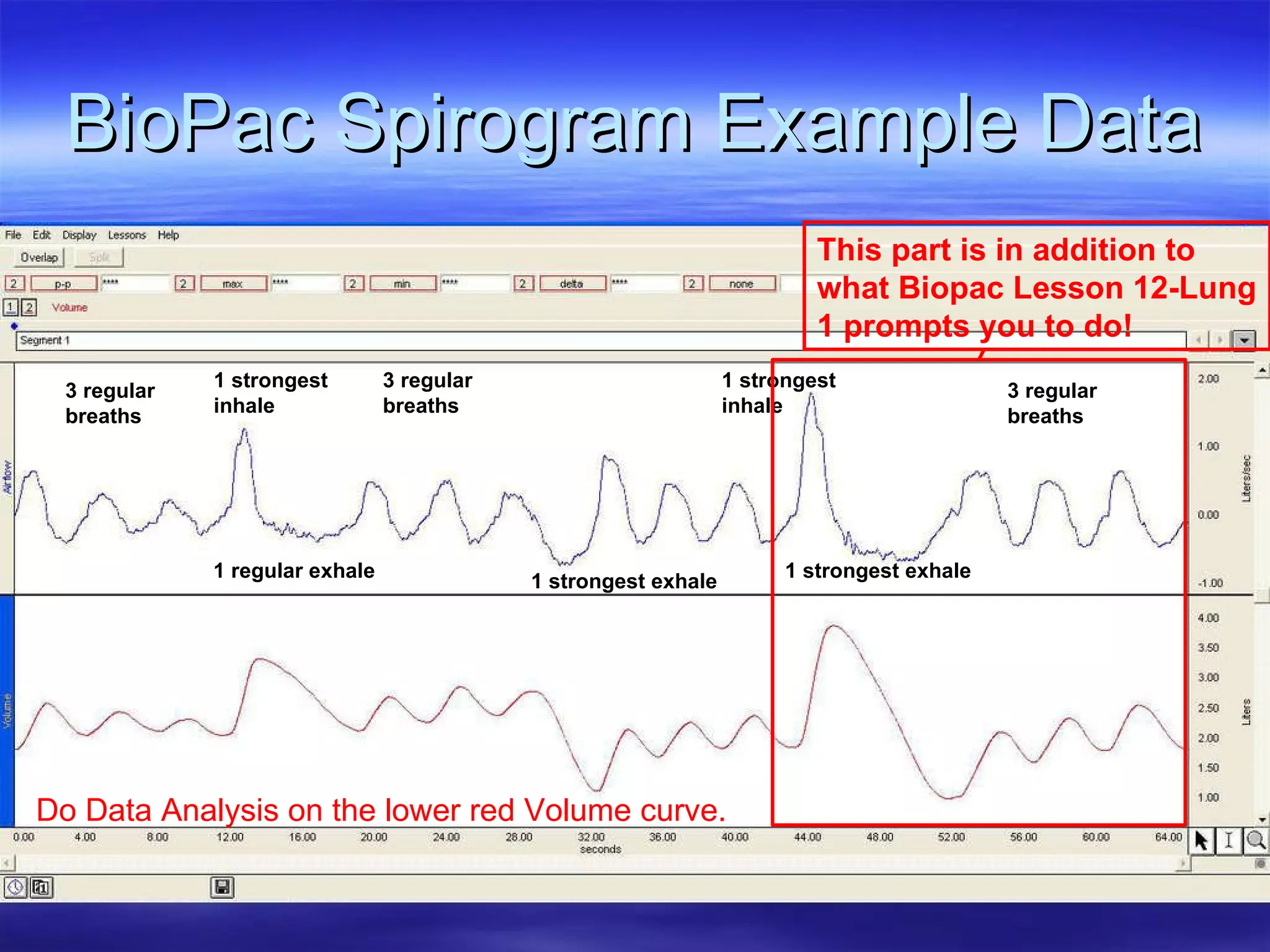
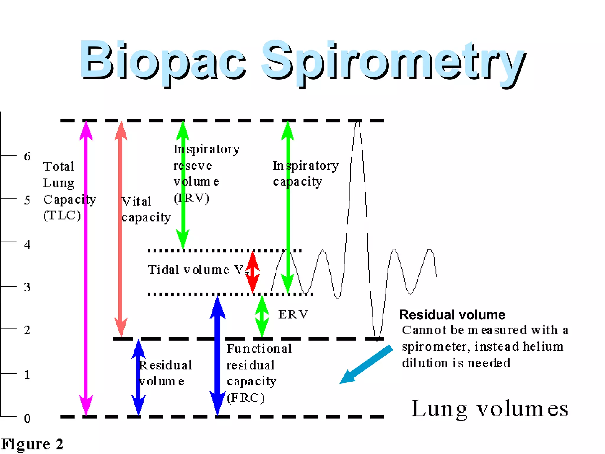
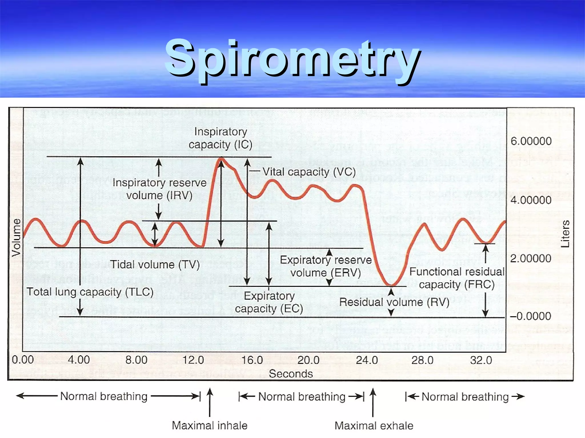
The Respiratory System & Spirometry Lab 6
Lab 6 Activities Identify parts of the respiratory system Respiratory tree, thoracic organs, and larynx models Fresh lung (if available) Examine histology of the: Trachea Lung Examine the operation of the lung model   Biopac L012-Lung-1:  Spirometry  Measure lung volumes and capacities
Respiratory System Structure Conduction zone: pathway for  pulmonary ventilation   Respiratory zone: membrane for gas exchange     external respiration Clinically, two parts: Upper respiratory tract Lower respiratory tract
Respiratory System Structure Histology reflects the different functions of the different parts of the system Position in  respiratory pathway determines cell type non-keratinized stratified squamous pseudostratified ciliated columnar cuboidal ciliated simple squamous / Type I Alveolar cells
 
Pseudostratified Columnar Epithelium Lamina Propria Hyaline Cartilage mucosa Submucosa
seudostratified epithelium = lamina propria ucous glands mooth muscle artilage
Gross Section Through Lung
Smoker’s Lungs Non-smoker  
Small Bronchiole
Lung Tissue slide  Respiratory Bronchiole Alveolar Duct Alveoli Alveolar Sac
Capillaries in Alveolar Wall
Spirometry
  SPIROMETRY –   RESPIRATORY VOLUMES  AND CAPACITIES Total Lung Capacity  = Inspiratory Reserve Volume (IRV) + Tidal Volume (TV) + Expiratory Reserve Volume (ERV) + Residual Volume (RV) Vital Capacity  = Inspiratory Reserve Volume (IRV) + Tidal Volume (TV) + Expiratory Reserve Volume (ERV) Inspiratory Capacity  = Inspiratory Reserve Volume (IRV) + Tidal Volume (TV) Functional Residual Capacity  = Expiratory Reserve Volume (ERV) + Residual Volume (RV)
Spirometry – Calibration Insert the calibration syringe/filter assembly into the side of the airflow transducer labeled " Inlet ."  Inlet Wait for 8 seconds of no air flow Inlet
Experimental Treatments 4 normal breaths Inhale as deeply as possible Exhale to normal depth 3 normal breaths Exhale as much as possible 3 normal breaths Inhale as much as possible Exhale as fast and completely as possible 4 normal breaths
BioPac Spirogram Example Data 3 regular breaths 3 regular breaths 3 regular breaths 1 strongest inhale 1 regular exhale 1 strongest inhale 1 strongest exhale 1 strongest exhale This part is in addition to what Biopac Lesson 12-Lung 1 prompts you to do! Do Data Analysis on the lower red Volume curve.
Summary of Class Data Link to Table
The End Lab 6
Biopac Spirometry Residual   volume
Spirometry

Lab 06 Spirometry

  • 1.
    The Respiratory System& Spirometry Lab 6
  • 2.
    Lab 6 ActivitiesIdentify parts of the respiratory system Respiratory tree, thoracic organs, and larynx models Fresh lung (if available) Examine histology of the: Trachea Lung Examine the operation of the lung model Biopac L012-Lung-1:  Spirometry Measure lung volumes and capacities
  • 3.
    Respiratory System StructureConduction zone: pathway for pulmonary ventilation Respiratory zone: membrane for gas exchange  external respiration Clinically, two parts: Upper respiratory tract Lower respiratory tract
  • 4.
    Respiratory System StructureHistology reflects the different functions of the different parts of the system Position in respiratory pathway determines cell type non-keratinized stratified squamous pseudostratified ciliated columnar cuboidal ciliated simple squamous / Type I Alveolar cells
  • 5.
  • 6.
    Pseudostratified Columnar EpitheliumLamina Propria Hyaline Cartilage mucosa Submucosa
  • 7.
    seudostratified epithelium =lamina propria ucous glands mooth muscle artilage
  • 8.
  • 9.
  • 10.
  • 11.
    Lung Tissue slide Respiratory Bronchiole Alveolar Duct Alveoli Alveolar Sac
  • 12.
  • 13.
  • 14.
    SPIROMETRY– RESPIRATORY VOLUMES AND CAPACITIES Total Lung Capacity = Inspiratory Reserve Volume (IRV) + Tidal Volume (TV) + Expiratory Reserve Volume (ERV) + Residual Volume (RV) Vital Capacity = Inspiratory Reserve Volume (IRV) + Tidal Volume (TV) + Expiratory Reserve Volume (ERV) Inspiratory Capacity = Inspiratory Reserve Volume (IRV) + Tidal Volume (TV) Functional Residual Capacity = Expiratory Reserve Volume (ERV) + Residual Volume (RV)
  • 15.
    Spirometry – CalibrationInsert the calibration syringe/filter assembly into the side of the airflow transducer labeled " Inlet ."  Inlet Wait for 8 seconds of no air flow Inlet
  • 16.
    Experimental Treatments 4normal breaths Inhale as deeply as possible Exhale to normal depth 3 normal breaths Exhale as much as possible 3 normal breaths Inhale as much as possible Exhale as fast and completely as possible 4 normal breaths
  • 17.
    BioPac Spirogram ExampleData 3 regular breaths 3 regular breaths 3 regular breaths 1 strongest inhale 1 regular exhale 1 strongest inhale 1 strongest exhale 1 strongest exhale This part is in addition to what Biopac Lesson 12-Lung 1 prompts you to do! Do Data Analysis on the lower red Volume curve.
  • 18.
    Summary of ClassData Link to Table
  • 19.
  • 20.
  • 21.