2. PULMONARY FUNCTION TEST
INTRODUCTION
Pulmonary function tests or lung function tests are useful in
assessing the functional status of the respiratory system both in
physiological and pathological condition
Lung function tests are based on the measurement of volume of air
breathed in and out in quiet breathing and forced breathing
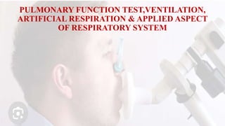
These tests are carried out mostly by using spirometer
3. TYPES OF LUNG FUNCTION TESTS
Lung function tests are of two types:
Static lung function tests
Dynamic lung function tests.
4. Static Lung Function Tests
Based on volume air that flows into or out of lungs
Not depend upon the rate of air flows
Include static lung volumes and static lung capacities
5. Dynamic Lung Function Tests
Rate at which air flows into or out of lungs
These tests are
1. Include forced vital capacity
2. Forced expiratory volume
3. Maximum ventilation volume
4. Peak expiratory flow
Dynamic lung function tests are useful in determining the
severity of obstructive and restrictive lung diseases
6. LUNG VOLUMES
Static lung volumes are the volumes of air breathed by an
individual
Static lung volumes are of four types
Tidal volume
Inspiratory reserve volume
Expiratory reserve volume
Residual volume
7. TIDAL VOLUME(TV)
volume of air breathed in and out of lungs in a single normal
quiet respiration.
Tidal volume signifies the normal depth of breathing.
Normal Value
500 mL (0.5 L).
8. INSPIRATORY RESERVE VOLUME (IRV)
Is an additional volume of air that can be inspired forcefully
after the end of normal inspiration
Normal Value
3,300 mL (3.3 L).
9. EXPIRATORY RESERVE VOLUME(ERV)
is the additional volume of air that can be expired out forcefully, after normal
expiration.
Normal Value
1,000 mL (1 L).
RESIDUAL VOLUME
Residual volume (RV) is the volume of air remaining in lungs even after forced
expiration.
Normally, lungs cannot be emptied completely even by forceful expiration.
significane
1. It helps to aerate the blood in between breathing
and during expiration
2. It maintains the contour of the lungs.
Normal Value
1,200 mL (1.2 L)
10. LUNG CAPACITIES
Static lung capacities are the combination of two or
more lung volumes.
Static lung capacities are of four types:
1. Inspiratory capacity
2. Vital capacity
3. Functional residual capacity
4. Total lung capacity.
11. INSPIRATORY CAPACITY
Inspiratory capacity (IC) is the maximum volume of air
that is inspired after normal expiration (end expiratory
position)
It includes tidal volume and inspiratory reserve volume
IC = TV + IRV
= 500 + 3,300 = 3,800 mL
12. VITAL CAPACITY (VC)
Vital capacity (VC) is the maximum volume of air that can be
expelled out forcefully after a deep (maximal) inspiration.
VC includes inspiratory reserve volume, tidal volume and
expiratory reserve volume.
VC = IRV + TV + ERV
= 3,300 + 500 + 1,000 = 4,800 mL
13. TOTAL LUNG CAPACITY
Total lung capacity (TLC) is the volume of air present in lungs after
a deep (maximal) inspiration
It includes all the volumes
TLC = IRV + TV + ERV + RV
= 3,300 + 500 + 1,000 + 1,200 = 6,000 mL
14. FUNCTIONAL RESIDUAL CAPACITY
Functional residual capacity (FRC) is the volume of air
remaining in lungs after normal expiration (after normal
tidal expiration)
Functional residual capacity includes
expiratory reserve volume and residual volume
FRC = ERV + RV
= 1,000 + 1,200 = 2,200 mL
15.
16.
17. MEASUREMENT OF LUNG VOLUMES AND
CAPACITIES
Spirometer
Respirometer.
Plethysmograph
18. SPIROMETER
Spirometer is made up of metal and it contains two chambers namely
outer chamber and inner chamber
Outer chamber is called the water chamber because it is filled with water.
A floating drum is immersed in the water in an inverted position
Drum is counter balanced by a weight
19. Weight is attached to the topofthe inverted drum by means of string or chain
A pen with ink is attached to the counter weight
Pen is made to write on a calibrated paper, which is fixed to a recording
device
Inner chamber is inverted and has a small hole at the top
A long metal tube passes through the inner chamber from the bottom
towards the top
20. Upper end of this tube reaches the top portion of the inner chamber
Then the tube passes through a hole at the top of inner chamber and
penetrates into outer water chamber above the level of water
A rubber tube is connected to the outer end of the metal tube
At the other end of this rubber tube, a mouthpiece is attached
Subject respires through this mouthpiece by closing the nose with a
nose clip
21. When the subject breathes with spirometer, during expiration, drum moves up and the
counter weight comes down.
Reverse of this occurs when the subject breathes the air from the spirometer, i.e. during
inspiration.
Upward and downward movements of the counter weight are recorded in the form of a
graph.
Upward deflection of the curve in the graph shows inspiration and the downward
deflection denotes expiration.
Spirometer is used only for a single breath.
Repeated cycles of respiration cannot be recorded by using this instrument because
carbon dioxide accumulates in the spirometer and oxygen or fresh air cannot be provided
to the subject
22. Respirometer
Respirometer is the modified spirometer.
It has provision for removal of carbon dioxide and supply of oxygen.
Carbon dioxide is removed by placing soda lime inside the instrument.
Oxygen is supplied to the instrument from the oxygen cylinder, by a suitable
valve system.
Oxygen is filled in the inverted drum above water level and the subject can
breathe in and out with instrument for about 6 minutes and recording can be
done continuously.
23.
24. Spirogram
Spirogram is the graphical record of lung volumes and capacities using
spirometer.
Upward deflection of the spirogram denotes inspiration and the
downward curve indicates expiration
In order to determine the lung volumes and capacities, following four
levels are to be noted in spirogram:
1. Normal end expiratory level
2. Normal end inspiratory level
3. Maximum expiratory level
4. Maximum inspiratory level.
25.
26. COMPUTERIZED SPIROMETER
Computerized spirometer is the solid state electronic equipment. It does
not contain a drum or water chamber. Subject has to respire into a
sophisticated transducer, which is connected to the instrument by means
of a cable.
Disadvantages of Spirometry
By using simple spirometer, respirometer or computerized spirometer,
not all the lung volumes and lung capacities can be measured.
28. VENTILATION
ventilation refers to circulation of replacement of air or gas in a space
In respiratory physiology, ventilation is the rate at which air enters or
leaves the lungs
Ventilation in respiratory physiology is of two types:
1. Pulmonary ventilation
2. Alveolar ventilation.
29. PULMONARY VENTILATION
DEFINITION
Pulmonary ventilation is defined as the volume of air moving in and
out of respiratory tract in a given unit of time during quiet breathing. It
is also called minute ventilation or respiratory minute volume (RMV).
Pulmonary ventilation is a cyclic process, by which fresh air enters the
lungs and an equal volume of air leaves the lungs.
30. NORMAL VALUE AND CALCULATION
Normal value of pulmonary ventilation is 6,000 mL(6 L)/minute.
It is the product of tidal volume (TV) and the rate of respiration
(RR).
It is calculated by the formula:
Pulmonary ventilation
= Tidal volume × Respiratory rate
= 500 mL × 12/minute
= 6,000 mL/minute.
31. ALVEOLAR VENTILATION
DEFINITION
1. Alveolar ventilation is the amount of air utilized for gaseous exchange every
minute.
2. Alveolar ventilation is different from pulmonary ventilation. In pulmonary
ventilation, 6 L of air moves in and out of respiratory tract every minute.
3. But the whole volume of air is not utilized for exchange of gases.
4. Volume of air subjected for exchange of gases is the alveolar ventilation.
5. Air trapped in the respiratory passage (dead space) does not take part in gaseous
exchange
32. NORMAL VALUE AND CALCULATION
Normal value of alveolar ventilation is 4,200 mL (4.2 L)/ minute.
It is calculated by the formula:
Alveolar ventilation = (Tidal volume – Dead space) x Respiratory rate
= (500 – 150) mL × 12/minute = 4,200 mL (4.2 L)/minute.
33. DEAD SPACE
DEFINITION
Dead space is defined as the part of the respiratory tract, where gaseous exchange does
not take place.
Air present in the dead space is called dead space air.
TYPES OF DEAD SPACE
Dead space is of two types:
1. Anatomical dead space
2. Physiological dead space.
34. WASTED VENTILATION ANDWASTEDAIR
Wasted ventilation is the volume of air that ventilates physiological
dead space.
Wasted air refers to air that is not utilized for gaseous exchange. Dead
space air is generally considered as wasted air.
35. NORMAL VALUE OF DEAD SPACE
Volume of normal dead space is 150 mL.
Under normal conditions, physiological dead space is equal to
anatomical dead space.
It is because, all the alveoli are functioning and all the alveoli receive
adequate blood flow in normal conditions.
Physiological dead space increases during res_x0002_piratory
diseases, which affect the pulmonary blood flow or the alveoli.
MEASUREMENT OF DEAD SPACE – NITROGEN
WASHOUT METHOD
36. ARTIFICIAL RESPIRATION
• CONDITIONS WHEN ARTIFICIAL RESPIRATION IS REQUIRED
• Artificial respiration is required whenever there is an arrest of
breathing, without cardiac failure.
• Arrest of breathing occurs in the following conditions:
• 1Accident
• 2. Drowning
• 3. Gas poisoning
• 4. Electric shock
• 5. Anesthesia.
37. Stoppage of oxygen supply for 5 minutes causes irreversible changes
in tissues of brain, particularly tissues of cerebral cortex.
So, artificial respiration (resuscitation) must be started quickly
without any delay, before the development of cardiac failure.
Purpose of artificial respiration is to ventilate the alveoli and to
stimulate the respiratory center
38. „METHODS OF ARTIFICIAL RESPIRATION
• Methods of artificial respiration are of two types:
• 1. Manual methods
• 2. Mechanical methods
39. „MANUAL METHODS
Manual methods of resuscitation can be applied quickly without waiting for the
availability of any mechanical aids.
Affected person must be provided with clear air.
Clothes around neck and chest regions must be loosened.
Mouth, face and throat should be cleared of mucus, saliva, foreign particles, etc.
Tongue must be drawn forward and it must be prevented from falling posteriorly,
which may cause airway obstruction.
Manual methods are of two types:
i. Mouth-to-mouth method
ii. Holger Nielsen method.
40. Mouth-to-mouth Method
The subject is kept in supine position and the resuscitator (person who give
resuscitation) kneels at the side of the subject.
By keeping the thumb on subject’s mouth, the lower jaw is pulled downwards.
Nostrils of the subject are closed with thumb and index finger of the other hand.
Resuscitator then takes a deep breath and exhales into the subject’s mouth
forcefully.
Volume of exhaled air must be twice the normal tidal volume. This expands the
subject’s lungs.
Then, the resuscitator removes his mouth from that of the subject.
41. Now, a passive expiration occurs in the subject due to elastic recoil of the lungs.
This procedure is repeated at a rate of 12 to 14 times a minute, till normal
respiration is restored.
Mouth-to-mouth method is the most effective manual method because, carbon
dioxide in expired air of the resuscitator can directly stimulate the respiratory
centers and facilitate the onset of respiration.
Only disadvantage is that the close contact between the mouths of resuscitator and
subject may not be accept_x0002_able for various reasons
42.
43.
44. •Holger Nielsen Method or Back PressureArm
Lift Method
Subject is placed in prone position with head turned to one side.
Hands are placed under the cheeks with flexion at elbow joint and
abduction of arms at the shoulders.
Resuscitator kneels beside the head of the subject.
By placing the palm of the hands over the back of the subject, the
resuscitator bends forward with straight arms (without flexion at
elbow) and applies pressure on the back of the subject.
45.
46. Weight of the resuscitator and pressure on back of the subject
compresses his chest and expels air from the lungs.
Later, the resuscitator leans back. At the same time, he draws the
subject’s arm forward by holding it just above elbow.
This procedure causes expansion of thoracic cage and flow of air into
the lungs.
The movements are repeated at the rate of 12 per minute, till the
normal respiration is restored
47. MECHANICAL METHODS
Mechanical methods of artificial respiration become necessary when
the subject needs artificial respiration for long periods.
It is essential during the respiratory failure due to paralysis of
respiratory muscles or any other cause.
Mechanical methods are of two types:
i. Drinker method
ii. Ventilation method
48. Drinker Method
The machine used in this method is called iron lung chamber or tank respirator.
The equipment has anairtight chamber, made of iron or steel. Subject is placed
inside this chamber with the head outside the chamber.
By means of some pumps, the pressure inside the chamber is made positive and
negative alternately.
During the negative pressure in the chamber, the subject’s thoracic cage expands
and inspiration occurs and during positive pressure the expiration occurs.
By using tank respirator, the patient can survive for a longer time, even up to the
period of one year till the natural respiratory functions are restored.
49.
50. Ventilation Method
A rubber tube is introduced into the trachea of the patient through the
mouth.
By using a pump, air or oxygen is pumped into the lungs with
pressure intermittently.
When air is pumped, inflation of lungs and inspiration occur. When it
is stopped, expiration occurs and the cycle is repeated.
Apparatus used for ventilation is called ventilator and it is mostly used
to treat acute respiratory failure.
51. Ventilator is of two types:
a. Volume ventilator
b. Pressure ventilator.
52. Volume ventilator
By volume ventilator, a constant volume of air is pumped
into the lungs of patients intermittently with minimum pressure.
Pressure ventilator
By pressure ventilator, air is pumped into the lungs of subject with
constant high pressure
53. APPLIED ASPECT OF RESPIRATORY
SYSTEM
• APNEA
Apnea is defined as the temporary arrest of breathing
• HYPOXIA
Hypoxia is defined as reduced availability of oxygen to the tissues.
54. • OXYGEN TOXICITY (POISONING)
Oxygen toxicity is the increased oxygen content in tissues, beyond
certain critical level
• HYPERCAPNEA
Hypercapnea is the increased carbon dioxide content
55. • HYPOCAPNEA
Hypocapnea is the decreased carbon dioxide content in blood.
• ASPHYXIA
Asphyxia is the condition characterized by combination of hypoxia and
hypercapnea, due to obstruction of air passage.