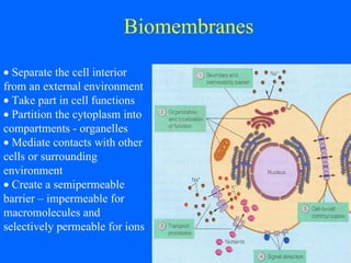
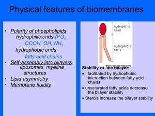
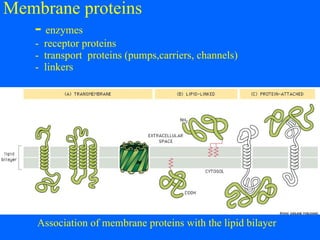
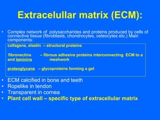
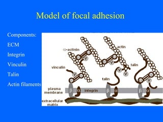
Membrane structure and mechanisms of cell adhesion and locomotion
   Separate the cell interior from an external environment     Take part in cell functions    Partition  the c y toplasm  into compartments - organelles    Mediate contacts with other cells or surrounding environment    Create a semipermeable barrier – impermeable for macromolecules and selectively permeable for ions Biomembranes
Biomembr anes Development of membrane concept 1890 : cell surface is of lipidic nature (Overton) 1926 : cellular membranes consist of lipid bilayers (Gorder and  Grendel) 1943:  the surface of lipid bilayer is  coated by proteins (Davson and  Danielli) 1960:  unit membrane (Robertson) 1972:  fluid mosaic model   (Singer a  Nicholson) 1980:  detailed structure of membrane  proteins  (Unwin a Henderson)
Chemical composition of membranes  membrane lipids   phospholipids, sfingolipids ,  sterols   membrane proteins  (glycoproteins)   Phospholipids: ( phosphoglycerides ) phosphatidyl-  cholin  ethanolamine serine   inositol Sphingolipids    -sfingosin Sterols     cholesterol    ergosterol
Physical features of biomembranes  Stabili ty   of  the  bila y er :    facilitated by hydrophobic interaction between fatty acid chains     unsaturated fatty acids decrease the bilayer stability     Sterols increase the bilayer stability Polarity of phospholipids hydrophilic ends  (PO 4 , ,  COOH, OH, NH 3 hydrophobic ends fatty acid chains   Self-assembly into bilayers   liposomes , myeline structures Lipid asymmetry Membrane fluidity
Mobility of membrane phospholipids Membrane fluidity rotation   lateral migration  flip-flop – transversal diffusion transition point
    Heterogeneity of membrane lipids    and their asymmetric distribution in bilayer. Rafts: small islands of sphingolipids and cholesterol creating a separate phase (50 nm in diameter) in outer leaflet of plasma membrane
Membrane proteins   -   enzymes  -  receptor proteins  -  transport  proteins (pumps,carriers, channels) -  linkers Association of membrane proteins with the lipid bilayer
Membrane proteins:    - have  both hydrophobic  and hydrophilic regions  - the hydrophobic region  extend  through the bilayer and is formed by hydrophobic  amino acids hydrophilic regios are exposed  to the aqueous environment on either side of the membrane   - Peripheral proteins are    attached to the bilayer by  lipid groups (dolichol)
Human red cells in scanning elektron  microscope
Membrane skeleton network of proteins under the plasma membrane. In red cells the membrane skeleton is formed by  spectrin, actin filaments and attachment proteins
Glycocalyx:   a coat of  poly- a oligosaccharides on the surface of plasma  membrane
Extracelullar matrix (ECM): Complex network of  polysaccharides and proteins produced by cells of connective tissue (fibroblasts, chondrocytes, osteocytes etc.) Main components: collagens, elastin   – structural proteins  fibronectins  – fibrous adhesive proteins interconnecting  ECM to a and  laminin s   meshwork    proteoglycans   – gly c oproteins forming a gel  ECM calcified in bone and teeth Ropelike in tendon Transparent in cornea Plant cell wall – specific type of extracellular matrix
Cellular interactions and cell adhesion carry out a structural role, important in cell migration, growth, immunological function, cell recognition, tissue repair, differentiation and embryogenesis
Cell-to-cell interaction: C ell adhesion molecules  ( CAM ):  cadherin, Ig superfamily CAM, mucin-like CAM, integrins selectins
Basal lamina T hin tough sheet of extracellular matrix . Composition :  collagen t y pe  IV  tensile strength laminin  provides adhesive sites for integri n  molecules in the plasma membrane of epithelial cells  other proteins
Structure and function of basal lamina
Keratinocyte crawling over surface
Model of focal adhesion   Components: ECM Integrin Vinculin Talin Actin filaments
Plasmamembr ane  a nd cell  migration Cell crawling   – formation of filopodia (thin stiff protrusions)  and lamelipodia (thin sheet-like extensions) – due to actin polymerization. The new position of the plasma membrane is fixed by focal adhesion complexes Contraction  of a part of the cytoplasm,  invagination  of the plasma membrane or invagination of cell layers due to the activity of actin-myosin I or actin-myosin II complexes: actin filaments are anchored to the plasma membrane. Actin filaments slide over each other, the sliding is mediated by myosin motors.
Key terms 1. Biomembranes – overview of the structure and functions  2. Development of membrane concept: lipid layer, lipid bilayer, phospholid bilayer, localization of membrane proteins 3. Structure of the phospholipid bilayer  4. Hydrophilic head and hydrophobic tails. Self-assembly of the bilayer. Lipid asymmetry. Membrane rafts. Liposomes. 5. Model of the fluid mosaic. I ntegr al a peripheral proteins. 6. Hydrophobic regions of integral proteins 7. Membrane glycoproteins  8. Glycocalyx  9. Extracelular matrix: main components: collagen, elastin, fibronectin, laminin, proteoglycans
Key terms – cont. 10. Basal lamina: components and function 12. Plasmamembrane and cell migration: cell crawling and contractile movements 13. Focal adhesion

L02 cell membrane_

  • 1.
    Membrane structure andmechanisms of cell adhesion and locomotion
  • 2.
    Separate the cell interior from an external environment  Take part in cell functions  Partition the c y toplasm into compartments - organelles  Mediate contacts with other cells or surrounding environment  Create a semipermeable barrier – impermeable for macromolecules and selectively permeable for ions Biomembranes
  • 3.
    Biomembr anes Developmentof membrane concept 1890 : cell surface is of lipidic nature (Overton) 1926 : cellular membranes consist of lipid bilayers (Gorder and Grendel) 1943: the surface of lipid bilayer is coated by proteins (Davson and Danielli) 1960: unit membrane (Robertson) 1972: fluid mosaic model (Singer a Nicholson) 1980: detailed structure of membrane proteins (Unwin a Henderson)
  • 4.
    Chemical composition ofmembranes membrane lipids phospholipids, sfingolipids , sterols membrane proteins (glycoproteins) Phospholipids: ( phosphoglycerides ) phosphatidyl- cholin ethanolamine serine inositol Sphingolipids -sfingosin Sterols cholesterol ergosterol
  • 5.
    Physical features ofbiomembranes Stabili ty of the bila y er :  facilitated by hydrophobic interaction between fatty acid chains  unsaturated fatty acids decrease the bilayer stability  Sterols increase the bilayer stability Polarity of phospholipids hydrophilic ends (PO 4 , , COOH, OH, NH 3 hydrophobic ends fatty acid chains Self-assembly into bilayers liposomes , myeline structures Lipid asymmetry Membrane fluidity
  • 6.
    Mobility of membranephospholipids Membrane fluidity rotation lateral migration flip-flop – transversal diffusion transition point
  • 7.
    Heterogeneity of membrane lipids and their asymmetric distribution in bilayer. Rafts: small islands of sphingolipids and cholesterol creating a separate phase (50 nm in diameter) in outer leaflet of plasma membrane
  • 8.
    Membrane proteins - enzymes - receptor proteins - transport proteins (pumps,carriers, channels) - linkers Association of membrane proteins with the lipid bilayer
  • 9.
    Membrane proteins: - have both hydrophobic and hydrophilic regions - the hydrophobic region extend through the bilayer and is formed by hydrophobic amino acids hydrophilic regios are exposed to the aqueous environment on either side of the membrane - Peripheral proteins are attached to the bilayer by lipid groups (dolichol)
  • 10.
    Human red cellsin scanning elektron microscope
  • 11.
    Membrane skeleton networkof proteins under the plasma membrane. In red cells the membrane skeleton is formed by spectrin, actin filaments and attachment proteins
  • 12.
    Glycocalyx: a coat of poly- a oligosaccharides on the surface of plasma membrane
  • 13.
    Extracelullar matrix (ECM):Complex network of polysaccharides and proteins produced by cells of connective tissue (fibroblasts, chondrocytes, osteocytes etc.) Main components: collagens, elastin – structural proteins fibronectins – fibrous adhesive proteins interconnecting ECM to a and laminin s meshwork proteoglycans – gly c oproteins forming a gel ECM calcified in bone and teeth Ropelike in tendon Transparent in cornea Plant cell wall – specific type of extracellular matrix
  • 14.
    Cellular interactions andcell adhesion carry out a structural role, important in cell migration, growth, immunological function, cell recognition, tissue repair, differentiation and embryogenesis
  • 15.
    Cell-to-cell interaction: Cell adhesion molecules ( CAM ): cadherin, Ig superfamily CAM, mucin-like CAM, integrins selectins
  • 16.
    Basal lamina Thin tough sheet of extracellular matrix . Composition : collagen t y pe IV tensile strength laminin provides adhesive sites for integri n molecules in the plasma membrane of epithelial cells other proteins
  • 17.
    Structure and functionof basal lamina
  • 18.
  • 19.
    Model of focaladhesion Components: ECM Integrin Vinculin Talin Actin filaments
  • 20.
    Plasmamembr ane a nd cell migration Cell crawling – formation of filopodia (thin stiff protrusions) and lamelipodia (thin sheet-like extensions) – due to actin polymerization. The new position of the plasma membrane is fixed by focal adhesion complexes Contraction of a part of the cytoplasm, invagination of the plasma membrane or invagination of cell layers due to the activity of actin-myosin I or actin-myosin II complexes: actin filaments are anchored to the plasma membrane. Actin filaments slide over each other, the sliding is mediated by myosin motors.
  • 21.
    Key terms 1.Biomembranes – overview of the structure and functions 2. Development of membrane concept: lipid layer, lipid bilayer, phospholid bilayer, localization of membrane proteins 3. Structure of the phospholipid bilayer 4. Hydrophilic head and hydrophobic tails. Self-assembly of the bilayer. Lipid asymmetry. Membrane rafts. Liposomes. 5. Model of the fluid mosaic. I ntegr al a peripheral proteins. 6. Hydrophobic regions of integral proteins 7. Membrane glycoproteins 8. Glycocalyx 9. Extracelular matrix: main components: collagen, elastin, fibronectin, laminin, proteoglycans
  • 22.
    Key terms –cont. 10. Basal lamina: components and function 12. Plasmamembrane and cell migration: cell crawling and contractile movements 13. Focal adhesion