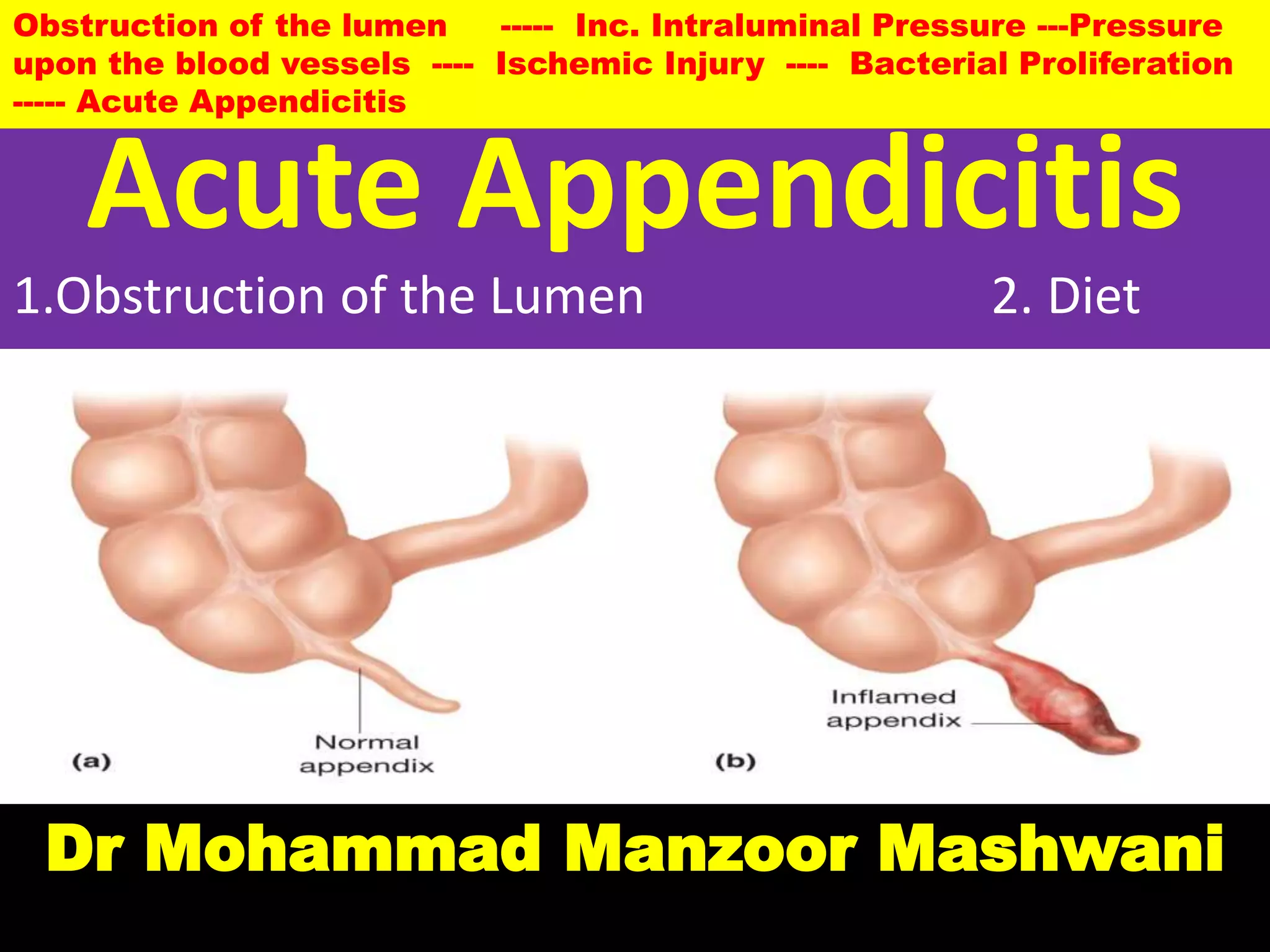
Acute Appendicitis
Dr Mohammad Manzoor Mashwani
1.Obstruction of the Lumen 2. Diet
Obstruction of the lumen ----- Inc. Intraluminal Pressure ---Pressure
upon the blood vessels ---- Ischemic Injury ---- Bacterial Proliferation
----- Acute Appendicitis
Appendix: NORMAL STRUCTURE
The appendix is a blind-ending tubular
diverticulum of the cecum, usually lying behind
the caecum and varies in length from 4 to 20
cm (average 7 cm).
The wall of the appendix consists of all the four
typical coats of the digestive tube: mucosa,
submucosa, muscularis externa & serosa.
A diverticulum is an outpouching of a hollow (or a fluid-filled) structure in the body.
Vermiform Appendix
CECUM
CECUM
Histology
The appendix is characterized by a thick wall but a relatively
narrow lumen which is stellate or irregular in
outline.
Intestinal villi are absent.
Mucosa: Epithelium, Lamina propria, muscularis
mucosa
Epithelium: The mucosa is lined mainly by absorptive cells
(enterocytes) which are columnar cells; only a
few/numerous goblet cells are present.
THICK WALL NARROW LUMEN
Histology
The lamina propria shows intestinal glands (crypts of Lieberkühn).
The intestinal glands in the appendix are less well developed, shorter,
and often spaced farther apart than those in the colon.
The lamina propria of the appendix is heavily infiltrated with lymphocytes and
contains numerous lymphoid nodules, which form the most
conspicuous histologic feature of this organ.
These lymphoid nodules show germinal centers and are not
confined to the lamina propria, but penetrate the muscularis
mucosae to extend into the submucosa (present often in the
submucosa).
Argentaffin and nonargentaffin endocrine cells are present in the base of mucosal glands just as in the small intestine.
Germinal centers are sites within secondary lymphoid organs where mature B lymphocytes proliferate,
differentiate, and mutate their antibody genes and switch the class of their antibodies (for example from IgM to IgG)
during a normal immune response to an infection. The primary lymphoid organs are bone marrow & thymus
& the secondary lymphoid organs are lymph nodes & spleen.
Histology
The submucosa has numerous blood vessels.
The connective tissue of the submucosa of appendix is characterized by the abundance of fat cells.
The submucosa has abundant lymphoid tissue.
The muscularis externa (propria)of the appendix is thin but has two
layers of smooth muscle cells (inner circular and outer
longitudinal) as elsewhere in the alimentary tract.
Teniae coli are absent.
The parasympathetic ganglia of the myenteric plexus are located between
the inner and outer smooth muscle layers of the muscularis externa.
Serosa forms the outermost coat of the appendix under which are seen
adipose cells.
Note:In the circular layer of muscularis externa the cells have spindle nuclei but in the
longitudinal layer the cells have rounded nuclei.
Summary of histology
I. Mucosa:
Epithelium: Columnar cells + goblet cells
Lamina propria: glands + lymphocytes+ lymphoid nodules.
Muscularis mucosae:
II. Submucosa: lymphoid tissues + blood vessels + fat cells.
III. Muscularis propria: thin, 2 layers of smooth muscle cells.
IV. Serosa: outermost coat under which are seen fat cells.
Serosa: The intraperitoneal organs or the organs suspended in the abdominal cavity have
serosa. Serosa is lined by mesothelial cells with some underlying connective tissue.
Adventitia: Retroperitoneal organs have adventia which is just connective tissue.
Serosa is for lubrication where as adventitia is to bind structures together.
Narrow &
Stellate
Lumen
circular layer with spindle nuclei
longitudinal layer with rounded nuclei
crypts of Lieberkühn
Thick Wall
Lymphoid Nodules
Lymphocytes
Fat cells
Acute Appendicitis
Acute Appendicitis
1.Obstruction of the Lumen 2. Diet
Obstruction of the lumen ----- Inc. Intraluminal Pressure ---Pressure
upon the blood vessels ---- Ischemic Injury ---- Bacterial Proliferation
----- Acute Appendicitis
Acute Appendicitis - Epidemiology
Acute inflammation of the appendix, acute appendicitis, is
the most common acute abdominal condition confronting the surgeon.
The condition is seen more commonly in older children and young
adults, and is uncommon at the extremes of age.
The disease is seen more frequently in the West and
in affluent societies which may be due to variation in diet—
a diet with low bulk or cellulose and high protein intake more
often causes appendicitis.
DIET
ETIOPATHOGENESIS
The most common mechanism is obstruction of the lumen
from various etiologic factors that leads to increased intraluminal pressure.
This presses upon the blood vessels to produce ischaemic injury
which in turn favours the bacterial proliferation and hence acute
appendicitis. Obstruction of the lumen ----- Inc. Intraluminal Pressure ---
Pressure upon the blood vessels ---- Ischemic Injury ---- Bacterial Proliferation -----
Acute Appendicitis
Acute appendicitis is thought to be initiated by progressive increases in intraluminal pressure
that compromises venous outflow. In 50% to 80% of cases, acute appendicitis is associated with
overt luminal obstruction, usually by a small, stone-like mass of stool, or fecalith, or, less
commonly, a gallstone, tumor, or mass of worms. Ischemic injury and stasis of luminal
contents, which favor bacterial proliferation, trigger inflammatory responses including tissue
edema and neutrophilic infiltration of the lumen, muscular wall, and periappendiceal soft
tissues.
FECALITH
Functions of Appendix
O
B
S
T
R
U
C
T
I
O
N
OBSTRUCTION
OBSTRUCTION
Tumors,
Gallstones.
Since it is a hollow organ
In children & Young
adults
Functions of Epithelium:
Mucosa:
1. Protection- mucus secretion
2. Absorption
3. Secretion
Mucous Secretion: 1. Protective
function- to prevent bacteria to enter
blood stream.
2. Lubrication.
Even it is plugged the appendix just keeps secreting mucus as
usual. When it happens fluid & mucus builds up which
increases pressure in the appendix just like a water balloon it
becomes bigger and physically presses the afferent viscera
fibers causing abdominal pain.
TLC
Along with that the intestinal
bacteria freely multiply and
this causes the immune
system recruit blood cells and
pus cells accumulate in the
appendix. This activation of
the immune system can be
seen in the lab as
leukocytosis.
Anorexia
Now that obstruction process the pressure in the appendix increases even more at certain point
that the pressure Keeps going and continues to swell up it pushes and compresses the small
blood vessels that supply blood in oxygen. Without the oxygen the cells in the wall of appendix
become ischemic & eventually die. Since these cells were responsible for secreting mucus &
keeping bacteria out now the growing colonies can invade the wall of appendix. As more cells
die the appendiceal wall becomes weaker & weaker and for a small proportion of patients it
Rupture of the infected appendix allow the bacteria to
escape the appendix & to get into the peritoneum &
patient often experiences peritonitis with rebound
tenderness. The most common complication with ruptured
appendix is pus & fluid getting out and forming an abscess
around the appendix called a periappendiceal abscess.
Sometimes subphrenic abscess may also form.
The common causes of appendicitis are as under:
A. Obstructive 50-80%
1. Faecolith/ Fecalith
2. Diffuse lymphoid hyperplasia,
especially in children.
3. Tumours
4. Calculi (Gallstones)
5. Foreign body
6. Worms (especially Enterobius vermicularis)
B. Non-obstructive
1. Haematogenous spread of generalised infection
2. Vascular occlusion
3. Inappropriate diet lacking roughage.
(Suppurative)
Phlegmonous Apostematous
Acute gangrenous Appendicitis
Phlegmon is a spreading diffuse inflammatory process with
formation of suppurative/purulent exudate or pus. This is the
result of acute purulent inflammation which may be related to
bacterial infection, however the term 'phlegmon' mostly refers
to a walled-off inflammatory mass without bacterial infection,
one that may be palpable on physical examination.
Aposteme:A swelling filled with purulent matter.
MORPHOLOGIC FEATURES
Grossly, the appearance depends upon the stage at
which the acutely-inflamed appendix is examined.
In early acute appendicitis, the organ is swollen and serosa
shows hyperaemia. The inflammatory reaction transforms the normal glistening serosa into a dull, granular
appearing, erythematous surface.
In welldeveloped acute inflammation called acute
suppurative appendicitis, the serosa is coated with
fibrinopurulent exudate and engorged vessels on the surface.
In further advanced cases called acute gangrenous
appendicitis, there is necrosis and ulcerations of mucosa
which extend
through the wall so that the appendix becomes soft and friable and the
surface is coated with greenish-black gangrenous necrosis.
On cut section
GROSS
Gross Examination
Swelling
Redness
Fibrinopurulent Exudate?
Prominent vessels
Gross description
● Lumen may contain blood-tinged pus
● Variable perforation, mucosal ulceration, fecalith
or other obstructing agent
On Cut section
Microscopy
Microscopically, the most important diagnostic histological criterion is the
neutrophilic infiltration of the muscularis propria.
In early stage, the other changes besides acute inflammatory changes, are
congestion and oedema of the appendiceal wall.
In later stages, the mucosa is sloughed off,
the wall becomes necrotic, the blood vessels may get
thrombosed
and there may be neutrophilic abscesses in the wall.
Diagnosis of acute appendicitis requires neutrophilic infiltration of the muscularis propria.
Microscopy
 Early:
● Mucosal ulceration
● Minimal (if early) to dense neutrophils in muscularis propria
 with necrosis,
 congestion,
 perivascular neutrophilic infiltrate
● Late:
 absent mucosa,
 necrotic wall,
 prominent fibrosis,
 granulation tissue,
 marked chronic inflammatory infiltrate in wall,
 thrombosed vessels
Acute appendicitis. Microscopic appearance showing diagnostic
neutrophilic infiltration into the muscularis propria. Other changes
present are necrosis of mucosa and periappendicitis.
Periappendicitis is defined as appendiceal serosal inflammation without mucosal involvement.
Lymphaticnodules
CLINICAL COURSE
The patient presents with features of acute abdomen
as under:
1. Colicky pain, initially around umbilicus but later localised to right iliac fossa
2. Nausea and vomiting
3. Anorexia
4. Pyrexia of mild grade
5. Abdominal tenderness
6. Increased pulse rate
7. Neutrophilic leucocytosis.
Both the appendix and the umbilicus
are innervated by segment T10 of the
spinal cord and hence the pain caused
by appendicitis is first felt in the region
of umbilicus (referred pain).
With increasing inflammation pain is
felt in the right iliac fossa due to
involvement of the parietal peritoneum
of the region which is sensitive to pain
in contrast to pain-insensitive visceral
peritoneum.
Leukocytes
TLC
T10
T10
Visceral Somatic reflex (Pain)
Clinical features
A classic physical finding is McBurney’s sign, deep
tenderness noted at a location two thirds of the
distance from the umbilicus to the right anterior
superior iliac spine (McBurney’s point).
Complications:
1. Peritonitis
2. Abscess
3. Adhesions
4. Portal Pylephlebitis
5. Mucocele
Appendiceal Abscess
COMPLICATIONS
If the condition is not adequately managed, the following
complications may occur:
1. Peritonitis. A perforated appendix as occurs in
gangrenous appendicitis may cause localised or
generalised peritonitis.
2. Appendiceal abscess. This is due to rupture of an
appendix giving rise to localised abscess in the right
iliac fossa. This abscess may spread to other sites such as
• between the liver and diaphragm (subphrenic abscess),
• into the pelvis between the urinary bladder and rectum,
and
• in the females may involve uterus and fallopian tubes.
3. Adhesions. Late complications of acute appendicitis are
fibrous adhesions to the greater omentum, small intestine
and other abdominal structures.
4. Portal pylephlebitis. Spread of infection into mesenteric
veins may produce septic phlebitis and liver abscess.
5. Mucocele. Distension of distal appendix by mucus
following recovery from an attack of acute appendicitis is
referred to as mucocele. It occurs generally due to proximal
obstruction but sometimes may be due to a benign or
malignant neoplasm in the appendix. An infected mucocele
may result in formation of empyema of the appendix.
Mucocele Empyema: A collection of
pus in body cavity.
TUMOURS OF APPENDIX
Tumours of the appendix are quite rare. These
include:
1. Carcinoid tumour (the most common),
2. Pseudomyxoma peritonei and
3. Adenocarcinoma.
CARCINOID TUMOUR
Both argentaffin and argyrophil types are encountered,
the former being more common.
Appendiceal carcinoids, occur more frequently in 3rd and 4th decades of life without any sex
predilection, are often solitary and behave as locally malignant tumours.
Morphology
Grossly, carcinoid tumour of the appendix is mostly situated
near the tip of the organ and appears as a
circumscribed nodule, usually less than 1 cm in
diameter, involving the wall but metastases are rare.
Histologically, carcinoid tumour of the appendix
resembles other carcinoids of the midgut.
Appendiceal carcinoids commonly involve the tip of the organ and are solitary
Tumors of Appendix
PSEUDOMYXOMA PERITONEI. Pseudomyxoma peritonei
is appearance of gelatinous mucinous material around the
appendix admixed with epithelial tumour cells.
It is generally due to mucinous collection from benign mucinous
cystadenoma of the ovary or mucin-secreting carcinoma of
the appendix.
ADENOCARCINOMA. It is an uncommon tumour in the
appendix and is morphologically similar to adenocarcinoma
elsewhere in the alimentary tract.
THANKS
Haveagoodday.

L acute appendicitis

  • 1.
    Acute Appendicitis Dr MohammadManzoor Mashwani 1.Obstruction of the Lumen 2. Diet Obstruction of the lumen ----- Inc. Intraluminal Pressure ---Pressure upon the blood vessels ---- Ischemic Injury ---- Bacterial Proliferation ----- Acute Appendicitis
  • 2.
    Appendix: NORMAL STRUCTURE Theappendix is a blind-ending tubular diverticulum of the cecum, usually lying behind the caecum and varies in length from 4 to 20 cm (average 7 cm). The wall of the appendix consists of all the four typical coats of the digestive tube: mucosa, submucosa, muscularis externa & serosa. A diverticulum is an outpouching of a hollow (or a fluid-filled) structure in the body. Vermiform Appendix CECUM CECUM
  • 3.
    Histology The appendix ischaracterized by a thick wall but a relatively narrow lumen which is stellate or irregular in outline. Intestinal villi are absent. Mucosa: Epithelium, Lamina propria, muscularis mucosa Epithelium: The mucosa is lined mainly by absorptive cells (enterocytes) which are columnar cells; only a few/numerous goblet cells are present. THICK WALL NARROW LUMEN
  • 4.
    Histology The lamina propriashows intestinal glands (crypts of Lieberkühn). The intestinal glands in the appendix are less well developed, shorter, and often spaced farther apart than those in the colon. The lamina propria of the appendix is heavily infiltrated with lymphocytes and contains numerous lymphoid nodules, which form the most conspicuous histologic feature of this organ. These lymphoid nodules show germinal centers and are not confined to the lamina propria, but penetrate the muscularis mucosae to extend into the submucosa (present often in the submucosa). Argentaffin and nonargentaffin endocrine cells are present in the base of mucosal glands just as in the small intestine. Germinal centers are sites within secondary lymphoid organs where mature B lymphocytes proliferate, differentiate, and mutate their antibody genes and switch the class of their antibodies (for example from IgM to IgG) during a normal immune response to an infection. The primary lymphoid organs are bone marrow & thymus & the secondary lymphoid organs are lymph nodes & spleen.
  • 5.
    Histology The submucosa hasnumerous blood vessels. The connective tissue of the submucosa of appendix is characterized by the abundance of fat cells. The submucosa has abundant lymphoid tissue. The muscularis externa (propria)of the appendix is thin but has two layers of smooth muscle cells (inner circular and outer longitudinal) as elsewhere in the alimentary tract. Teniae coli are absent. The parasympathetic ganglia of the myenteric plexus are located between the inner and outer smooth muscle layers of the muscularis externa. Serosa forms the outermost coat of the appendix under which are seen adipose cells. Note:In the circular layer of muscularis externa the cells have spindle nuclei but in the longitudinal layer the cells have rounded nuclei.
  • 6.
    Summary of histology I.Mucosa: Epithelium: Columnar cells + goblet cells Lamina propria: glands + lymphocytes+ lymphoid nodules. Muscularis mucosae: II. Submucosa: lymphoid tissues + blood vessels + fat cells. III. Muscularis propria: thin, 2 layers of smooth muscle cells. IV. Serosa: outermost coat under which are seen fat cells. Serosa: The intraperitoneal organs or the organs suspended in the abdominal cavity have serosa. Serosa is lined by mesothelial cells with some underlying connective tissue. Adventitia: Retroperitoneal organs have adventia which is just connective tissue. Serosa is for lubrication where as adventitia is to bind structures together.
  • 7.
    Narrow & Stellate Lumen circular layerwith spindle nuclei longitudinal layer with rounded nuclei crypts of Lieberkühn Thick Wall Lymphoid Nodules Lymphocytes Fat cells
  • 8.
    Acute Appendicitis Acute Appendicitis 1.Obstructionof the Lumen 2. Diet Obstruction of the lumen ----- Inc. Intraluminal Pressure ---Pressure upon the blood vessels ---- Ischemic Injury ---- Bacterial Proliferation ----- Acute Appendicitis
  • 9.
    Acute Appendicitis -Epidemiology Acute inflammation of the appendix, acute appendicitis, is the most common acute abdominal condition confronting the surgeon. The condition is seen more commonly in older children and young adults, and is uncommon at the extremes of age. The disease is seen more frequently in the West and in affluent societies which may be due to variation in diet— a diet with low bulk or cellulose and high protein intake more often causes appendicitis. DIET
  • 10.
    ETIOPATHOGENESIS The most commonmechanism is obstruction of the lumen from various etiologic factors that leads to increased intraluminal pressure. This presses upon the blood vessels to produce ischaemic injury which in turn favours the bacterial proliferation and hence acute appendicitis. Obstruction of the lumen ----- Inc. Intraluminal Pressure --- Pressure upon the blood vessels ---- Ischemic Injury ---- Bacterial Proliferation ----- Acute Appendicitis Acute appendicitis is thought to be initiated by progressive increases in intraluminal pressure that compromises venous outflow. In 50% to 80% of cases, acute appendicitis is associated with overt luminal obstruction, usually by a small, stone-like mass of stool, or fecalith, or, less commonly, a gallstone, tumor, or mass of worms. Ischemic injury and stasis of luminal contents, which favor bacterial proliferation, trigger inflammatory responses including tissue edema and neutrophilic infiltration of the lumen, muscular wall, and periappendiceal soft tissues. FECALITH
  • 11.
  • 12.
  • 13.
    Functions of Epithelium: Mucosa: 1.Protection- mucus secretion 2. Absorption 3. Secretion Mucous Secretion: 1. Protective function- to prevent bacteria to enter blood stream. 2. Lubrication. Even it is plugged the appendix just keeps secreting mucus as usual. When it happens fluid & mucus builds up which increases pressure in the appendix just like a water balloon it becomes bigger and physically presses the afferent viscera fibers causing abdominal pain.
  • 14.
    TLC Along with thatthe intestinal bacteria freely multiply and this causes the immune system recruit blood cells and pus cells accumulate in the appendix. This activation of the immune system can be seen in the lab as leukocytosis.
  • 15.
    Anorexia Now that obstructionprocess the pressure in the appendix increases even more at certain point that the pressure Keeps going and continues to swell up it pushes and compresses the small blood vessels that supply blood in oxygen. Without the oxygen the cells in the wall of appendix become ischemic & eventually die. Since these cells were responsible for secreting mucus & keeping bacteria out now the growing colonies can invade the wall of appendix. As more cells die the appendiceal wall becomes weaker & weaker and for a small proportion of patients it
  • 16.
    Rupture of theinfected appendix allow the bacteria to escape the appendix & to get into the peritoneum & patient often experiences peritonitis with rebound tenderness. The most common complication with ruptured appendix is pus & fluid getting out and forming an abscess around the appendix called a periappendiceal abscess. Sometimes subphrenic abscess may also form.
  • 17.
    The common causesof appendicitis are as under: A. Obstructive 50-80% 1. Faecolith/ Fecalith 2. Diffuse lymphoid hyperplasia, especially in children. 3. Tumours 4. Calculi (Gallstones) 5. Foreign body 6. Worms (especially Enterobius vermicularis) B. Non-obstructive 1. Haematogenous spread of generalised infection 2. Vascular occlusion 3. Inappropriate diet lacking roughage.
  • 18.
  • 19.
    Phlegmon is aspreading diffuse inflammatory process with formation of suppurative/purulent exudate or pus. This is the result of acute purulent inflammation which may be related to bacterial infection, however the term 'phlegmon' mostly refers to a walled-off inflammatory mass without bacterial infection, one that may be palpable on physical examination. Aposteme:A swelling filled with purulent matter.
  • 20.
    MORPHOLOGIC FEATURES Grossly, theappearance depends upon the stage at which the acutely-inflamed appendix is examined. In early acute appendicitis, the organ is swollen and serosa shows hyperaemia. The inflammatory reaction transforms the normal glistening serosa into a dull, granular appearing, erythematous surface. In welldeveloped acute inflammation called acute suppurative appendicitis, the serosa is coated with fibrinopurulent exudate and engorged vessels on the surface. In further advanced cases called acute gangrenous appendicitis, there is necrosis and ulcerations of mucosa which extend through the wall so that the appendix becomes soft and friable and the surface is coated with greenish-black gangrenous necrosis. On cut section GROSS
  • 21.
  • 23.
    Gross description ● Lumenmay contain blood-tinged pus ● Variable perforation, mucosal ulceration, fecalith or other obstructing agent On Cut section
  • 24.
    Microscopy Microscopically, the mostimportant diagnostic histological criterion is the neutrophilic infiltration of the muscularis propria. In early stage, the other changes besides acute inflammatory changes, are congestion and oedema of the appendiceal wall. In later stages, the mucosa is sloughed off, the wall becomes necrotic, the blood vessels may get thrombosed and there may be neutrophilic abscesses in the wall. Diagnosis of acute appendicitis requires neutrophilic infiltration of the muscularis propria.
  • 25.
    Microscopy  Early: ● Mucosalulceration ● Minimal (if early) to dense neutrophils in muscularis propria  with necrosis,  congestion,  perivascular neutrophilic infiltrate ● Late:  absent mucosa,  necrotic wall,  prominent fibrosis,  granulation tissue,  marked chronic inflammatory infiltrate in wall,  thrombosed vessels
  • 26.
    Acute appendicitis. Microscopicappearance showing diagnostic neutrophilic infiltration into the muscularis propria. Other changes present are necrosis of mucosa and periappendicitis. Periappendicitis is defined as appendiceal serosal inflammation without mucosal involvement. Lymphaticnodules
  • 28.
    CLINICAL COURSE The patientpresents with features of acute abdomen as under: 1. Colicky pain, initially around umbilicus but later localised to right iliac fossa 2. Nausea and vomiting 3. Anorexia 4. Pyrexia of mild grade 5. Abdominal tenderness 6. Increased pulse rate 7. Neutrophilic leucocytosis. Both the appendix and the umbilicus are innervated by segment T10 of the spinal cord and hence the pain caused by appendicitis is first felt in the region of umbilicus (referred pain). With increasing inflammation pain is felt in the right iliac fossa due to involvement of the parietal peritoneum of the region which is sensitive to pain in contrast to pain-insensitive visceral peritoneum. Leukocytes TLC T10 T10
  • 29.
  • 30.
    Clinical features A classicphysical finding is McBurney’s sign, deep tenderness noted at a location two thirds of the distance from the umbilicus to the right anterior superior iliac spine (McBurney’s point). Complications: 1. Peritonitis 2. Abscess 3. Adhesions 4. Portal Pylephlebitis 5. Mucocele Appendiceal Abscess
  • 31.
    COMPLICATIONS If the conditionis not adequately managed, the following complications may occur: 1. Peritonitis. A perforated appendix as occurs in gangrenous appendicitis may cause localised or generalised peritonitis. 2. Appendiceal abscess. This is due to rupture of an appendix giving rise to localised abscess in the right iliac fossa. This abscess may spread to other sites such as • between the liver and diaphragm (subphrenic abscess), • into the pelvis between the urinary bladder and rectum, and • in the females may involve uterus and fallopian tubes.
  • 32.
    3. Adhesions. Latecomplications of acute appendicitis are fibrous adhesions to the greater omentum, small intestine and other abdominal structures. 4. Portal pylephlebitis. Spread of infection into mesenteric veins may produce septic phlebitis and liver abscess. 5. Mucocele. Distension of distal appendix by mucus following recovery from an attack of acute appendicitis is referred to as mucocele. It occurs generally due to proximal obstruction but sometimes may be due to a benign or malignant neoplasm in the appendix. An infected mucocele may result in formation of empyema of the appendix. Mucocele Empyema: A collection of pus in body cavity.
  • 33.
    TUMOURS OF APPENDIX Tumoursof the appendix are quite rare. These include: 1. Carcinoid tumour (the most common), 2. Pseudomyxoma peritonei and 3. Adenocarcinoma. CARCINOID TUMOUR Both argentaffin and argyrophil types are encountered, the former being more common. Appendiceal carcinoids, occur more frequently in 3rd and 4th decades of life without any sex predilection, are often solitary and behave as locally malignant tumours.
  • 34.
    Morphology Grossly, carcinoid tumourof the appendix is mostly situated near the tip of the organ and appears as a circumscribed nodule, usually less than 1 cm in diameter, involving the wall but metastases are rare. Histologically, carcinoid tumour of the appendix resembles other carcinoids of the midgut. Appendiceal carcinoids commonly involve the tip of the organ and are solitary
  • 35.
    Tumors of Appendix PSEUDOMYXOMAPERITONEI. Pseudomyxoma peritonei is appearance of gelatinous mucinous material around the appendix admixed with epithelial tumour cells. It is generally due to mucinous collection from benign mucinous cystadenoma of the ovary or mucin-secreting carcinoma of the appendix. ADENOCARCINOMA. It is an uncommon tumour in the appendix and is morphologically similar to adenocarcinoma elsewhere in the alimentary tract.
  • 36.