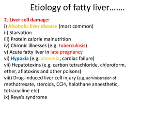
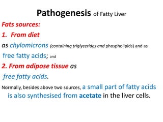
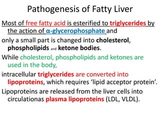
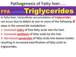
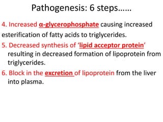
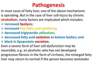
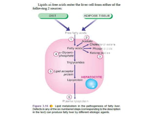
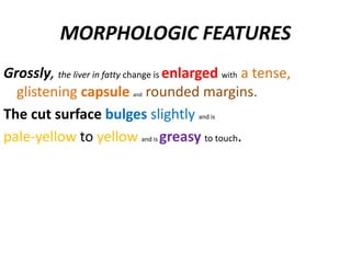
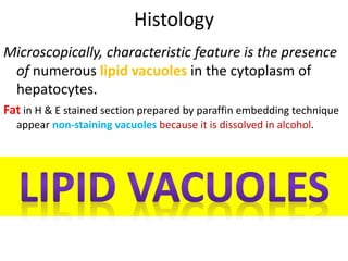
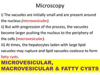
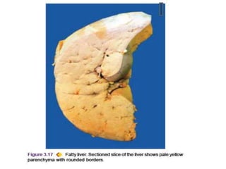
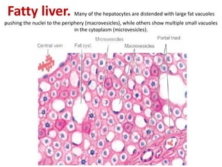
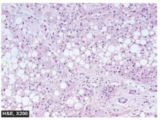
Fatty Change
Fatty liver
Dr Mohammad Manzoor Mashwani
Morphologic forms of reversible cell injury are:
1. Hydropic change (cloudy swelling, or vacuolar degeneration)
2. Fatty change
3. Hyaline change
4. Mucoid change
FATTY CHANGE (STEATOSIS)
Fatty change, steatosis or fatty metamorphosis is the
intracellular accumulation of neutral fat within
parenchymal cells.
It includes the older, now abandoned, terms of fatty degeneration and fatty
infiltration because fatty change neither necessarily involves degeneration nor
infiltration.
The deposit is in the cytosol and represents an absolute
increase in the intracellular lipids.
It is especially common in the liver but may occur in
other non-fatty tissues like the heart, skeletal muscle,
kidneys (lipoid nephrosis or minimum change disease) and other organs.
Fatty Metamorphosis
Fatty Liver
Liver is the commonest site for accumulation of fat
because it plays central role in fat metabolism.
Depending upon the cause and amount of accumulation, fatty change may be mild
and reversible, or severe producing irreversible cell injury and cell death.
Morphologic forms of reversible cell injury are:
1. Hydropic change (cloudy swelling, or vacuolar degeneration)
2. Fatty change
3. Hyaline change
4. Mucoid change
ETIOLOGY
Fatty change in the liver may result from one of the two types
of causes:
1. Conditions with excess fat (hyperlipidameia), exceeding the
capacity of the liver to metabolise it.
2. Liver cell damage, when fat cannot be metabolised in it.
These causes are listed below:
1. Conditions with excess fat:
i) Obesity
ii) Diabetes mellitus
iii) Congenital hyperlipidaemia
Etiology of fatty liver…….
2. Liver cell damage:
i) Alcoholic liver disease (most common)
ii) Starvation
iii) Protein calorie malnutrition
iv) Chronic illnesses (e.g. tuberculosis)
v) Acute fatty liver in late pregnancy
vi) Hypoxia (e.g. anaemia, cardiac failure)
vii) Hepatotoxins (e.g. carbon tetrachloride, chloroform,
ether, aflatoxins and other poisons)
viii) Drug-induced liver cell injury (e.g. administration of
methotrexate, steroids, CCl4, halothane anaesthetic,
tetracycline etc)
ix) Reye’s syndrome
PATHOGENESIS
Mechanism of fatty liver depends upon the
stage at which the etiologic
agent acts in the normal fat transport and
metabolism.
Hence, pathogenesis of fatty liver is best understood
in the light of normal fat metabolism in the liver.
Pathogenesis of Fatty Liver
Fats sources:
1. From diet
as chylomicrons (containing triglycerides and phospholipids) and as
free fatty acids; and
2. From adipose tissue as
free fatty acids.
Normally, besides above two sources, a small part of fatty acids
is also synthesised from acetate in the liver cells.
Pathogenesis of Fatty Liver
Most of free fatty acid is esterified to triglycerides by
the action of α-glycerophosphate and
only a small part is changed into cholesterol,
phospholipids and ketone bodies.
While cholesterol, phospholipids and ketones are
used in the body,
intracellular triglycerides are converted into
lipoproteins, which requires ‘lipid acceptor protein’.
Lipoproteins are released from the liver cells into
circulationas plasma lipoproteins (LDL, VLDL).
Pathogenesis of Fatty liver…..
In fatty liver, intracellular accumulation of triglycerides
can occur due to defect at one or more of the following 6
steps in the normal fat metabolism :
1. Increased entry of free fatty acids into the liver.
2. Increased synthesis of fatty acids by the liver.
3. Decreased conversion of fatty acids into ketone bodies
resulting in increased esterification of fatty acids to
triglycerides.
FFA Triglycerides
Pathogenesis: 6 steps……
4. Increased α-glycerophosphate causing increased
esterification of fatty acids to triglycerides.
5. Decreased synthesis of ‘lipid acceptor protein’
resulting in decreased formation of lipoprotein from
triglycerides.
6. Block in the excretion of lipoprotein from the liver
into plasma.
Pathogenesis
In most cases of fatty liver, one of the above mechanisms
is operating. But in the case of liver cell injury by chronic
alcoholism, many factors are implicated which includes:
 increased lipolysis;
 increased free fatty acid synthesis;
 decreased triglyceride utilisation;
 decreased fatty acid oxidation to ketone bodies; and
 block in lipoprotein excretion.
Even a severe form of liver cell dysfunction may be
reversible; e.g. an alcoholic who has not developed
progressive fibrosis in the form of cirrhosis, the enlarged fatty
liver may return to normal if the person becomes teetotaler.
MORPHOLOGIC FEATURES
Grossly, the liver in fatty change is enlarged with a tense,
glistening capsule and rounded margins.
The cut surface bulges slightly and is
pale-yellow to yellow and is greasy to touch.
Histology
Microscopically, characteristic feature is the presence
of numerous lipid vacuoles in the cytoplasm of
hepatocytes.
Fat in H & E stained section prepared by paraffin embedding technique
appear non-staining vacuoles because it is dissolved in alcohol.
Microscopy
i) The vacuoles are initially small and are present around
the nucleus (microvesicular).
ii) But with progression of the process, the vacuoles
become larger pushing the nucleus to the periphery of
the cells (macrovesicular).
iii) At times, the hepatocytes laden with large lipid
vacuoles may rupture and lipid vacuoles coalesce to form
fatty cysts.
MICROVESICULAR,
MACROVESICULAR & FATTY CYSTS
iv) Infrequently, lipogranulomas may appear
consisting of collections of lymphocytes,
macrophages, and some multinucleated giant cells.
v) Fat can be demonstrated in fresh unfixed tissue by
frozen section followed by fat stains such as Sudan
dyes (Sudan III, IV, Sudan black) and oil red O.
Alternatively, osmic acid which is a fixative as well as
a stain can be used to demonstrate fat in the tissue.
Fatty liver. Many of the hepatocytes are distended with large fat vacuoles
pushing the nuclei to the periphery (macrovesicles), while others show multiple small vacuoles
in the cytoplasm (microvesicles).
THANKs

Fatty liver

  • 1.
    Fatty Change Fatty liver DrMohammad Manzoor Mashwani Morphologic forms of reversible cell injury are: 1. Hydropic change (cloudy swelling, or vacuolar degeneration) 2. Fatty change 3. Hyaline change 4. Mucoid change
  • 2.
    FATTY CHANGE (STEATOSIS) Fattychange, steatosis or fatty metamorphosis is the intracellular accumulation of neutral fat within parenchymal cells. It includes the older, now abandoned, terms of fatty degeneration and fatty infiltration because fatty change neither necessarily involves degeneration nor infiltration. The deposit is in the cytosol and represents an absolute increase in the intracellular lipids. It is especially common in the liver but may occur in other non-fatty tissues like the heart, skeletal muscle, kidneys (lipoid nephrosis or minimum change disease) and other organs. Fatty Metamorphosis
  • 3.
    Fatty Liver Liver isthe commonest site for accumulation of fat because it plays central role in fat metabolism. Depending upon the cause and amount of accumulation, fatty change may be mild and reversible, or severe producing irreversible cell injury and cell death. Morphologic forms of reversible cell injury are: 1. Hydropic change (cloudy swelling, or vacuolar degeneration) 2. Fatty change 3. Hyaline change 4. Mucoid change
  • 4.
    ETIOLOGY Fatty change inthe liver may result from one of the two types of causes: 1. Conditions with excess fat (hyperlipidameia), exceeding the capacity of the liver to metabolise it. 2. Liver cell damage, when fat cannot be metabolised in it. These causes are listed below: 1. Conditions with excess fat: i) Obesity ii) Diabetes mellitus iii) Congenital hyperlipidaemia
  • 5.
    Etiology of fattyliver……. 2. Liver cell damage: i) Alcoholic liver disease (most common) ii) Starvation iii) Protein calorie malnutrition iv) Chronic illnesses (e.g. tuberculosis) v) Acute fatty liver in late pregnancy vi) Hypoxia (e.g. anaemia, cardiac failure) vii) Hepatotoxins (e.g. carbon tetrachloride, chloroform, ether, aflatoxins and other poisons) viii) Drug-induced liver cell injury (e.g. administration of methotrexate, steroids, CCl4, halothane anaesthetic, tetracycline etc) ix) Reye’s syndrome
  • 6.
    PATHOGENESIS Mechanism of fattyliver depends upon the stage at which the etiologic agent acts in the normal fat transport and metabolism. Hence, pathogenesis of fatty liver is best understood in the light of normal fat metabolism in the liver.
  • 7.
    Pathogenesis of FattyLiver Fats sources: 1. From diet as chylomicrons (containing triglycerides and phospholipids) and as free fatty acids; and 2. From adipose tissue as free fatty acids. Normally, besides above two sources, a small part of fatty acids is also synthesised from acetate in the liver cells.
  • 8.
    Pathogenesis of FattyLiver Most of free fatty acid is esterified to triglycerides by the action of α-glycerophosphate and only a small part is changed into cholesterol, phospholipids and ketone bodies. While cholesterol, phospholipids and ketones are used in the body, intracellular triglycerides are converted into lipoproteins, which requires ‘lipid acceptor protein’. Lipoproteins are released from the liver cells into circulationas plasma lipoproteins (LDL, VLDL).
  • 9.
    Pathogenesis of Fattyliver….. In fatty liver, intracellular accumulation of triglycerides can occur due to defect at one or more of the following 6 steps in the normal fat metabolism : 1. Increased entry of free fatty acids into the liver. 2. Increased synthesis of fatty acids by the liver. 3. Decreased conversion of fatty acids into ketone bodies resulting in increased esterification of fatty acids to triglycerides. FFA Triglycerides
  • 10.
    Pathogenesis: 6 steps…… 4.Increased α-glycerophosphate causing increased esterification of fatty acids to triglycerides. 5. Decreased synthesis of ‘lipid acceptor protein’ resulting in decreased formation of lipoprotein from triglycerides. 6. Block in the excretion of lipoprotein from the liver into plasma.
  • 11.
    Pathogenesis In most casesof fatty liver, one of the above mechanisms is operating. But in the case of liver cell injury by chronic alcoholism, many factors are implicated which includes:  increased lipolysis;  increased free fatty acid synthesis;  decreased triglyceride utilisation;  decreased fatty acid oxidation to ketone bodies; and  block in lipoprotein excretion. Even a severe form of liver cell dysfunction may be reversible; e.g. an alcoholic who has not developed progressive fibrosis in the form of cirrhosis, the enlarged fatty liver may return to normal if the person becomes teetotaler.
  • 13.
    MORPHOLOGIC FEATURES Grossly, theliver in fatty change is enlarged with a tense, glistening capsule and rounded margins. The cut surface bulges slightly and is pale-yellow to yellow and is greasy to touch.
  • 14.
    Histology Microscopically, characteristic featureis the presence of numerous lipid vacuoles in the cytoplasm of hepatocytes. Fat in H & E stained section prepared by paraffin embedding technique appear non-staining vacuoles because it is dissolved in alcohol.
  • 15.
    Microscopy i) The vacuolesare initially small and are present around the nucleus (microvesicular). ii) But with progression of the process, the vacuoles become larger pushing the nucleus to the periphery of the cells (macrovesicular). iii) At times, the hepatocytes laden with large lipid vacuoles may rupture and lipid vacuoles coalesce to form fatty cysts. MICROVESICULAR, MACROVESICULAR & FATTY CYSTS
  • 16.
    iv) Infrequently, lipogranulomasmay appear consisting of collections of lymphocytes, macrophages, and some multinucleated giant cells. v) Fat can be demonstrated in fresh unfixed tissue by frozen section followed by fat stains such as Sudan dyes (Sudan III, IV, Sudan black) and oil red O. Alternatively, osmic acid which is a fixative as well as a stain can be used to demonstrate fat in the tissue.
  • 18.
    Fatty liver. Manyof the hepatocytes are distended with large fat vacuoles pushing the nuclei to the periphery (macrovesicles), while others show multiple small vacuoles in the cytoplasm (microvesicles).
  • 20.