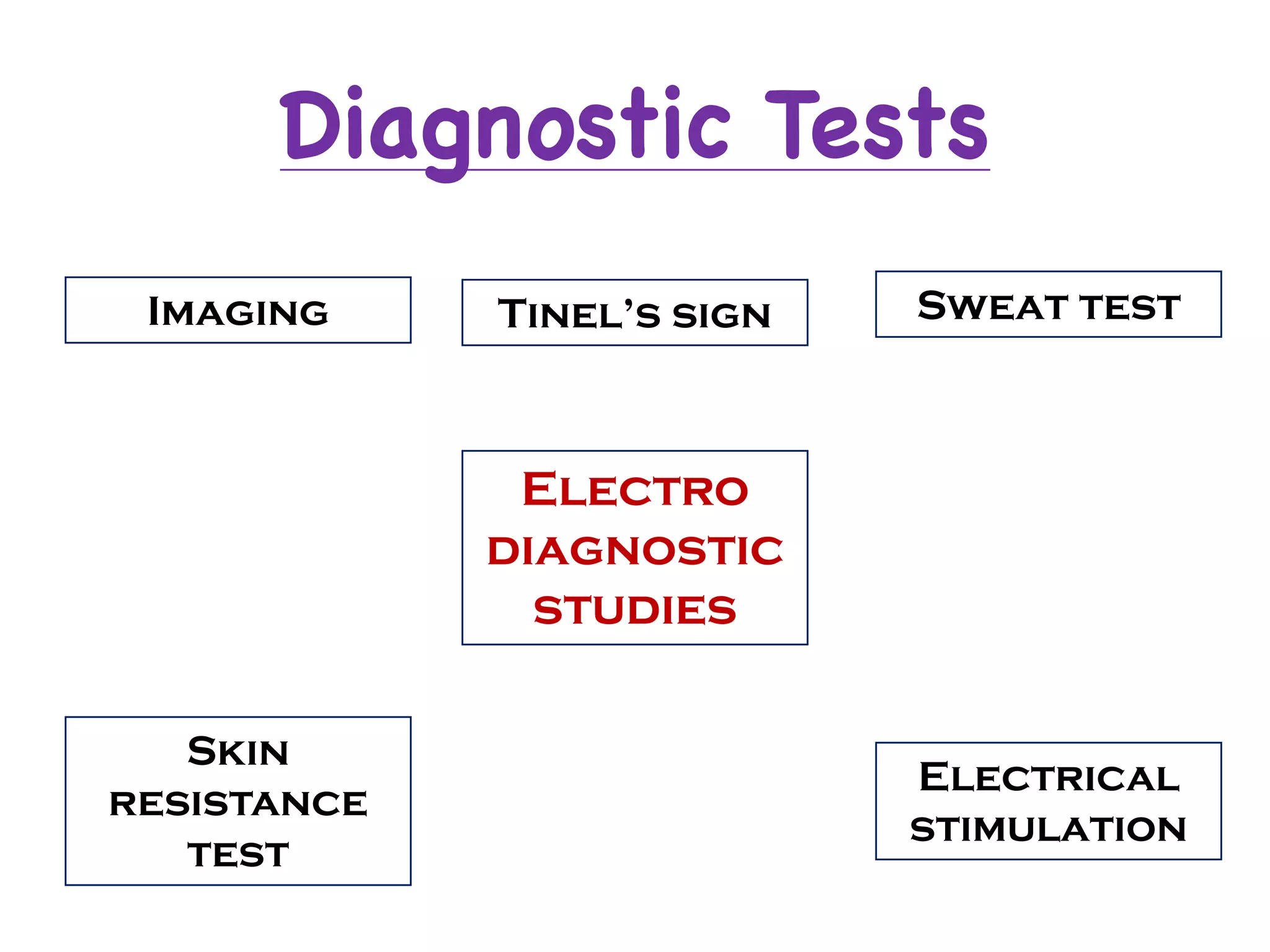
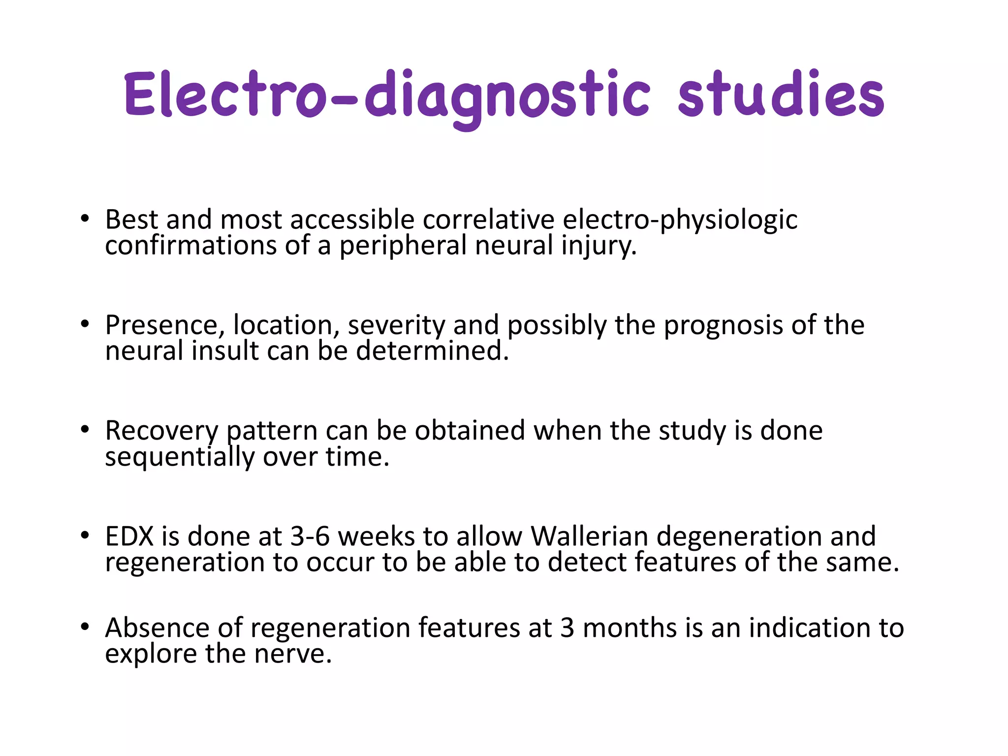
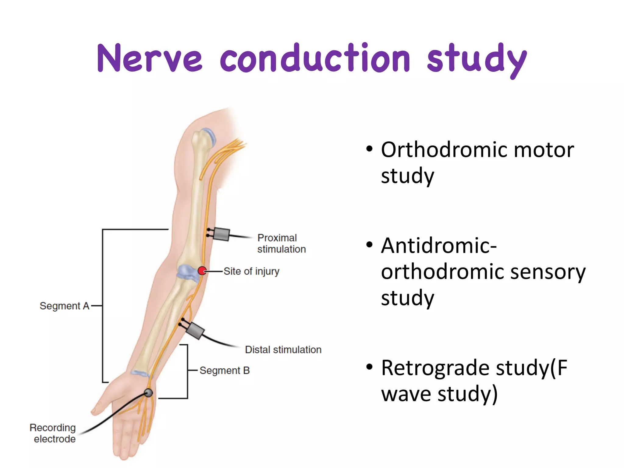
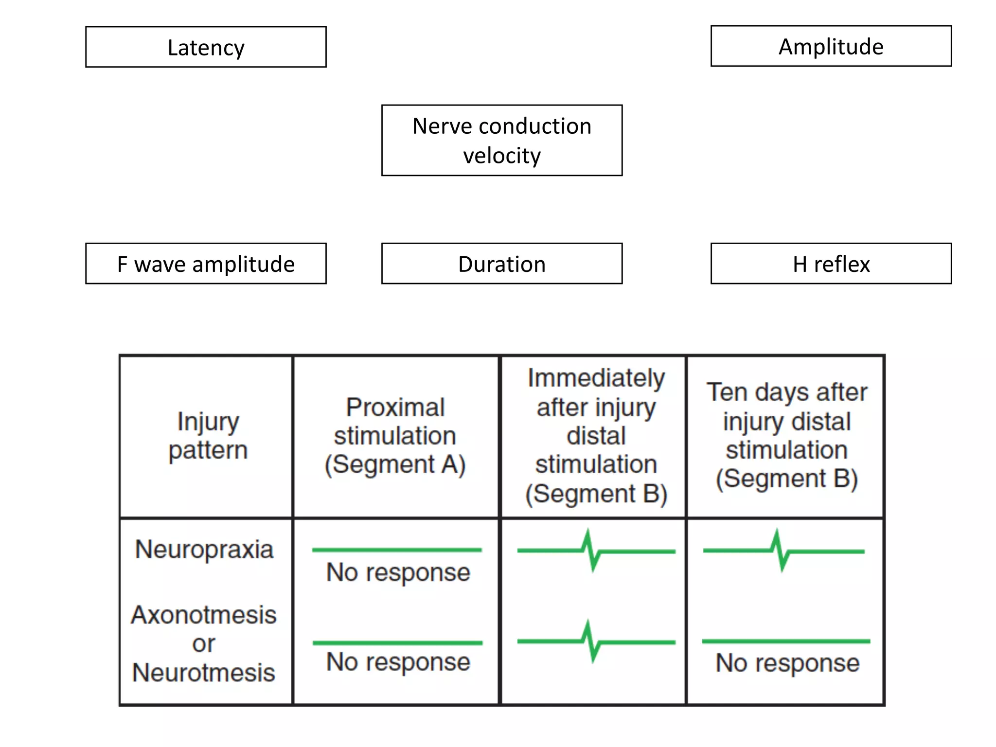
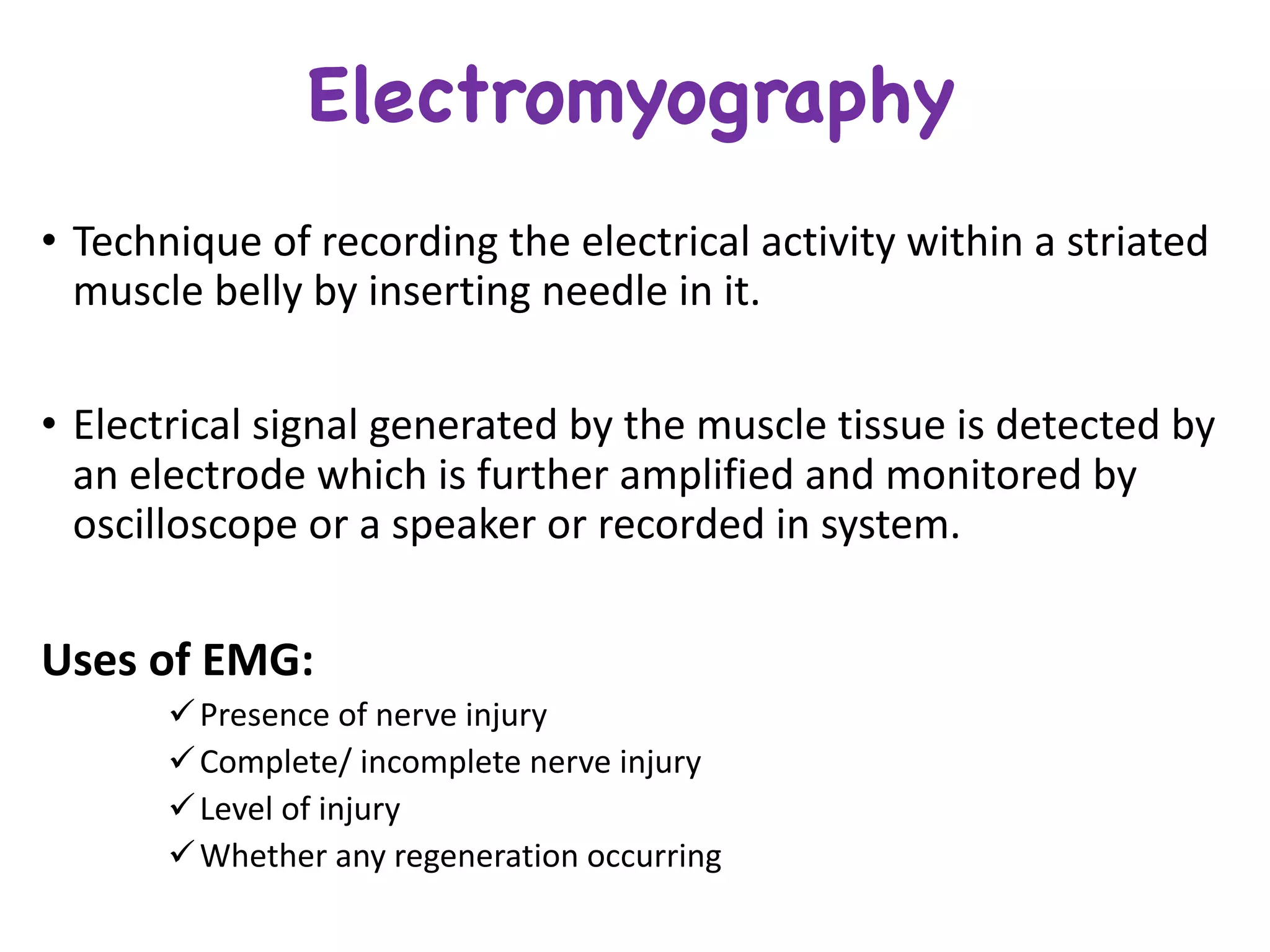
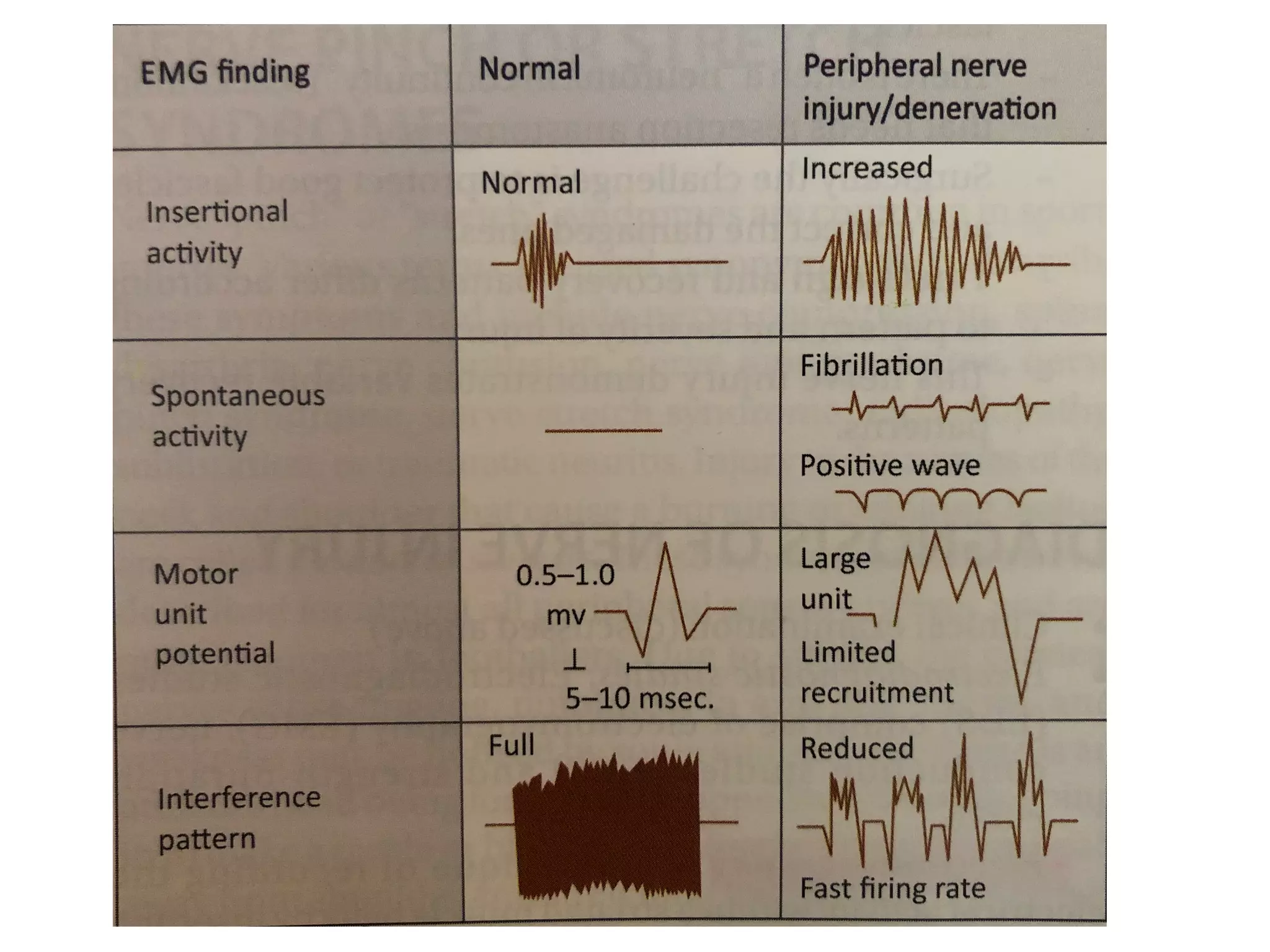
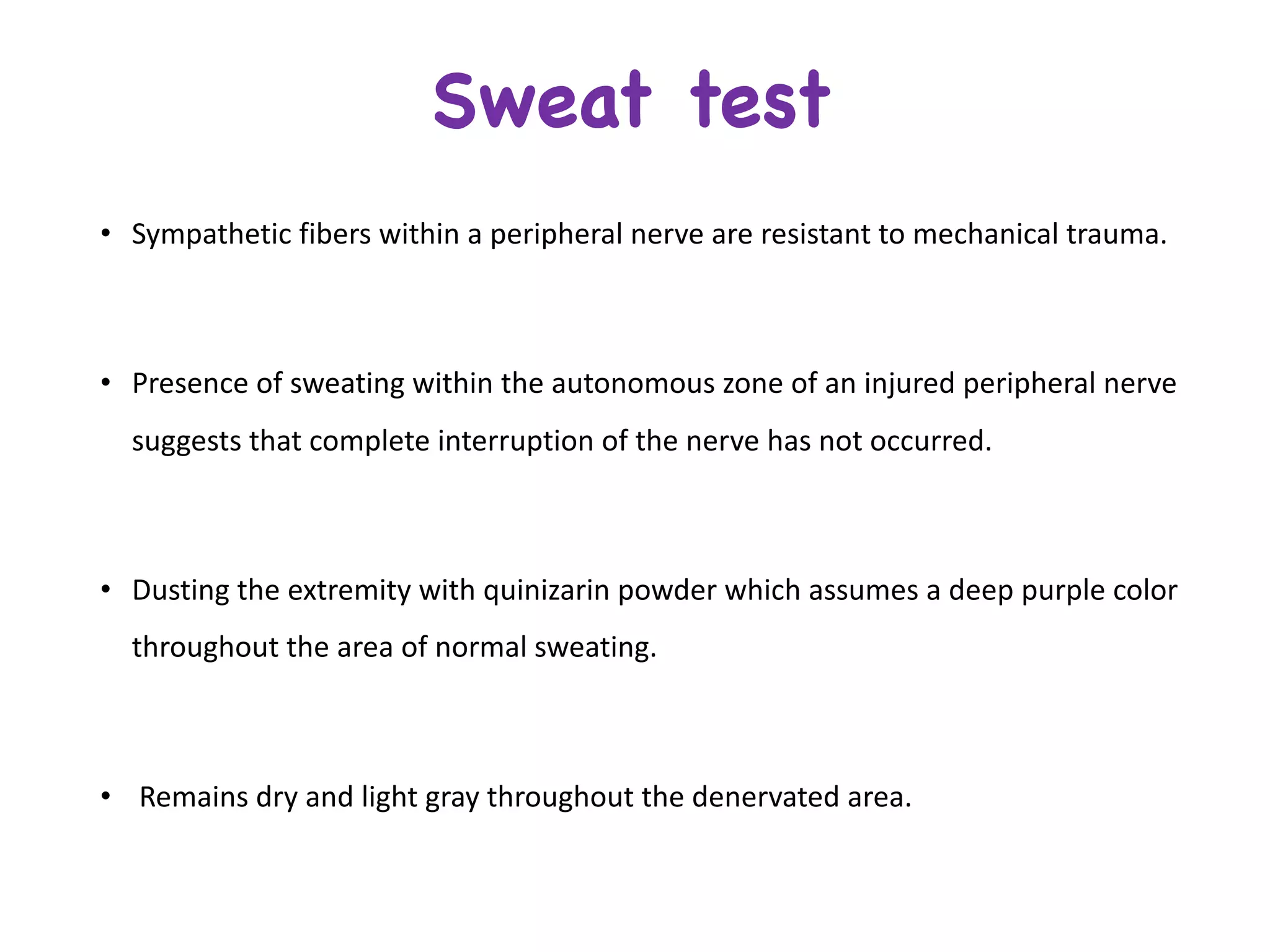
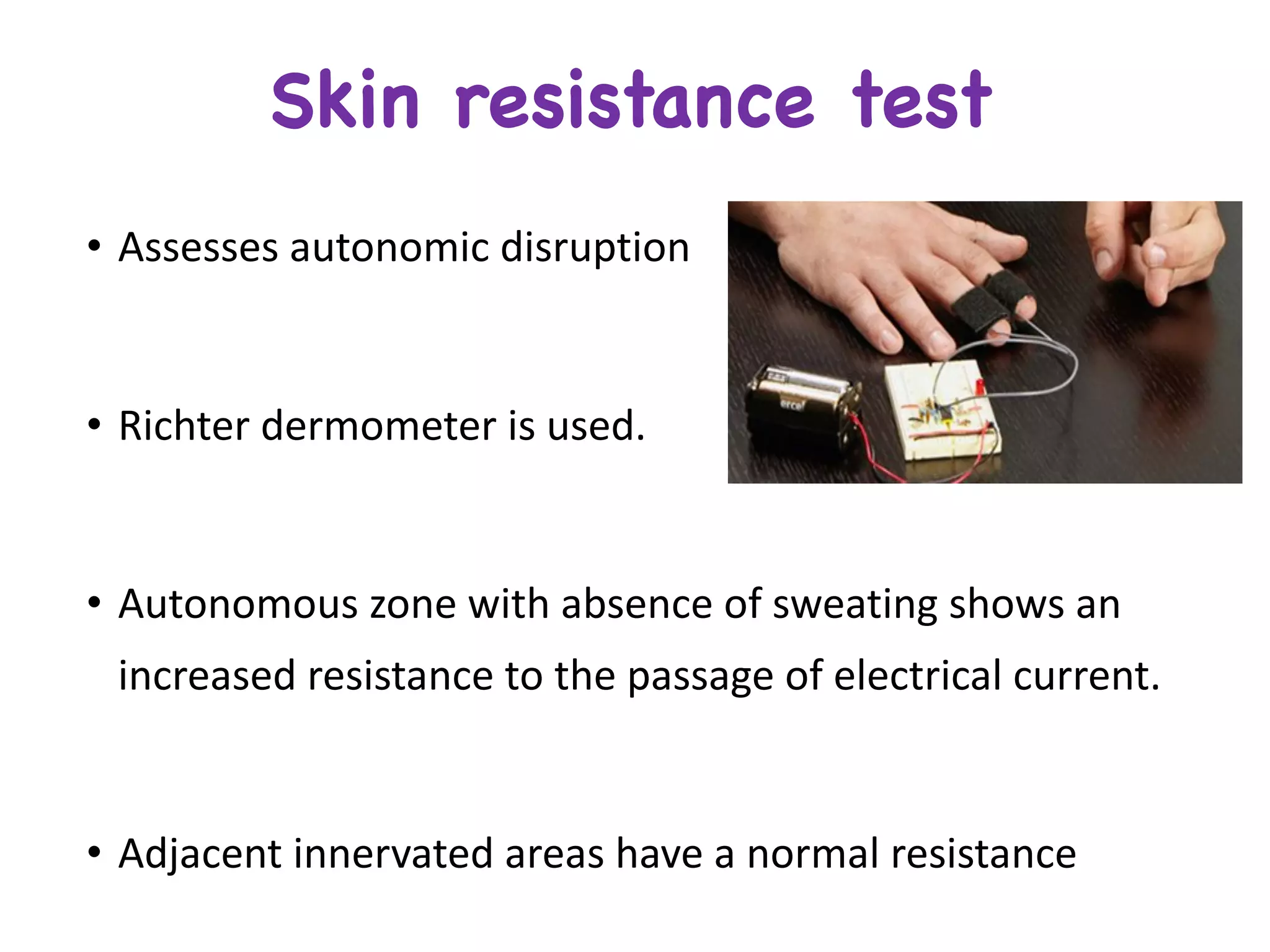
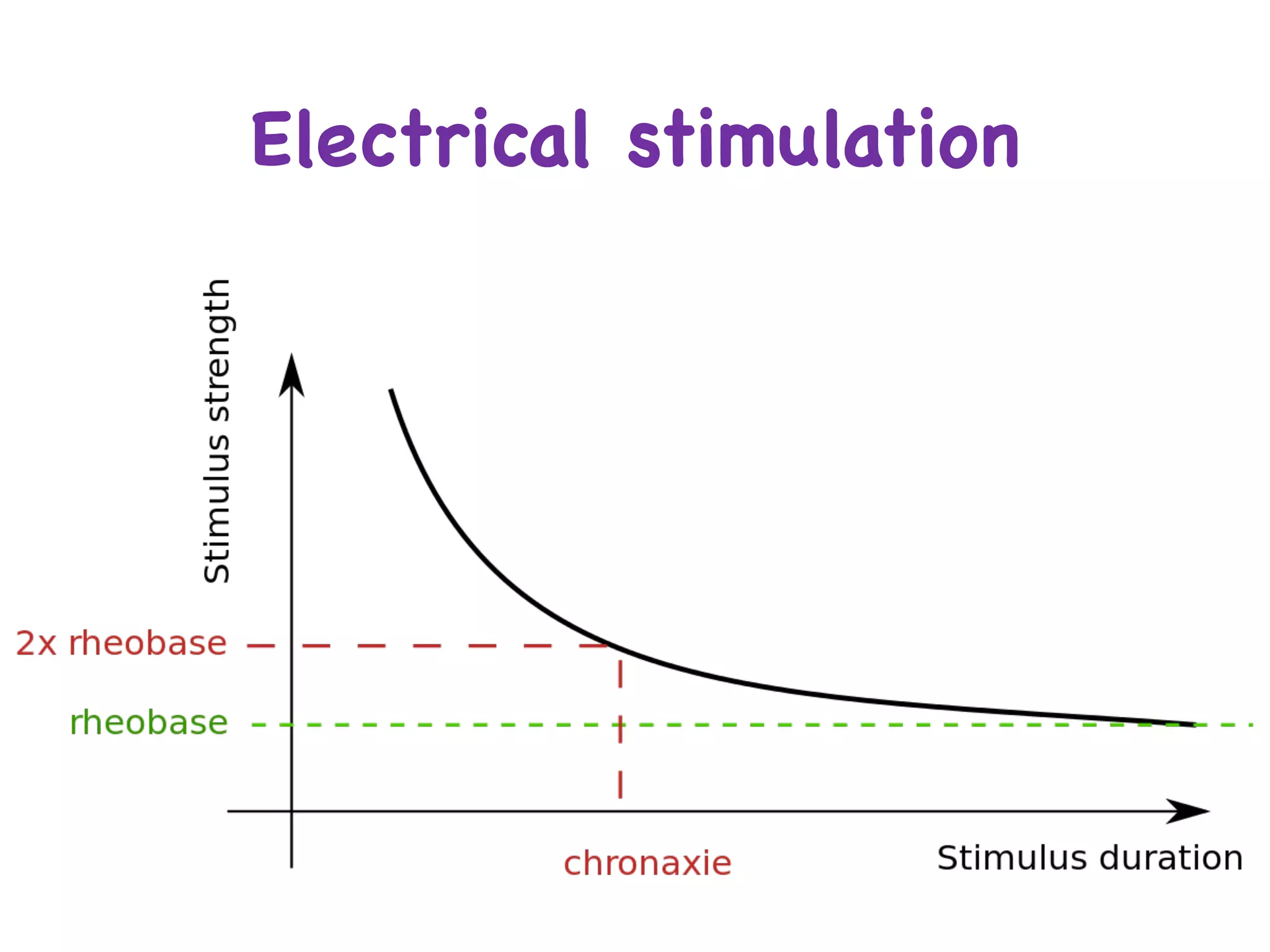
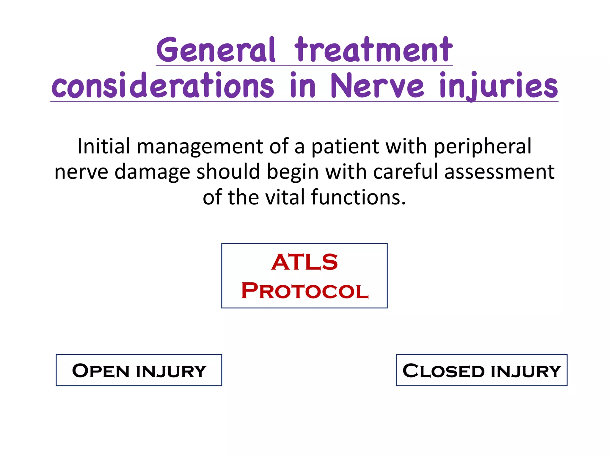
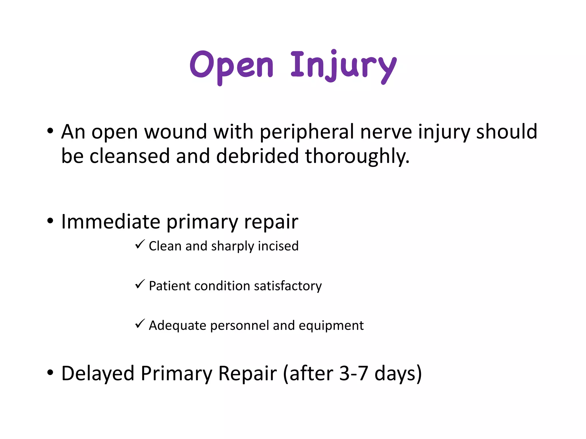
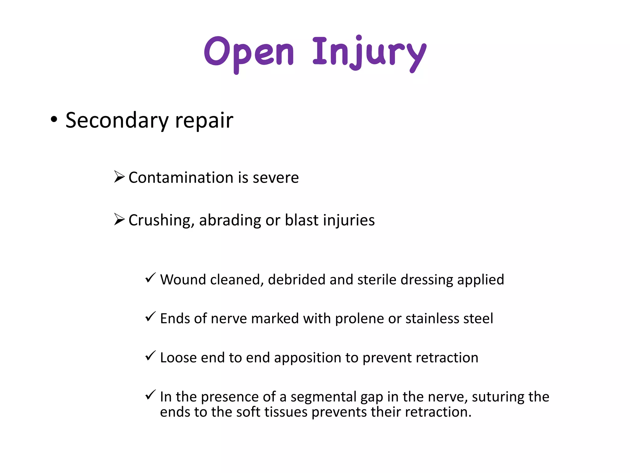
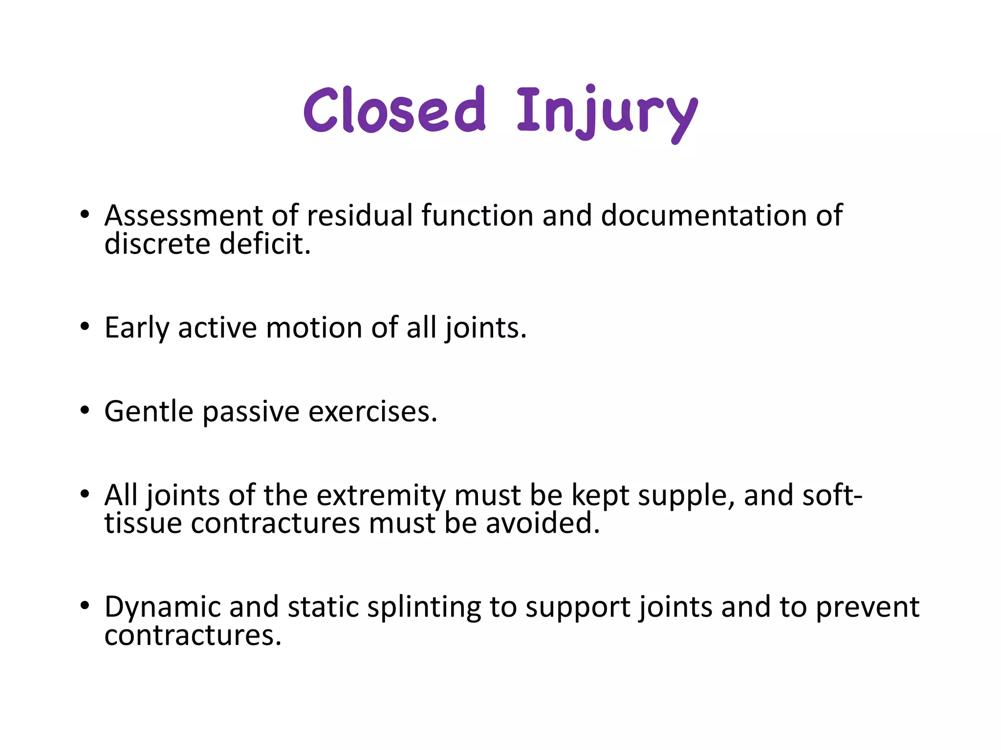
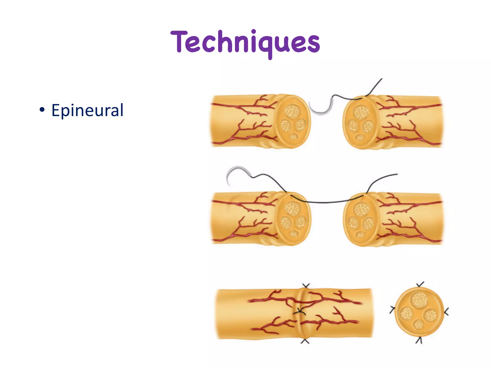
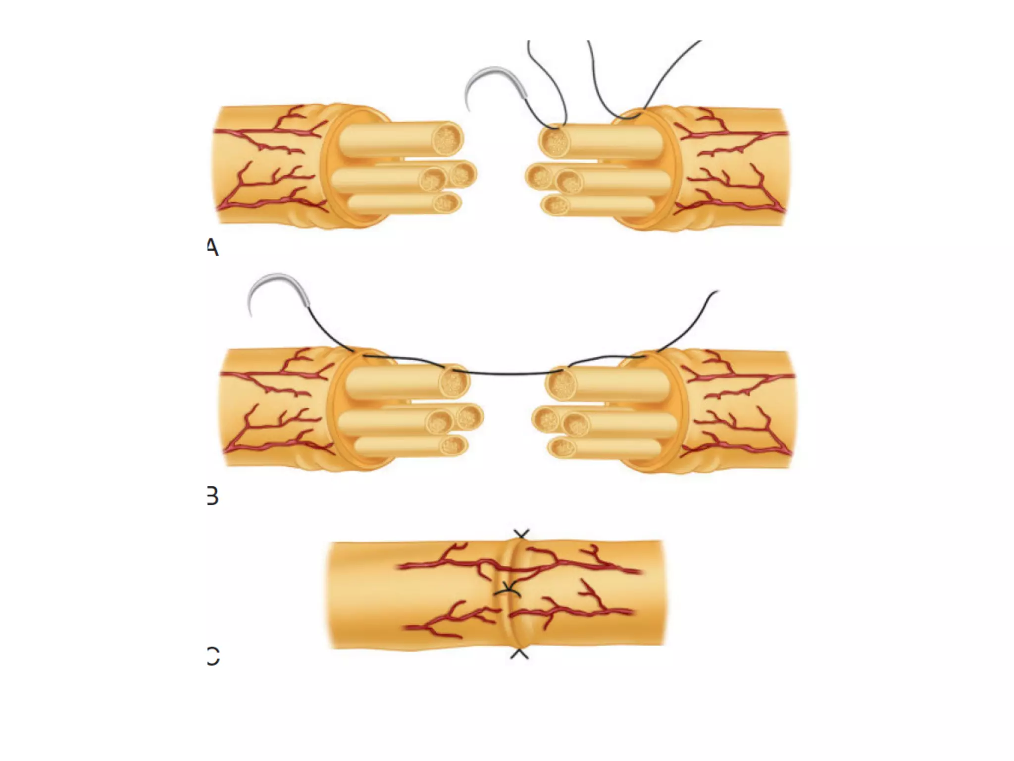
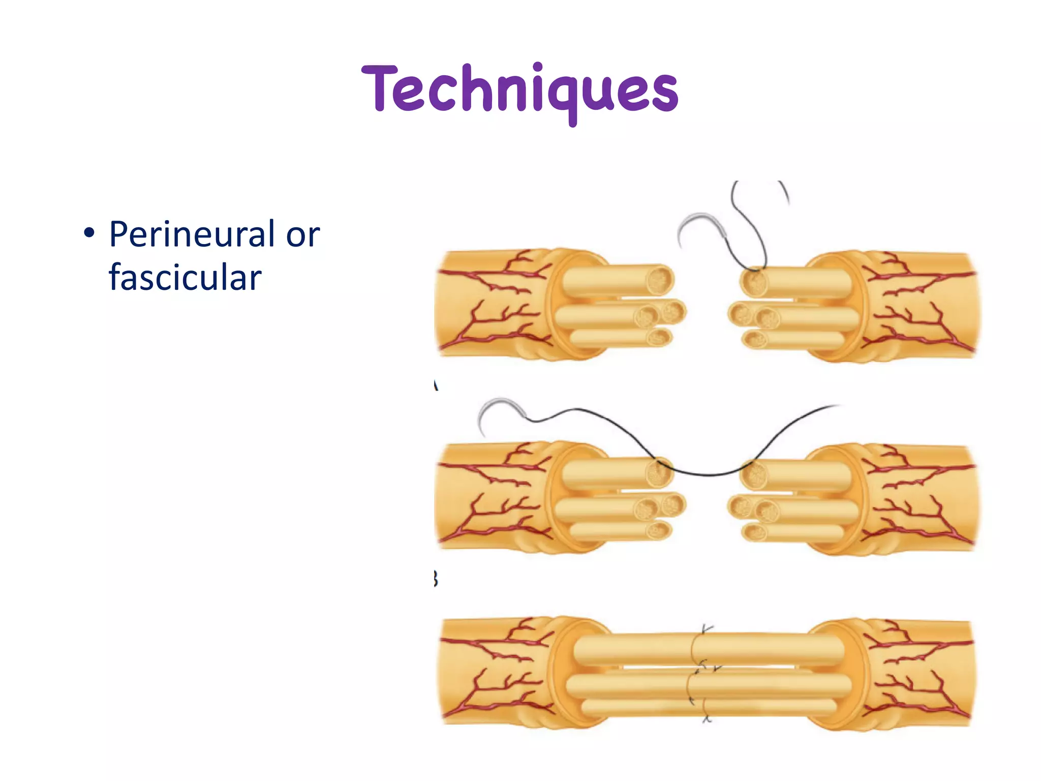
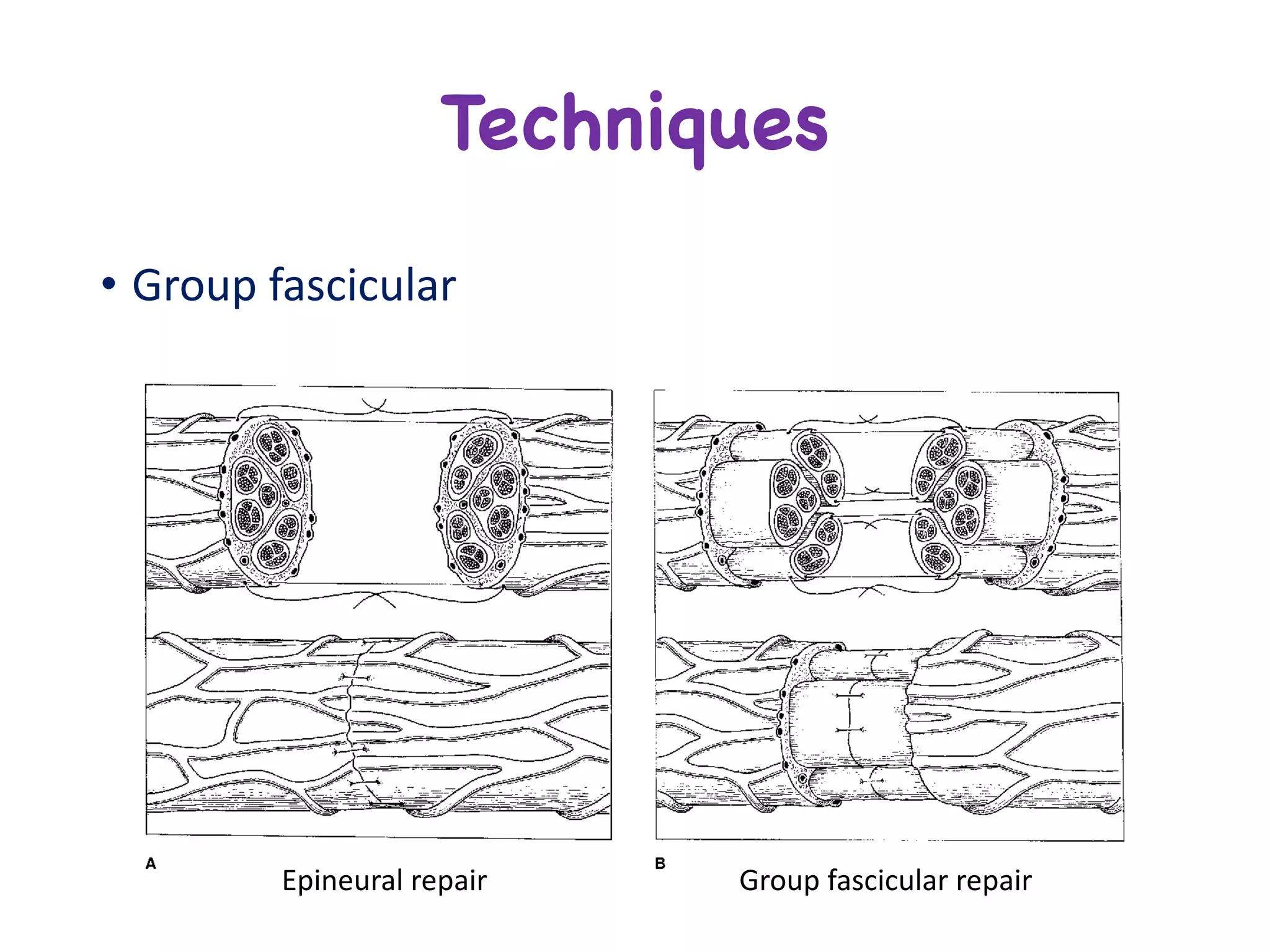
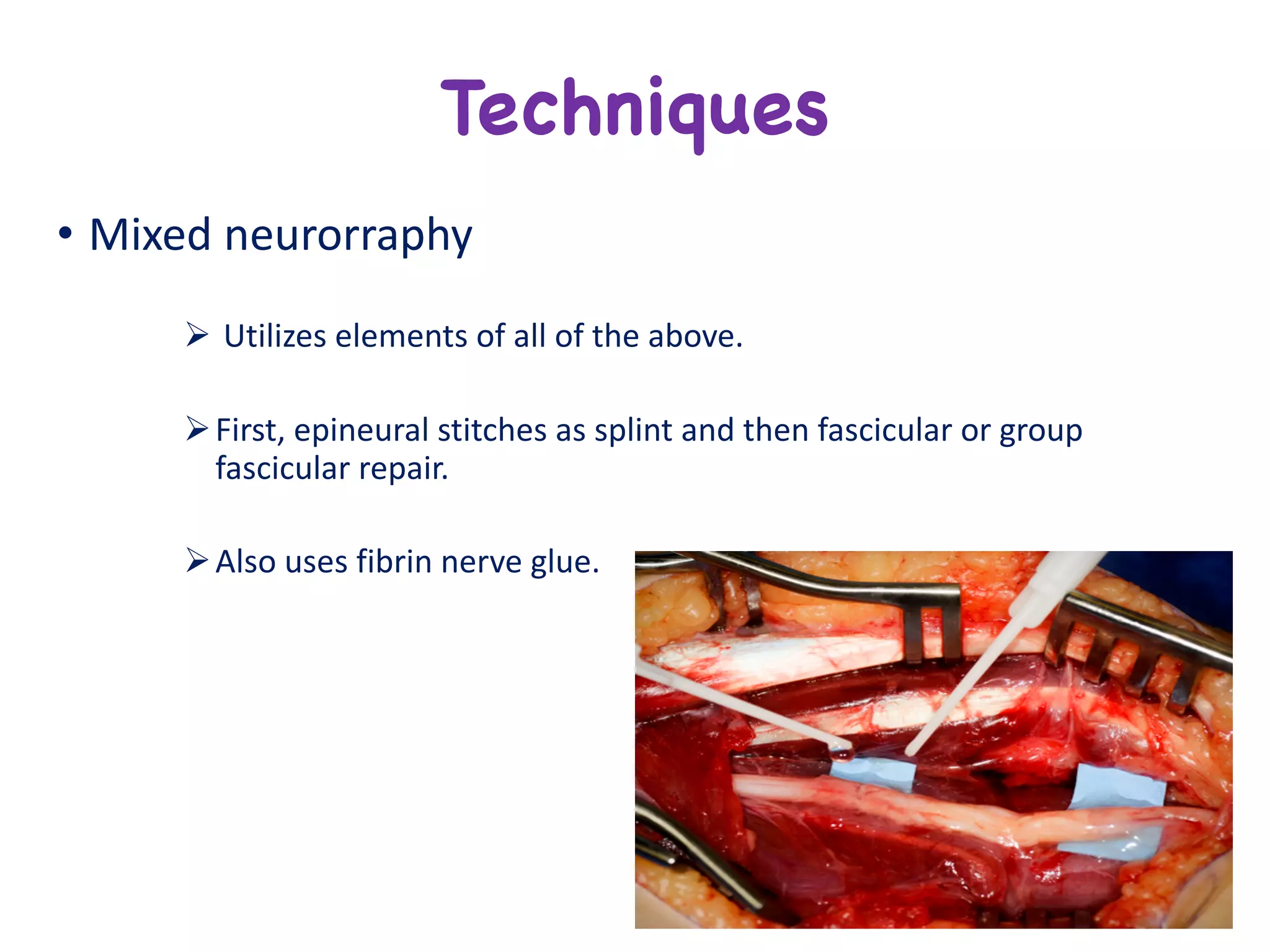
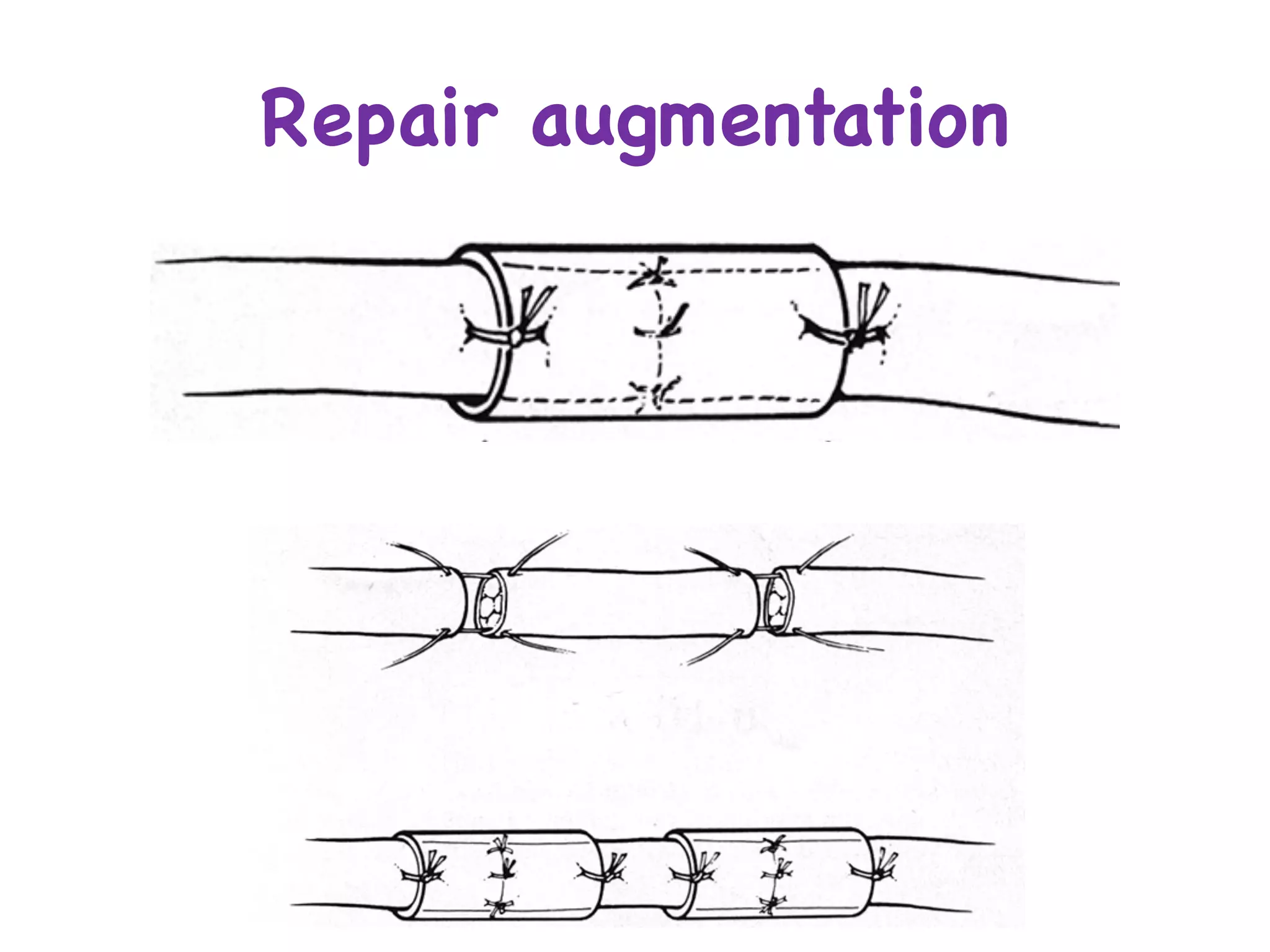
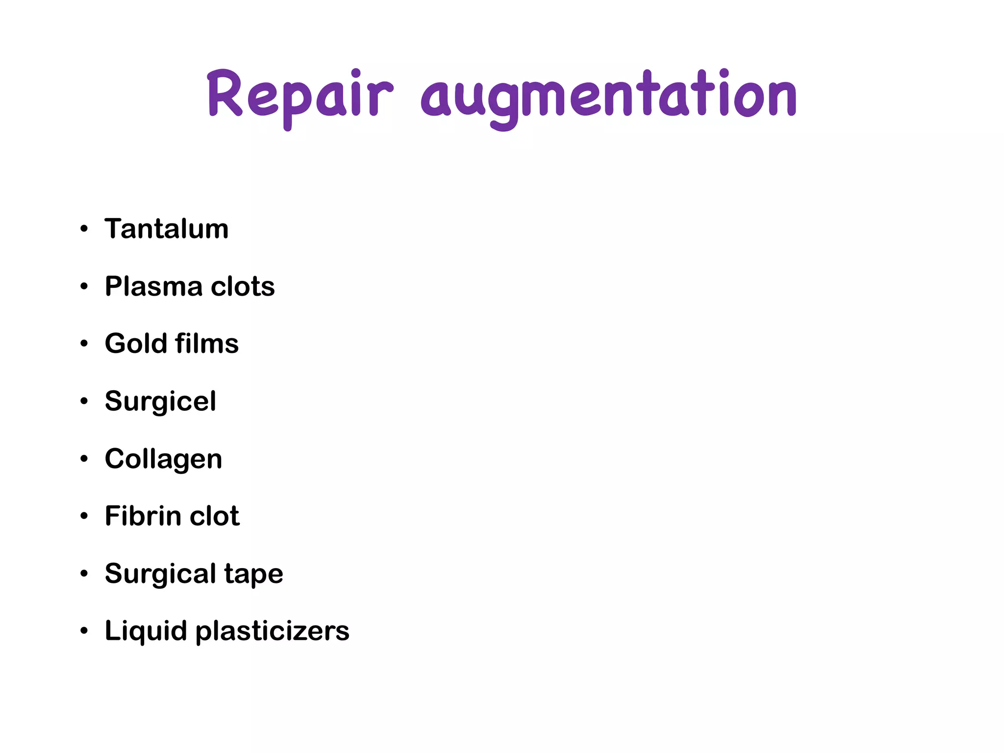
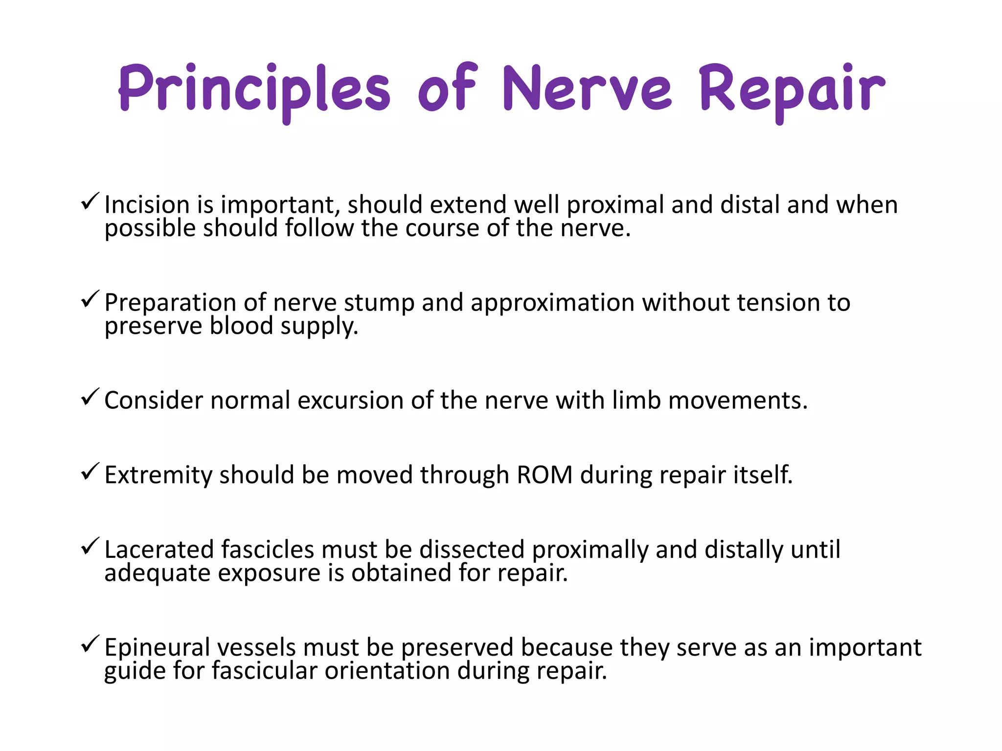
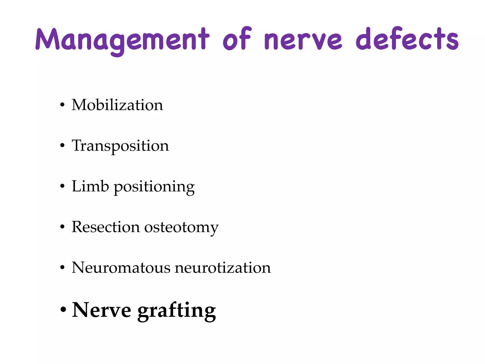
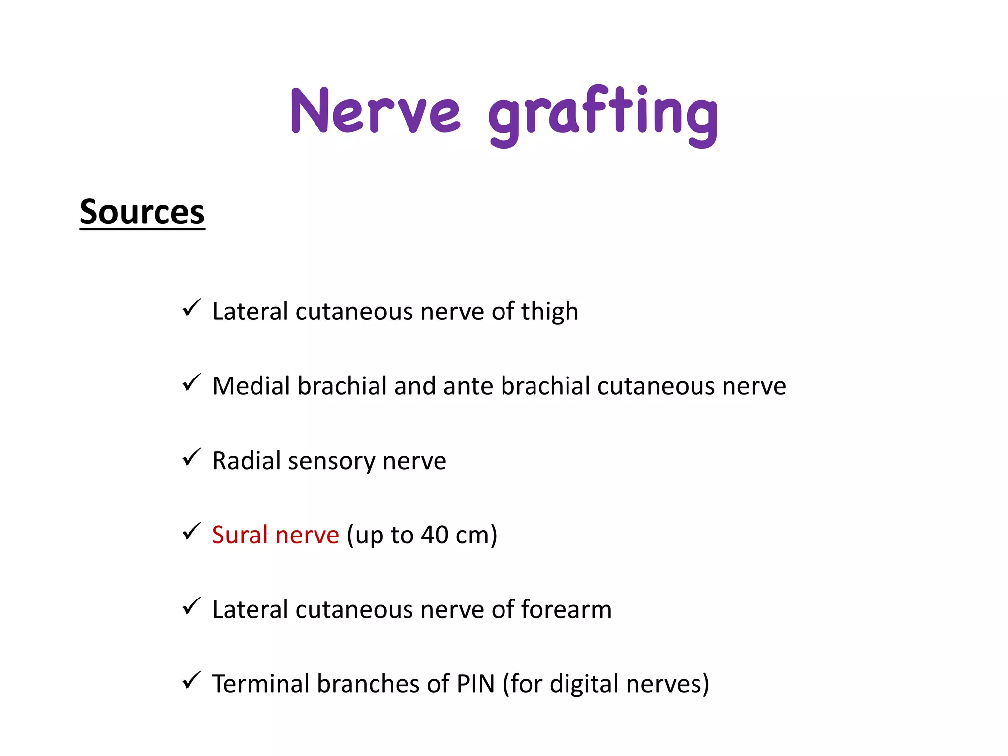
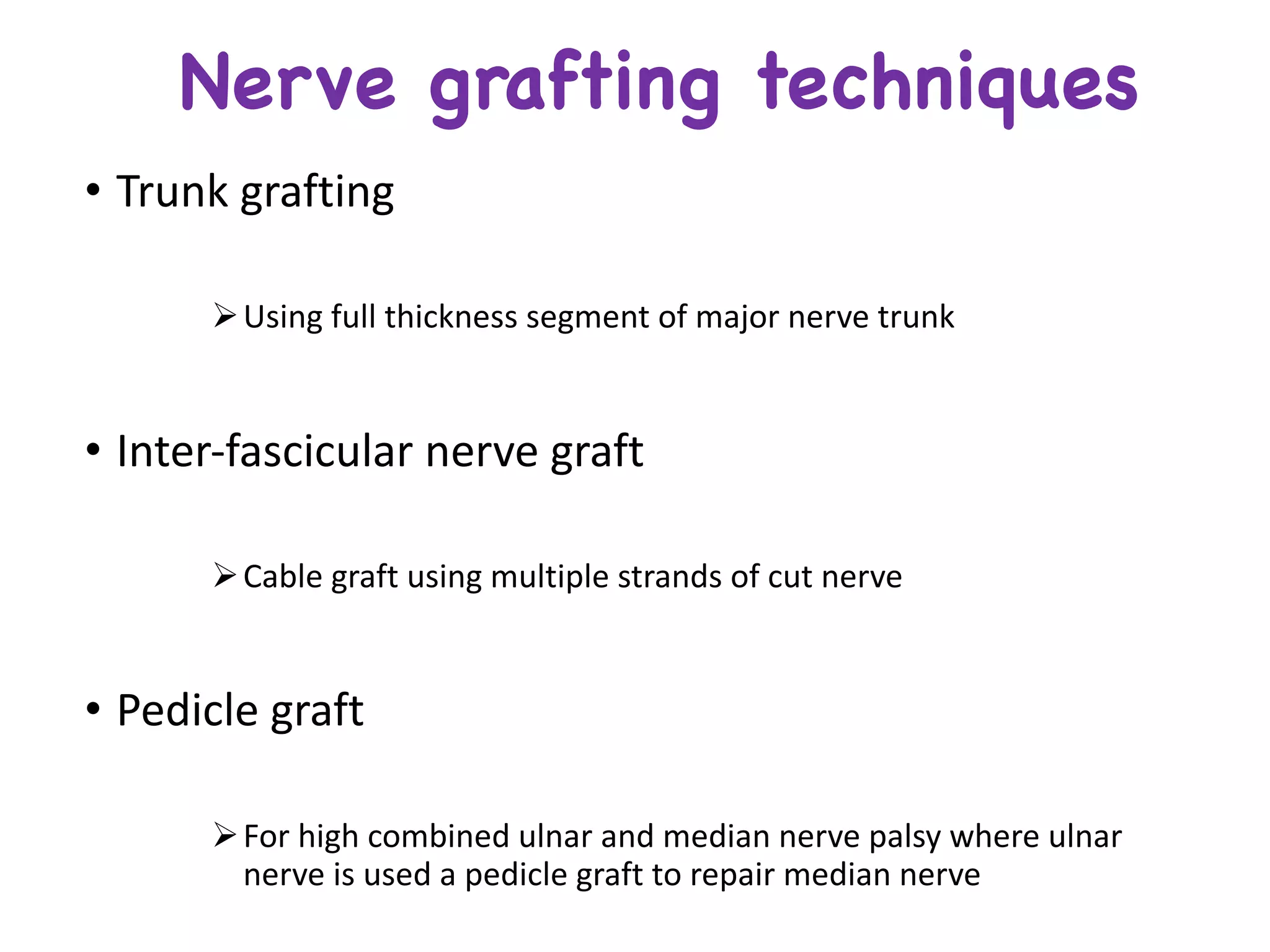
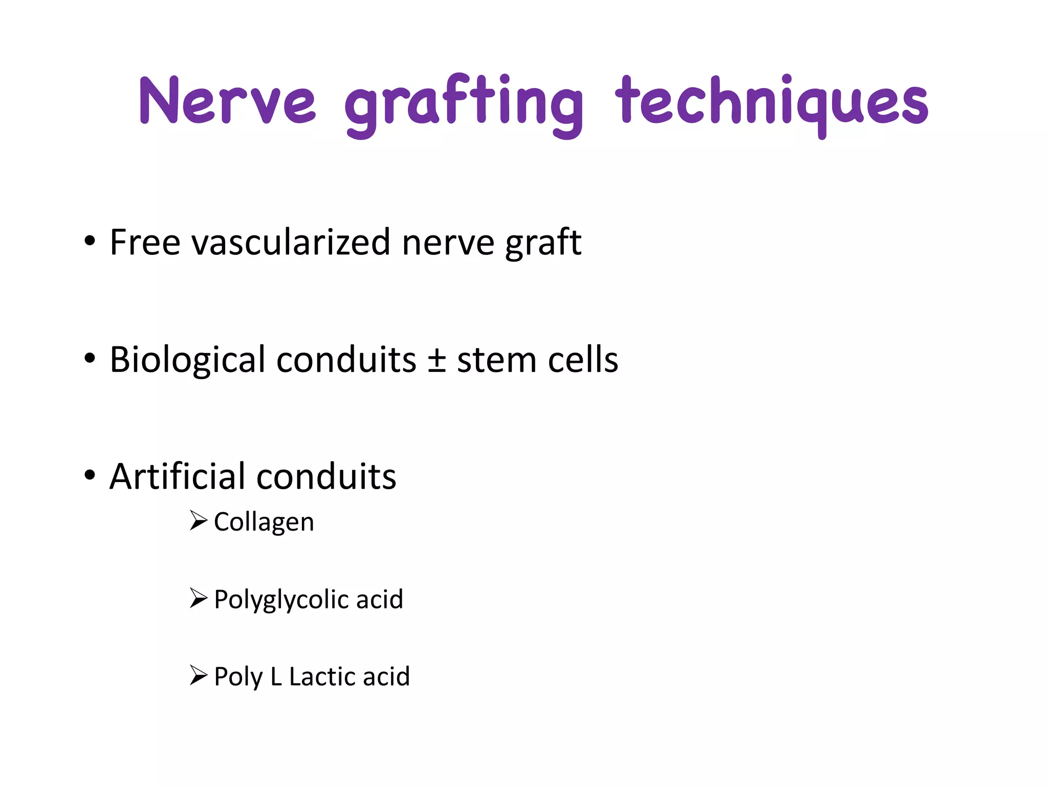
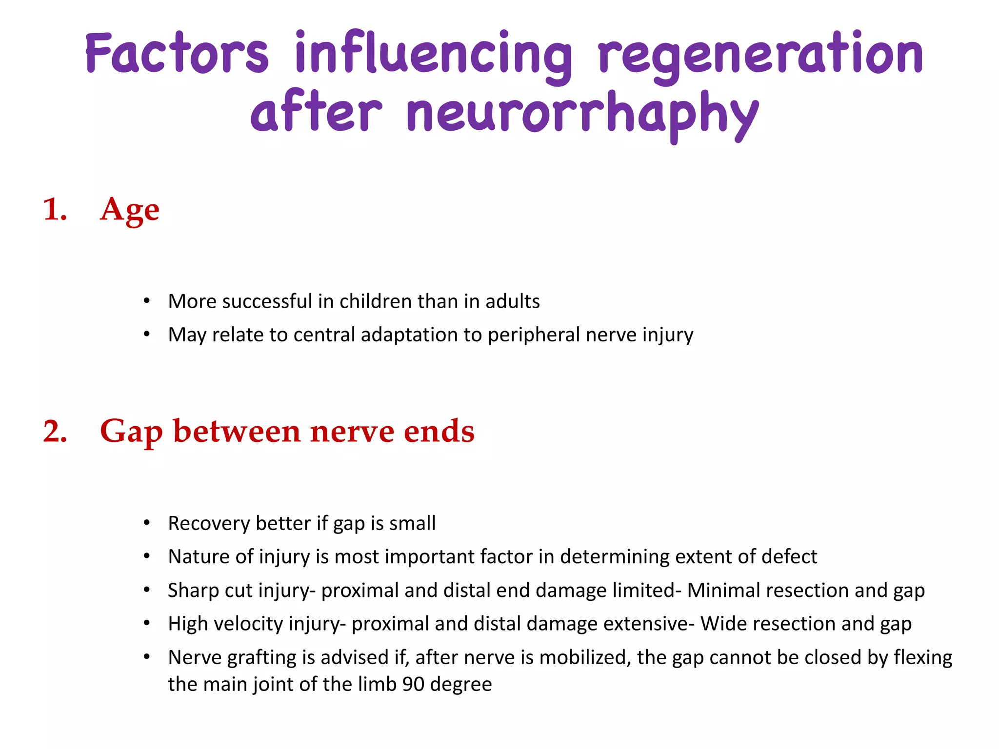
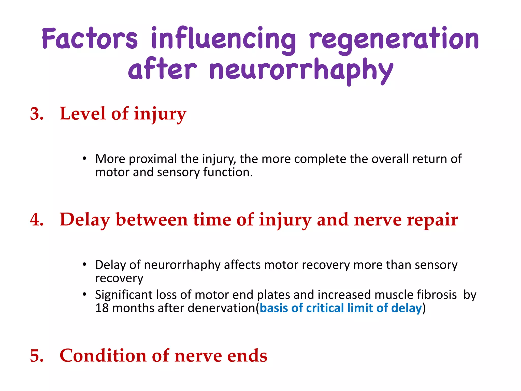
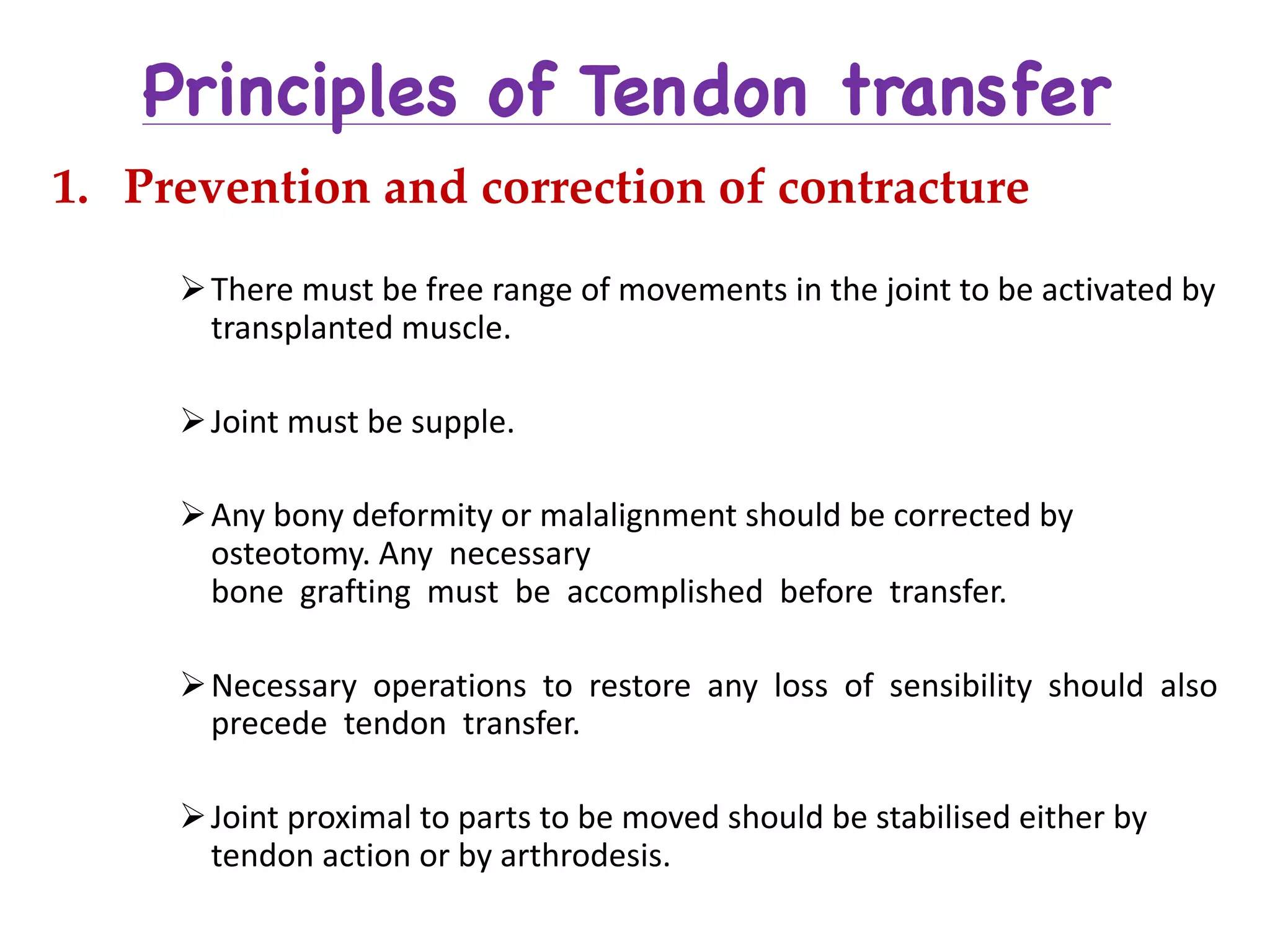
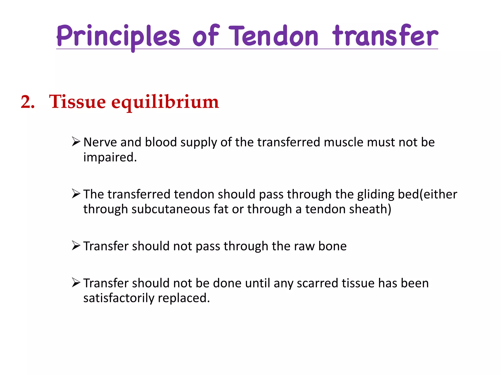
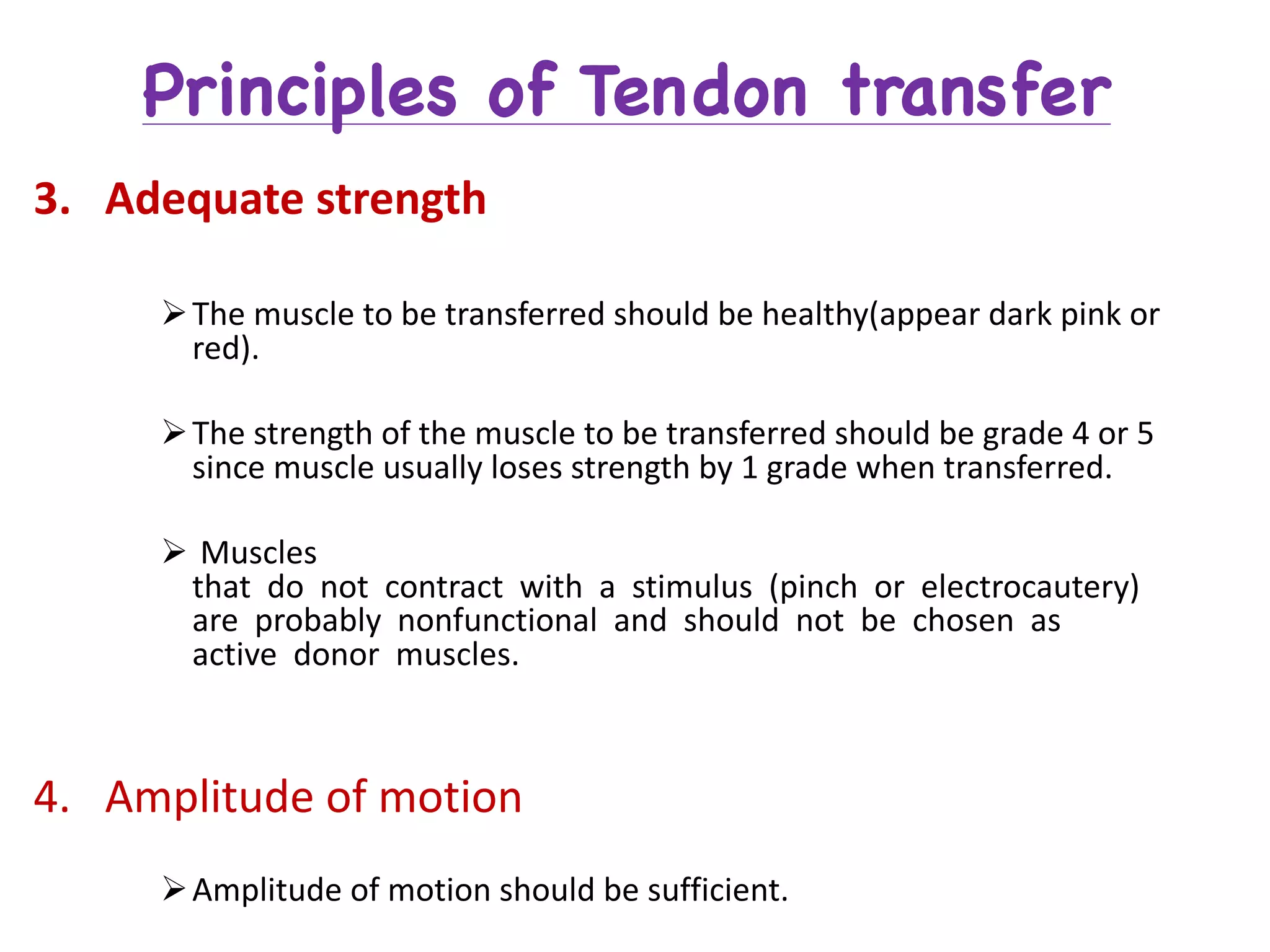
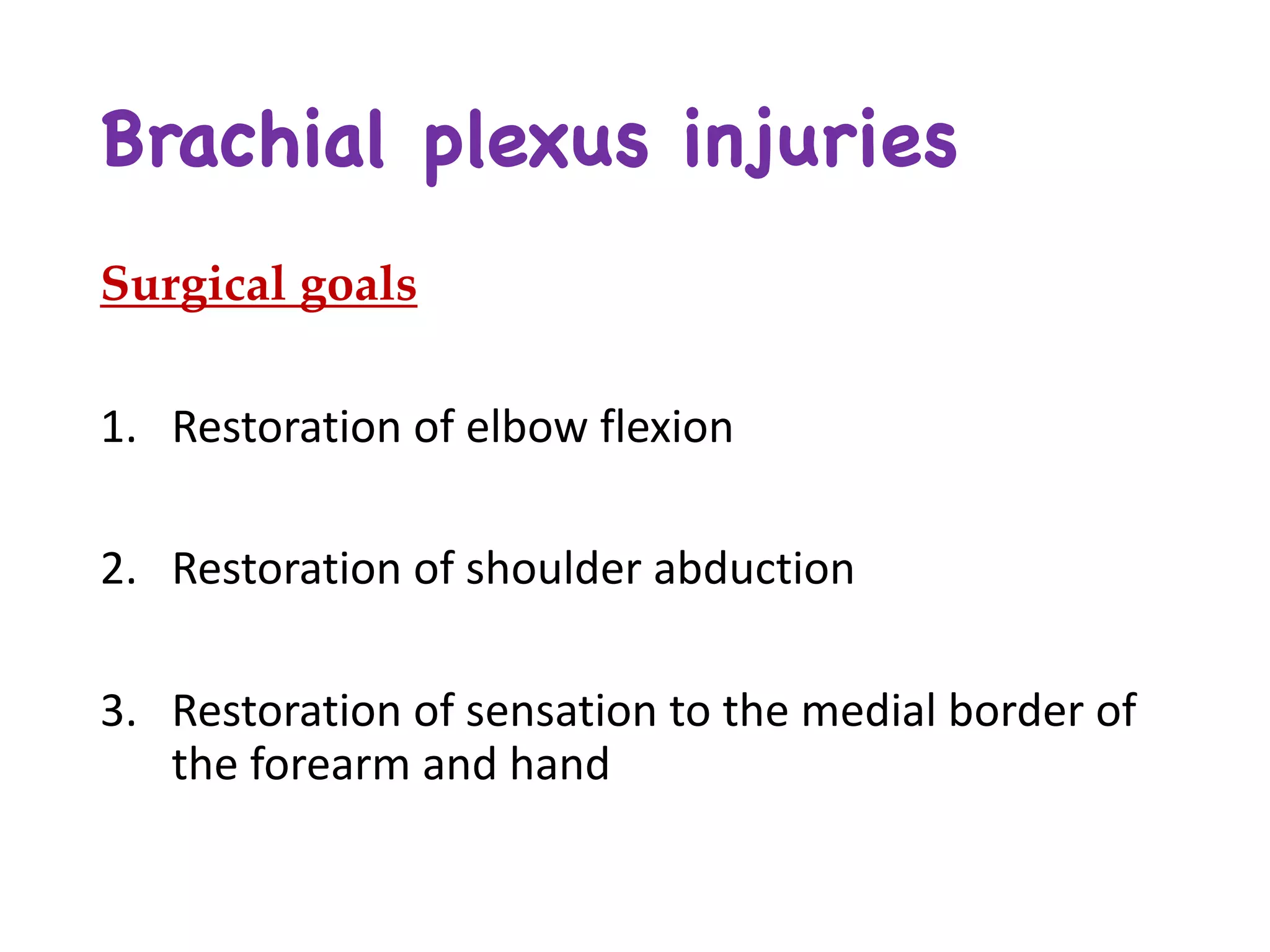
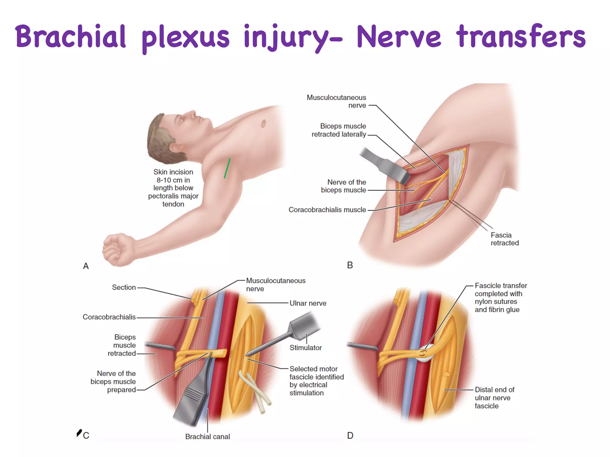
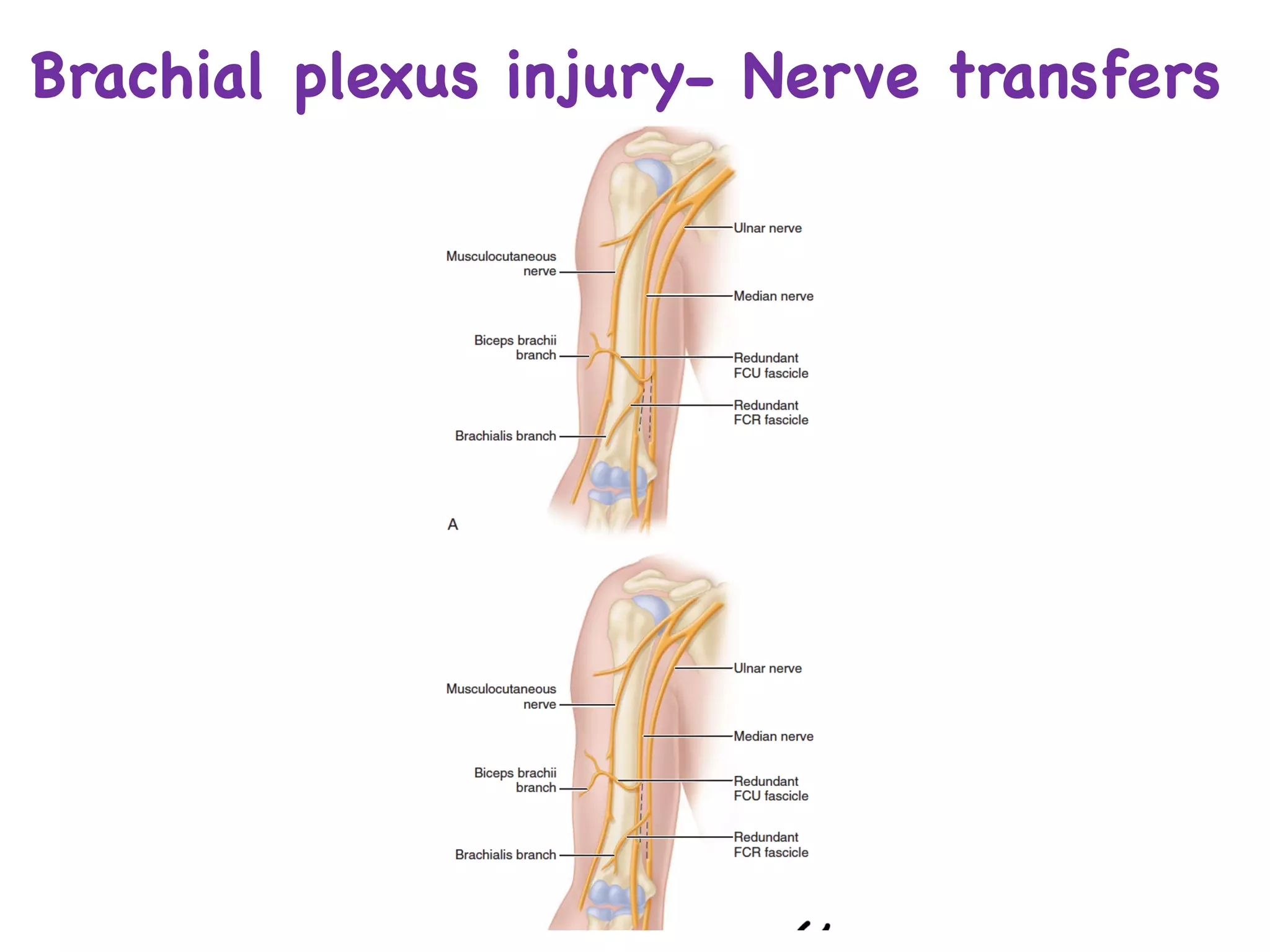
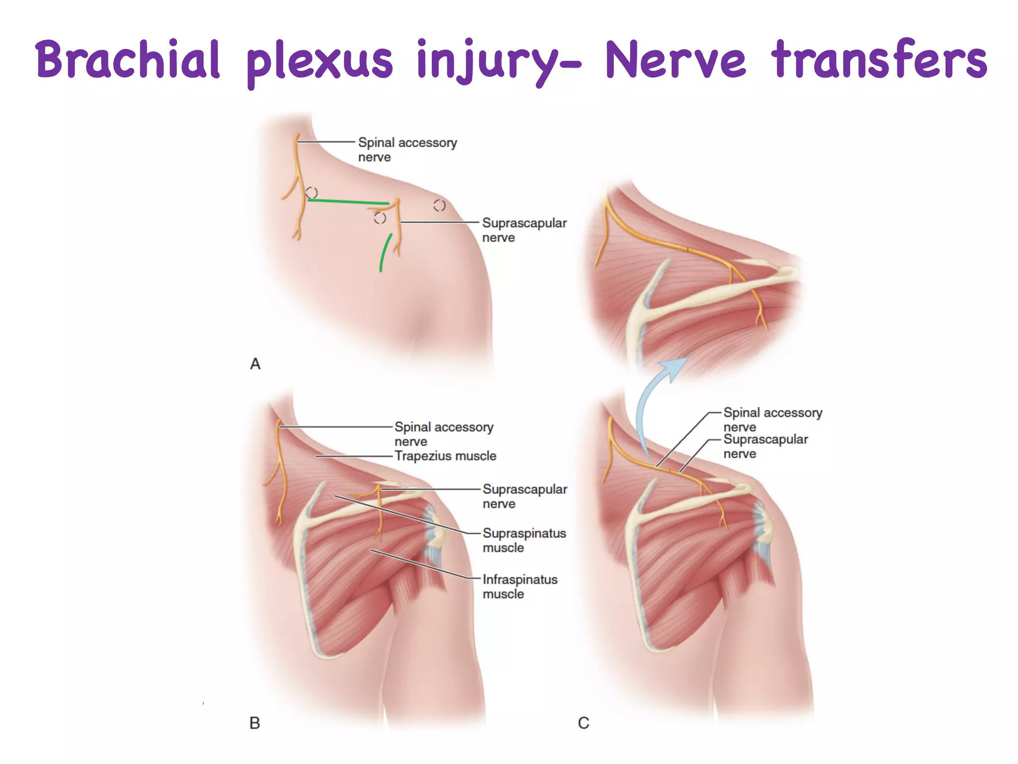
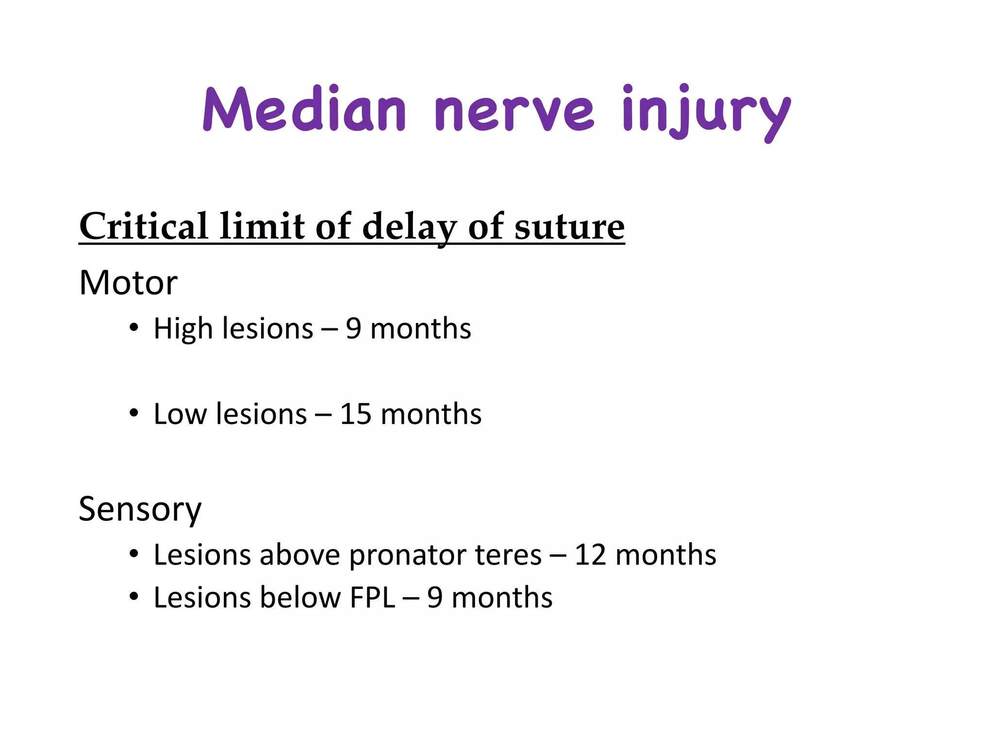
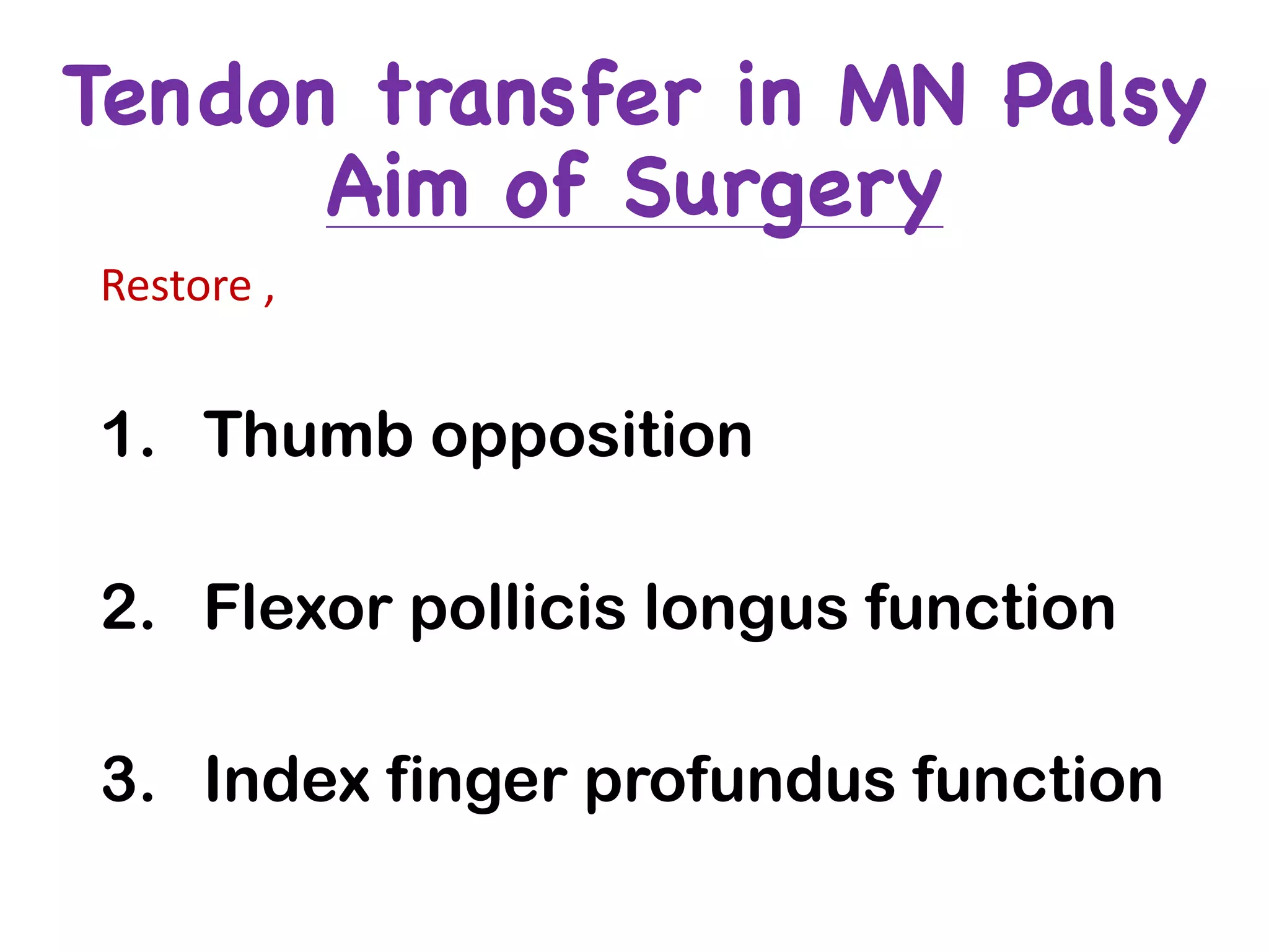
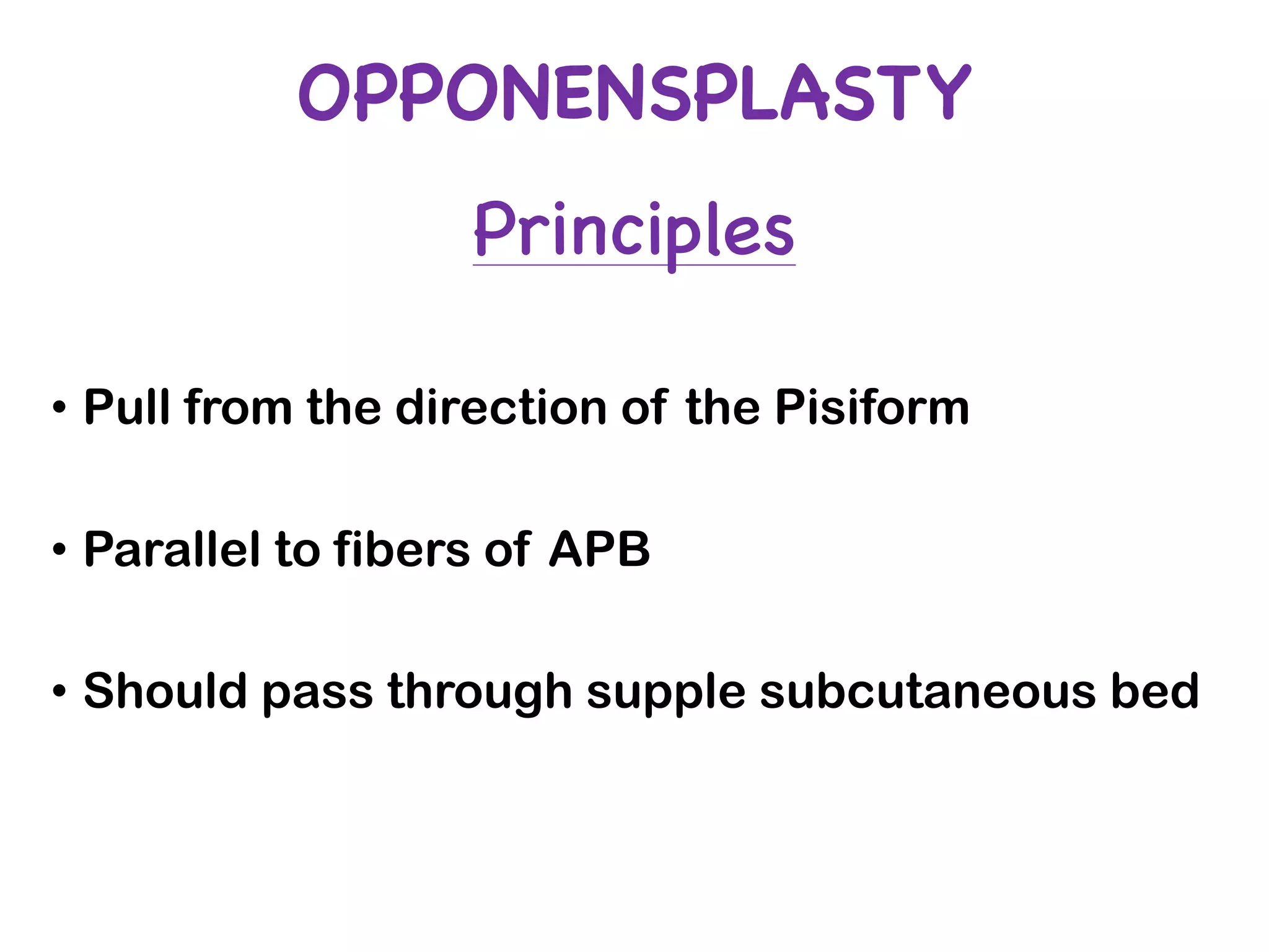
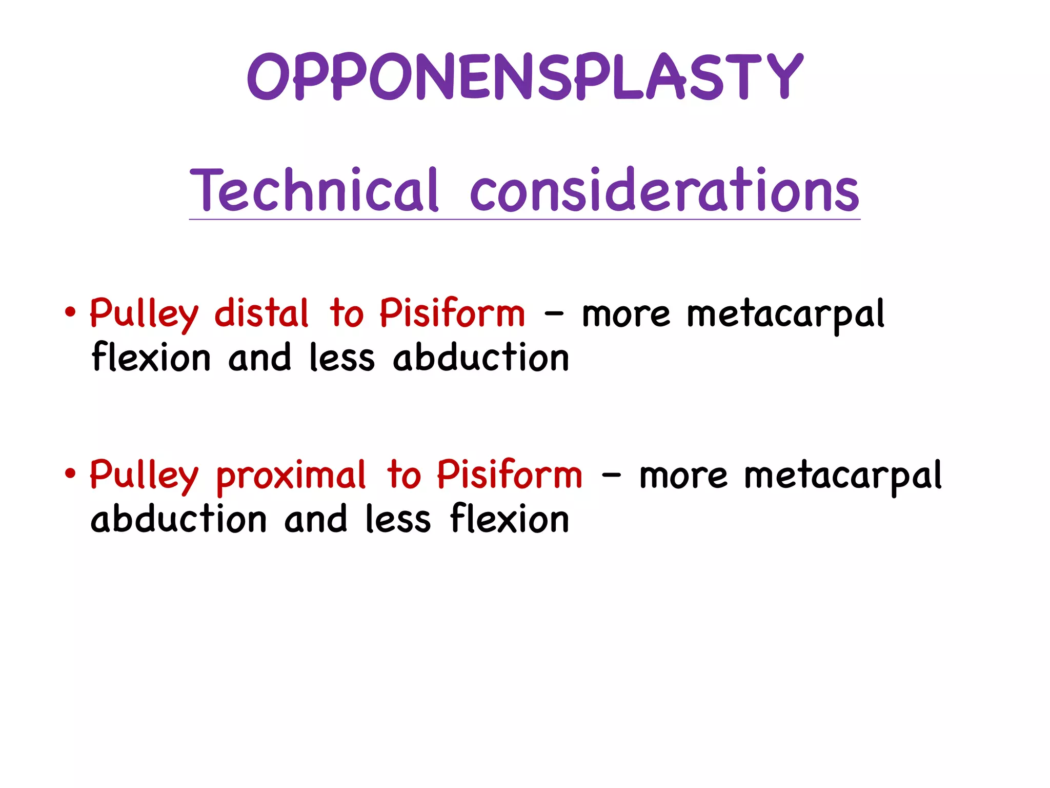
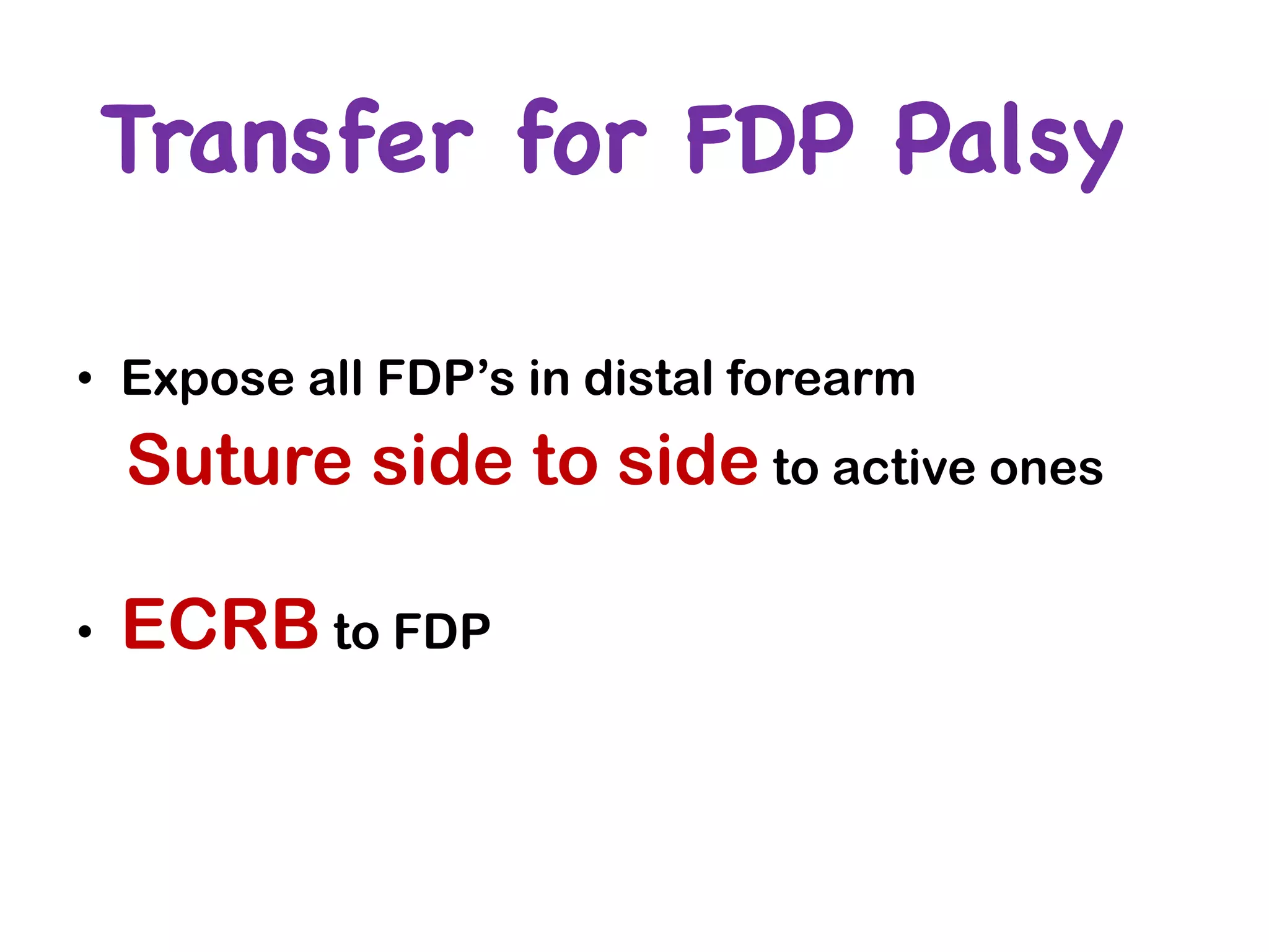
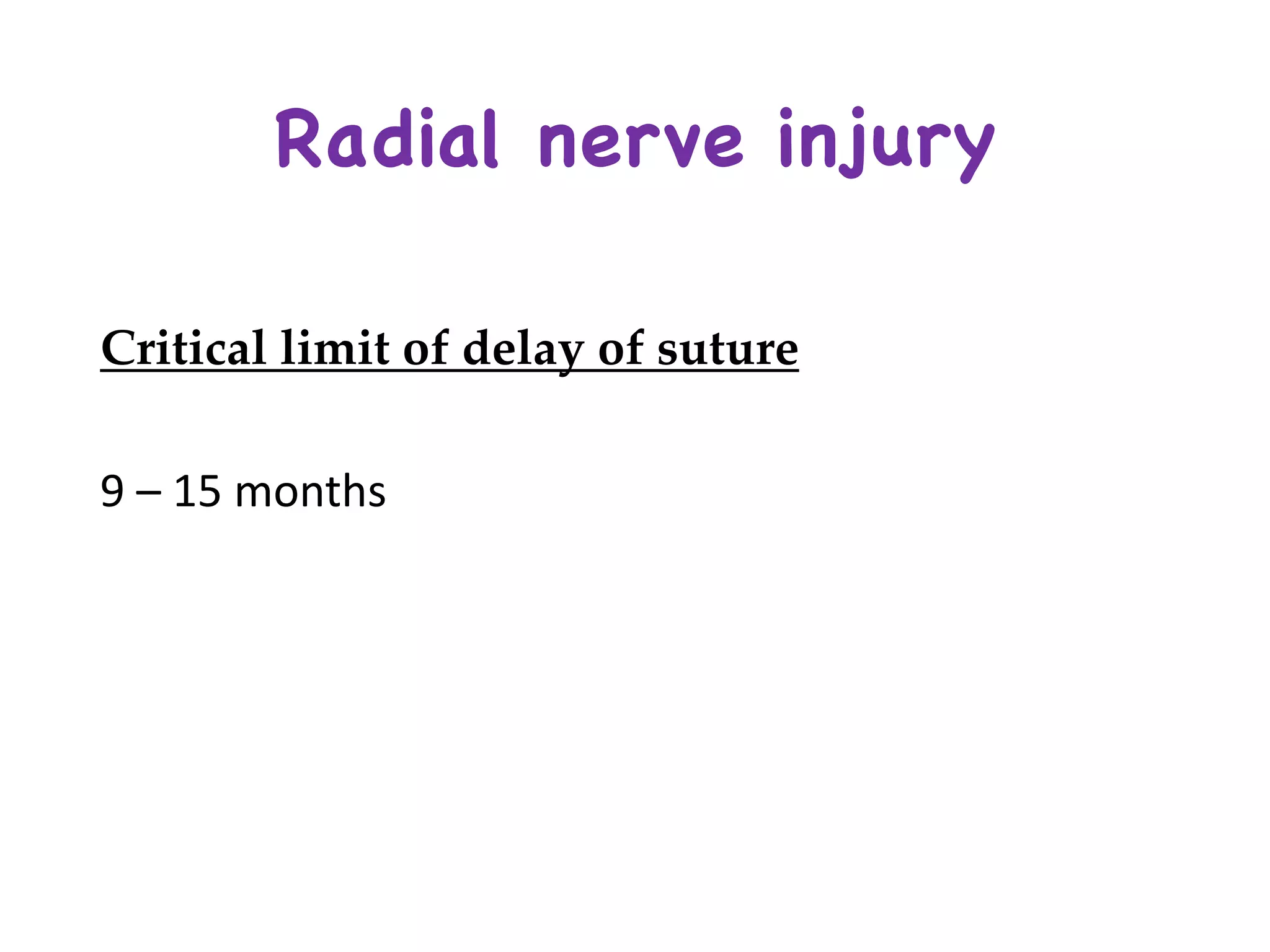
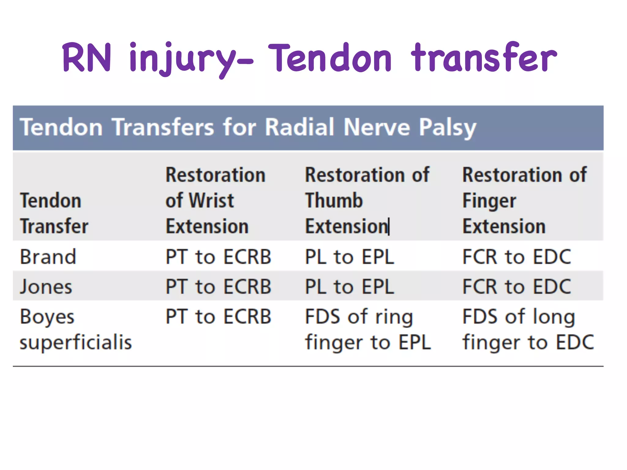
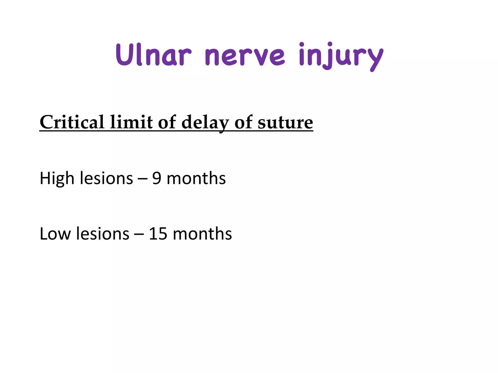
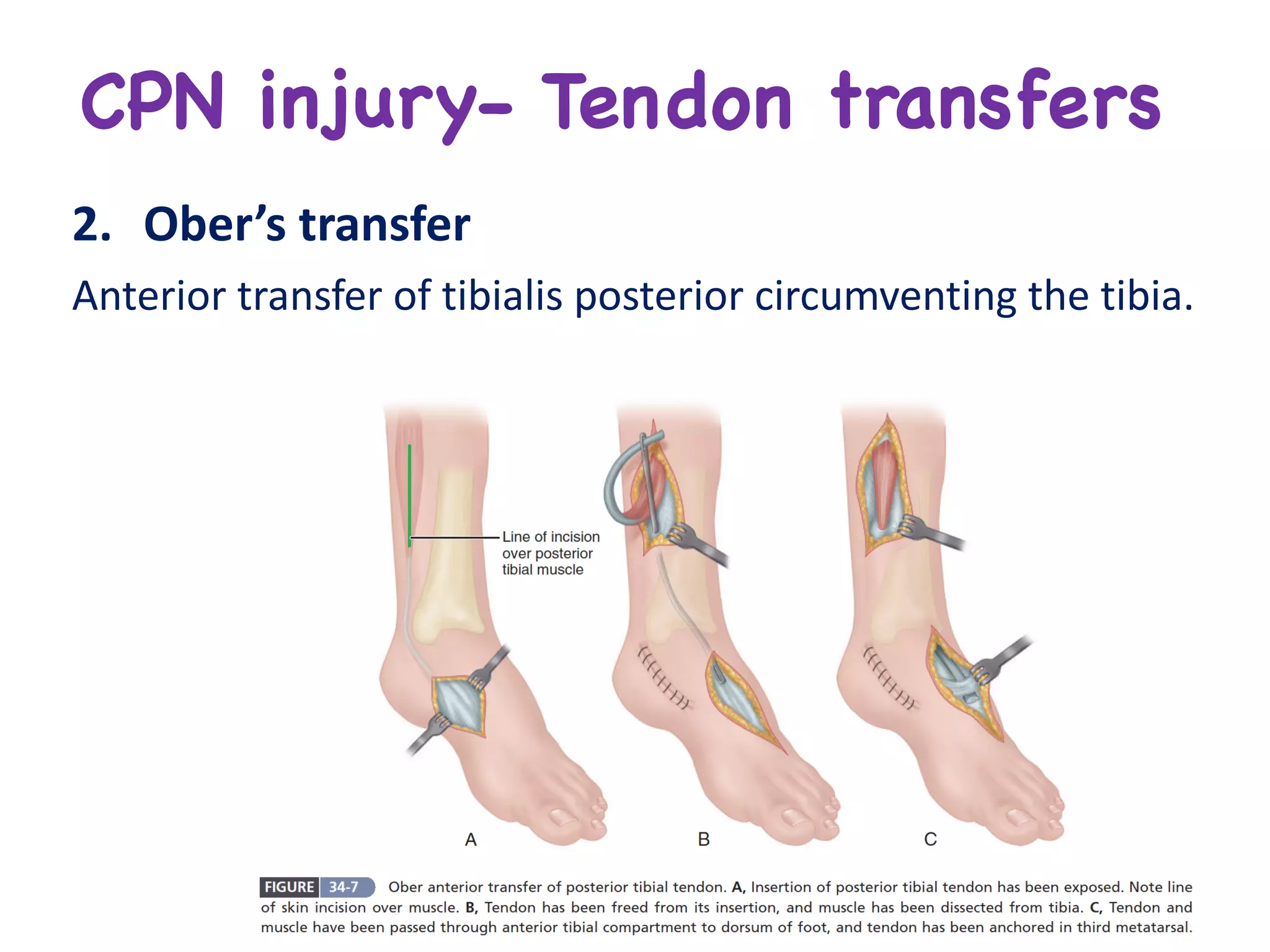
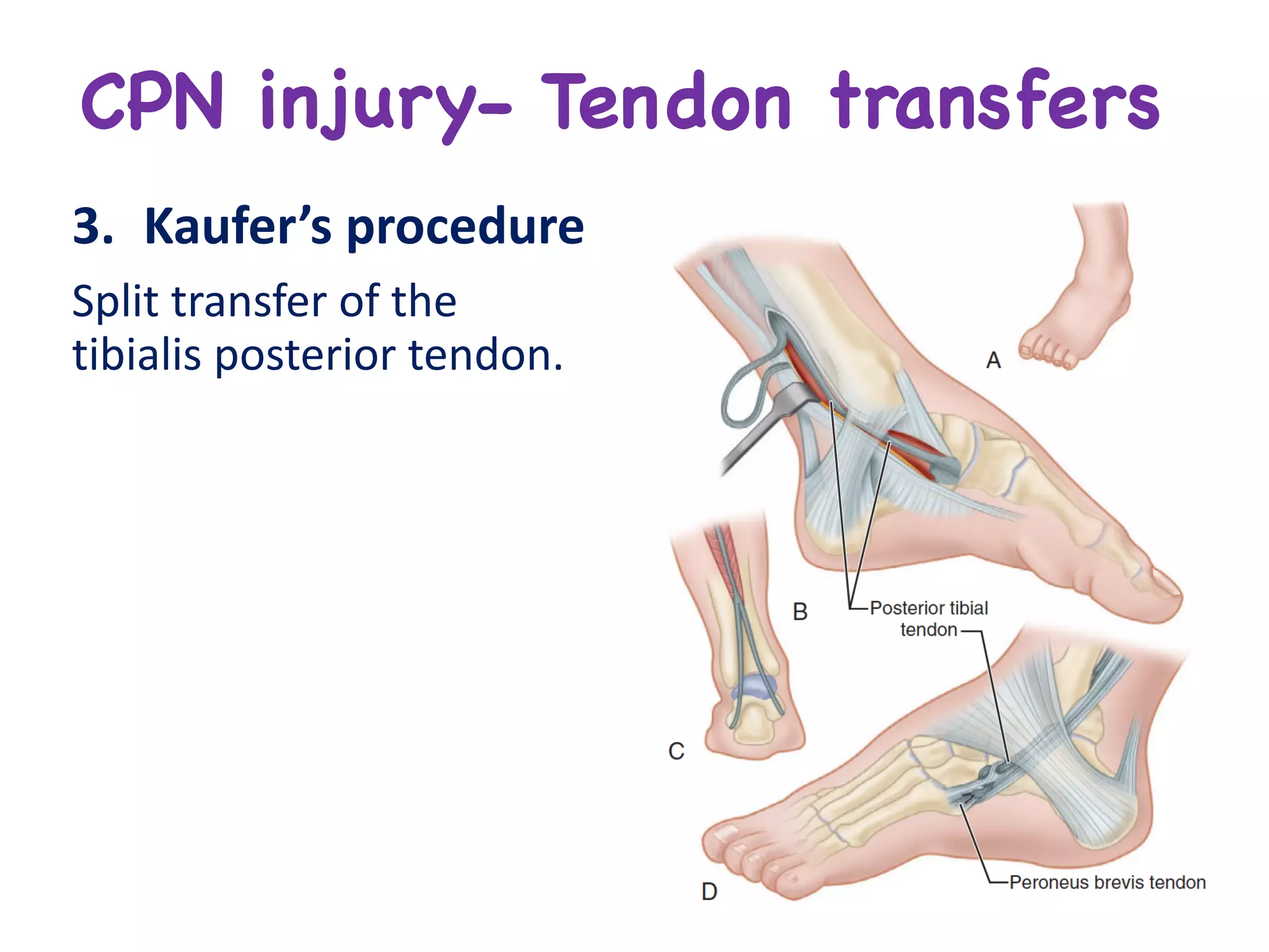
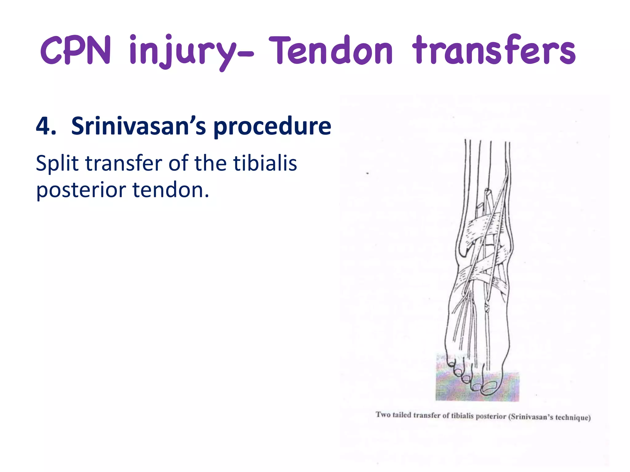
Management in
Peripheral Nerve Injuries
Dr. Jose Austine
Resident, Dept. of Orthopaedic surgery,
Kasturba Medical College, Mangalore
Moderators
Dr. Deepak Pinto
Dr. Sunil Murthy
Ø Diagnostic tests
Ø General treatment considerations in
Nerve injury
Ø Nerve Repair
Ø Tendon transfer
Ø Management in specific peripheral
nerve injury
Diagnostic Tests
Diagnostic Tests
Imaging
Electro
diagnostic
studies
Tinel’s sign Sweat test
Skin
resistance
test
Electrical
stimulation
Electro-diagnostic studies
• Best	and	most	accessible	correlative	electro-physiologic	
confirmations	of	a	peripheral	neural	injury.
• Presence,	location,	severity	and	possibly	the	prognosis	of	the	
neural	insult	can	be	determined.
• Recovery	pattern	can	be	obtained	when	the	study	is	done	
sequentially	over	time.
• EDX	is	done	at	3-6	weeks	to	allow	Wallerian	degeneration	and	
regeneration	to	occur	to	be	able	to	detect	features	of	the	same.
• Absence	of	regeneration	features	at	3	months	is	an	indication	to	
explore	the	nerve.
Nerve conduction study
• Orthodromic motor	
study
• Antidromic-
orthodromic sensory	
study	
• Retrograde	study(F	
wave	study)
Nerve	conduction	
velocity	
Amplitude	Latency
DurationF	wave	amplitude H	reflex
Electromyography
• Technique	of	recording	the	electrical	activity	within	a	striated	
muscle	belly	by	inserting	needle	in	it.
• Electrical	signal	generated	by	the	muscle	tissue	is	detected	by	
an	electrode	which	is	further	amplified	and	monitored	by	
oscilloscope	or	a	speaker or	recorded	in	system.
Uses	of	EMG:
üPresence	of	nerve	injury
üComplete/	incomplete	nerve	injury
üLevel	of	injury
üWhether	any	regeneration	occurring
• Initial	post	injury	phase- Normal	unless	prior	injury
• At	10-14	days- Positive	sharp	waves
• At	14-18	days- Fibrillations;	Voluntary	motor	unit	
potentials	have	attenuated	amplitudes
• At	3	months- Motor	unit	potential	amplitude	
progressively	increases;	Polyphasic waves	(Nerve	
regeneration	process)
• Between	2-6	months- Larger	than	normal	appearing	
potentials	till	nerve	regeneration	is	complete
• Positive	sharp	waves	and	Fibrillations	=	Wallerian	degeneration	occurring	
• Polyphasic waves	=	Nerve	regeneration	process
• EMG	recording	looked	for	in	specific	muscles-
ØBrachioradialis - Radial	Nerve
ØAbductor	Pollicis Brevis- Median	Nerve
ØAbductor	digiti minimi- Ulnar	Nerve
Tinel’s sign
• Gentle	percussion	along	the	course	of	an	injured	nerve.
• Distal	to	proximal
• Transient	tingling	sensation	felt	in	the	distribution	of	the	injured	nerve	rather	
than	at	the	area	percussed,	and	the	sensation	should	persist	for	several	
seconds	after	stimulation.
• Positive	Tinel sign	is	presumptive	evidence	that	regenerating	axonal	sprouts	
that	have	not	obtained	complete	myelinization are	progressing	along	the	
endoneurial tube.
• Neurapraxia – Absent	
• Axontmesis- Present	and	progressive	
• Neurotmesis- Present	and	non-progressive
• Strong	response	at	site- Poor	prognosis
• Fading	response	at	site- Good	prognosis
• Persistent	response	at	site- Unequivocal	
Tinel’s sign
• Sympathetic	fibers	within	a	peripheral	nerve	are	resistant	to	mechanical	trauma.
• Presence	of	sweating	within	the	autonomous	zone	of	an	injured	peripheral	nerve	
suggests	that	complete	interruption	of	the	nerve	has	not	occurred.
• Dusting	the	extremity	with	quinizarin powder	which	assumes	a	deep	purple	color	
throughout	the	area	of	normal	sweating.
• Remains	dry	and	light	gray	throughout	the	denervated area.	
Sweat test
• Assesses	autonomic	disruption
• Richter	dermometer is	used.
• Autonomous	zone	with	absence	of	sweating	shows	an	
increased	resistance	to	the	passage	of	electrical	current.
• Adjacent	innervated	areas	have	a	normal	resistance
Skin resistance test
Electrical stimulation
General treatment
considerations in
Nerve injuries
General treatment
considerations in Nerve injuries
Initial	management	of	a	patient	with	peripheral	
nerve	damage	should	begin	with	careful	assessment	
of	the	vital	functions.
ATLS
Protocol
Open injury Closed injury
Open Injury
• An	open	wound	with	peripheral	nerve	injury	should	
be	cleansed	and	debrided	thoroughly.
• Immediate	primary	repair
ü Clean	and	sharply	incised	
ü Patient	condition	satisfactory
ü Adequate	personnel	and	equipment
• Delayed	Primary	Repair	(after	3-7	days)
• Secondary	repair
ØContamination	is	severe
ØCrushing,	abrading	or	blast	injuries	
ü Wound	cleaned,	debrided	and	sterile	dressing	applied	
ü Ends	of	nerve	marked	with	prolene or	stainless	steel
ü Loose	end	to	end	apposition	to	prevent	retraction
ü In	the	presence	of	a	segmental	gap	in	the	nerve,	suturing	the	
ends	to	the	soft	tissues	prevents	their	retraction.
Open Injury
Closed Injury
• Assessment	of	residual	function	and	documentation	of	
discrete	deficit.
• Early	active	motion	of	all	joints.
• Gentle	passive	exercises.
• All	joints	of	the	extremity	must	be	kept	supple,	and	soft-
tissue	contractures	must	be	avoided.
• Dynamic	and	static	splinting	to	support	joints	and	to	prevent	
contractures.
• Early	surgical	exploration	is	avoided	unless	open	reduction	planned.
• Awaiting	re-innervation	seems	reasonable.
• Early	ultrasound	imaging	of	the	involved	nerve	can	determine	the	extent	of	
injury.
• Periodic	EMG,	nerve	conduction	velocity	studies,	and	frequent	clinical	
evaluation.
• If	no	evidence	of	regeneration	then	nerve	exploration.
If	the	nerve	deficit	follows	manipulation	or	casting	of	a	closed	fracture	in	the	
absence	of	a	prior	nerve	deficit,	early	exploration	of	the	nerve	is	indicated.
Closed Injury with Fracture
Nerve repair
Methods	of	nerve	repair
1. Primary	– within	hours
2. Delayed	primary	– within	3-7	days
3. Secondary	– more	than	7	days
Nerve repair (Neurorrhaphy)
• Epineural
Techniques
• Perineural or	
fascicular
Techniques
• Group	fascicular
Techniques
Epineural repair Group	fascicular	repair
• Mixed	neurorraphy
Ø Utilizes	elements	of	all	of	the	above.
ØFirst,	epineural stitches	as	splint	and	then	fascicular	or	group	
fascicular	repair.
ØAlso	uses	fibrin	nerve	glue.
Techniques
Repair augmentation
Repair augmentation
• Tantalum
• Plasma clots
• Gold films
• Surgicel
• Collagen
• Fibrin clot
• Surgical tape
• Liquid plasticizers
Principles of Nerve Repair
üIncision	is	important,	should	extend	well	proximal	and	distal	and	when	
possible	should	follow	the	course	of	the	nerve.
üPreparation	of	nerve	stump	and	approximation	without	tension	to	
preserve	blood	supply.
üConsider	normal	excursion	of	the	nerve	with	limb	movements.	
üExtremity	should	be	moved	through	ROM	during	repair	itself.	
üLacerated	fascicles	must	be	dissected	proximally	and	distally	until	
adequate	exposure	is	obtained	for	repair.
üEpineural vessels	must	be	preserved	because	they	serve	as	an	important	
guide	for	fascicular	orientation	during	repair.
Management of nerve defects
• Mobilization
• Transposition
• Limb positioning
• Resection osteotomy
• Neuromatous neurotization
• Nerve grafting
Nerve grafting
Sources
ü Lateral	cutaneous	nerve	of	thigh
ü Medial	brachial	and	ante	brachial	cutaneous	nerve	
ü Radial	sensory	nerve	
ü Sural	nerve	(up	to	40	cm)	
ü Lateral	cutaneous	nerve	of	forearm	
ü Terminal	branches	of	PIN	(for	digital	nerves)
Nerve grafting techniques
• Trunk	grafting	
ØUsing	full	thickness	segment	of	major	nerve	trunk
• Inter-fascicular	nerve	graft	
ØCable	graft	using	multiple	strands	of	cut	nerve
• Pedicle	graft	
ØFor	high	combined	ulnar	and	median	nerve	palsy	where	ulnar	
nerve	is	used	a	pedicle	graft	to	repair	median	nerve
Nerve grafting techniques
• Free	vascularized	nerve	graft
• Biological	conduits	± stem	cells	
• Artificial	conduits		
ØCollagen
ØPolyglycolic acid	
ØPoly	L	Lactic	acid
Factors influencing regeneration
after neurorrhaphy
1. Age
• More	successful	in	children	than	in	adults	
• May	relate	to	central	adaptation	to	peripheral	nerve	injury
2. Gap between nerve ends
• Recovery	better	if	gap	is	small
• Nature	of	injury	is	most	important	factor	in	determining	extent	of	defect
• Sharp	cut	injury- proximal	and	distal	end	damage	limited- Minimal	resection	and	gap
• High	velocity	injury- proximal	and	distal	damage	extensive- Wide	resection	and	gap
• Nerve	grafting	is	advised	if,	after	nerve	is	mobilized,	the	gap	cannot	be	closed	by	flexing	
the	main	joint	of	the	limb	90	degree
Factors influencing regeneration
after neurorrhaphy
3. Level of injury
• More	proximal	the	injury,	the	more	complete	the	overall	return	of	
motor	and	sensory	function.	
4. Delay between time of injury and nerve repair
• Delay	of	neurorrhaphy affects	motor	recovery	more	than	sensory	
recovery
• Significant	loss	of	motor	end	plates	and	increased	muscle	fibrosis		by	
18	months	after	denervation(basis	of	critical	limit	of	delay)
5. Condition of nerve ends
Tendon transfers
Principles of Tendon transfer
1. Prevention and correction of contracture
ØThere	must	be	free	range	of	movements	in	the	joint	to	be	activated	by	
transplanted	muscle.
ØJoint	must	be	supple.
ØAny	bony	deformity	or	malalignment	should	be	corrected	by	
osteotomy.	Any necessary	
bone grafting must be accomplished before transfer.
ØNecessary operations to restore any loss of sensibility should also	
precede tendon transfer.
ØJoint	proximal	to	parts	to	be	moved	should	be	stabilised either	by	
tendon	action	or	by	arthrodesis.
Principles of Tendon transfer
2. Tissue equilibrium
ØNerve	and	blood	supply	of	the	transferred	muscle	must	not	be	
impaired.
ØThe	transferred	tendon	should	pass	through	the	gliding	bed(either	
through	subcutaneous	fat	or	through	a	tendon	sheath)
ØTransfer	should	not	pass	through	the	raw	bone
ØTransfer	should	not	be	done	until	any	scarred	tissue	has	been	
satisfactorily	replaced.
Principles of Tendon transfer
3. Adequate	strength
ØThe	muscle	to	be	transferred	should	be	healthy(appear	dark	pink	or	
red).
ØThe	strength	of	the	muscle	to	be	transferred	should	be	grade	4	or	5	
since	muscle	usually	loses	strength	by	1	grade	when	transferred.
Ø Muscles	
that do not contract with a stimulus (pinch or electrocautery)	
are probably nonfunctional and should not be chosen as	
active donor muscles.
4. Amplitude	of	motion
ØAmplitude	of	motion	should	be	sufficient.
Principles of Tendon transfer
5. Straight line of pull
Ø The	origin	and	the	newly	transferred	insertion	should	be	in	a	straight	line.
Ø Whenever	acute	angle	is	used,	a	pulley	should	be	used.
Ø Transferred	tendon	should	retain	its	own	sheath	or	should	be	inserted	into	
sheath	of	another	tendon.
Ø Tendon	should	be	attached	under	moderate	tension.
Ø When	tendon	is	split	to	provide	insertion	to	various	points,	tension	should	be	
equal	to	all	points.
Ø Freed	end	of	the	transferred	tendon	should	be	attached	as	close	to	the	
insertion	of	paralysed tendon.
Ø Muscle	detached	from	its	insertion	some	time	before	transfer	will	have	
developed	a	contracture;	its	tendon	should	be	anchored	under	more	tension	
than	usual	because	it	will	stretch	and	regain	some	of	its	excursion.
Principles of Tendon transfer
6. Synergism
ØIt	is	desirable	to	use	a	synergistic	muscle	as	it	is	easier	to	
rehabilitate	the	muscle	after	surgery.	
ØAgonists	are	preferred	to	antagonists.
7. One tendon- One function
8. Expendable donor
Management in
specific peripheral
nerve injuries
Brachial plexus injuries
Surgical goals
1. Restoration	of	elbow	flexion
2. Restoration	of	shoulder	abduction	
3. Restoration	of	sensation	to	the	medial	border	of	
the	forearm	and	hand
Brachial plexus injuries
Various surgical techniques employed
1. Primary	neurorrhaphy
2. Neurolysis
3. Nerve	grafting	
4. Neurotization
5. Tendon	transfers	
6. Shoulder	arthrodesis
Brachial plexus injury- Nerve transfers
Critical limit of delay of suture
Motor
• High	lesions	– 9	months	
• Low	lesions	– 15	months	
Sensory
• Lesions	above	pronator	teres – 12	months
• Lesions	below	FPL	– 9	months
Median nerve injury
Tendon transfer in MN Palsy
Aim of Surgery
Restore	,	
1. Thumb opposition
2. Flexor pollicis longus function
3. Index finger profundus function
OPPONENSPLASTY
Principles
• Pull from the direction of the Pisiform
• Parallel to fibers of APB
• Should pass through supple subcutaneous bed
• Pulley distal to Pisiform – more metacarpal
flexion and less abduction
• Pulley proximal to Pisiform – more metacarpal
abduction and less flexion
OPPONENSPLASTY
Technical considerations
Tendon transfer for Opposition
ØBurkhalter technique- Transfer	of	EIP
ØRiordan/ Brand technique- Transfer	of	FDS of	ring	finger
ØCamitz technique- Transfer	of	palmaris	longus
ØGroves and Goldner- FCU combined	with	FDS transfer
ØLittler and Cooley- Transfer	of	abductor	digiti quinti
Transfer for FPL Palsy
•Thumb IP joint fusion is more reliable
•Brachioradialis to FPL
Transfer for FPL Palsy
• Brachioradialis to FPL
Transfer for FDP Palsy
• Expose all FDP’s in distal forearm
Suture side to side to active ones
• ECRB to FDP
Critical limit of delay of suture
9	– 15	months	
Radial nerve injury
RN injury- Tendon transfer
Critical limit of delay of suture
High	lesions	– 9	months	
Low	lesions	– 15	months	
Ulnar nerve injury
UN injury- Tendon transfer
Restoration of Thumb adduction
Ø Boyes technique- Transfer	of	brachioradialis or	
radial	wrist	extensor
Ø Smith	technique- Transfer	of	ECRB
Ø Modified	Royle Thompson	technique- Transfer	for	
FDS	for	both	thumb	adduction	and	opposition
RESTORATION OF INTRINSIC FUNCTION OF FINGERS
Static	 Dynamic	
Prevent hyperextension of MP joints by
either shortening their palmar capsules
or creating “checkrein” ligaments or
tenodeses
Must be positive for Bouvier manoeuvre
• Zancolli’s volar capsulorrhaphy and
flexor pulley advancement(Bunnel-
Palande)
• Riordan static tenodesis
• Dermadesis and flexor pulley
advancement
• Srinivasan’s extensor diversion
grafting
• Michael’s bone block procedure
• Parkes static tenodesis
• Fowler’s wrist tenodesis technique
Involve transferring functional muscle
tendon units to restore another by
transferring the working unit to a new
location.
• Stiles-Bunnell- Brand’s 4 tailed
FDS transfer
• Zancolli’s Lasso procedure
• EIP and EDM transfers(Fowler’s)
• Brand’s ECRL and ECRB transfer
• Wrist motor transfers
• Riordan’s FCR transfer
• Palmaris longus transfer
Critical limit of delay of suture
12	months	
Peroneal nerve injury
CPN injury- Tendon transfers
1. Barr’s	transfer	
Anterior	transfer	of	tibialis	
posterior	through	the	anterior	
interosseous	membrane.
CPN injury- Tendon transfers
2. Ober’s	transfer	
Anterior	transfer	of	tibialis	posterior	circumventing	the	tibia.
CPN injury- Tendon transfers
3. Kaufer’s procedure
Split	transfer	of	the	
tibialis	posterior	tendon.
CPN injury- Tendon transfers
4. Srinivasan’s	procedure
Split	transfer	of	the	tibialis	
posterior	tendon.
Jose Austine- Management in peripheral nerve injuries-seminar version

Jose Austine- Management in peripheral nerve injuries-seminar version

  • 1.
    Management in Peripheral NerveInjuries Dr. Jose Austine Resident, Dept. of Orthopaedic surgery, Kasturba Medical College, Mangalore Moderators Dr. Deepak Pinto Dr. Sunil Murthy
  • 2.
    Ø Diagnostic tests ØGeneral treatment considerations in Nerve injury Ø Nerve Repair Ø Tendon transfer Ø Management in specific peripheral nerve injury
  • 3.
  • 4.
    Diagnostic Tests Imaging Electro diagnostic studies Tinel’s signSweat test Skin resistance test Electrical stimulation
  • 5.
    Electro-diagnostic studies • Best and most accessible correlative electro-physiologic confirmations of a peripheral neural injury. •Presence, location, severity and possibly the prognosis of the neural insult can be determined. • Recovery pattern can be obtained when the study is done sequentially over time. • EDX is done at 3-6 weeks to allow Wallerian degeneration and regeneration to occur to be able to detect features of the same. • Absence of regeneration features at 3 months is an indication to explore the nerve.
  • 6.
    Nerve conduction study •Orthodromic motor study • Antidromic- orthodromic sensory study • Retrograde study(F wave study)
  • 7.
  • 8.
  • 10.
    • Initial post injury phase- Normal unless prior injury •At 10-14 days- Positive sharp waves • At 14-18 days- Fibrillations; Voluntary motor unit potentials have attenuated amplitudes • At 3 months- Motor unit potential amplitude progressively increases; Polyphasic waves (Nerve regeneration process) • Between 2-6 months- Larger than normal appearing potentials till nerve regeneration is complete
  • 11.
    • Positive sharp waves and Fibrillations = Wallerian degeneration occurring • Polyphasicwaves = Nerve regeneration process • EMG recording looked for in specific muscles- ØBrachioradialis - Radial Nerve ØAbductor Pollicis Brevis- Median Nerve ØAbductor digiti minimi- Ulnar Nerve
  • 12.
    Tinel’s sign • Gentle percussion along the course of an injured nerve. •Distal to proximal • Transient tingling sensation felt in the distribution of the injured nerve rather than at the area percussed, and the sensation should persist for several seconds after stimulation. • Positive Tinel sign is presumptive evidence that regenerating axonal sprouts that have not obtained complete myelinization are progressing along the endoneurial tube.
  • 13.
    • Neurapraxia –Absent • Axontmesis- Present and progressive • Neurotmesis- Present and non-progressive • Strong response at site- Poor prognosis • Fading response at site- Good prognosis • Persistent response at site- Unequivocal Tinel’s sign
  • 14.
    • Sympathetic fibers within a peripheral nerve are resistant to mechanical trauma. • Presence of sweating within the autonomous zone of an injured peripheral nerve suggests that complete interruption of the nerve has not occurred. •Dusting the extremity with quinizarin powder which assumes a deep purple color throughout the area of normal sweating. • Remains dry and light gray throughout the denervated area. Sweat test
  • 15.
    • Assesses autonomic disruption • Richter dermometeris used. • Autonomous zone with absence of sweating shows an increased resistance to the passage of electrical current. • Adjacent innervated areas have a normal resistance Skin resistance test
  • 16.
  • 17.
  • 18.
    General treatment considerations inNerve injuries Initial management of a patient with peripheral nerve damage should begin with careful assessment of the vital functions. ATLS Protocol Open injury Closed injury
  • 19.
    Open Injury • An open wound with peripheral nerve injury should be cleansed and debrided thoroughly. •Immediate primary repair ü Clean and sharply incised ü Patient condition satisfactory ü Adequate personnel and equipment • Delayed Primary Repair (after 3-7 days)
  • 20.
    • Secondary repair ØContamination is severe ØCrushing, abrading or blast injuries ü Wound cleaned, debrided and sterile dressing applied üEnds of nerve marked with prolene or stainless steel ü Loose end to end apposition to prevent retraction ü In the presence of a segmental gap in the nerve, suturing the ends to the soft tissues prevents their retraction. Open Injury
  • 21.
    Closed Injury • Assessment of residual function and documentation of discrete deficit. •Early active motion of all joints. • Gentle passive exercises. • All joints of the extremity must be kept supple, and soft- tissue contractures must be avoided. • Dynamic and static splinting to support joints and to prevent contractures.
  • 22.
    • Early surgical exploration is avoided unless open reduction planned. • Awaiting re-innervation seems reasonable. •Early ultrasound imaging of the involved nerve can determine the extent of injury. • Periodic EMG, nerve conduction velocity studies, and frequent clinical evaluation. • If no evidence of regeneration then nerve exploration. If the nerve deficit follows manipulation or casting of a closed fracture in the absence of a prior nerve deficit, early exploration of the nerve is indicated. Closed Injury with Fracture
  • 23.
  • 24.
    Methods of nerve repair 1. Primary – within hours 2.Delayed primary – within 3-7 days 3. Secondary – more than 7 days Nerve repair (Neurorrhaphy)
  • 25.
  • 27.
  • 28.
  • 29.
    • Mixed neurorraphy Ø Utilizes elements of all of the above. ØFirst, epineuralstitches as splint and then fascicular or group fascicular repair. ØAlso uses fibrin nerve glue. Techniques
  • 30.
  • 31.
    Repair augmentation • Tantalum •Plasma clots • Gold films • Surgicel • Collagen • Fibrin clot • Surgical tape • Liquid plasticizers
  • 32.
    Principles of NerveRepair üIncision is important, should extend well proximal and distal and when possible should follow the course of the nerve. üPreparation of nerve stump and approximation without tension to preserve blood supply. üConsider normal excursion of the nerve with limb movements. üExtremity should be moved through ROM during repair itself. üLacerated fascicles must be dissected proximally and distally until adequate exposure is obtained for repair. üEpineural vessels must be preserved because they serve as an important guide for fascicular orientation during repair.
  • 33.
    Management of nervedefects • Mobilization • Transposition • Limb positioning • Resection osteotomy • Neuromatous neurotization • Nerve grafting
  • 34.
    Nerve grafting Sources ü Lateral cutaneous nerve of thigh üMedial brachial and ante brachial cutaneous nerve ü Radial sensory nerve ü Sural nerve (up to 40 cm) ü Lateral cutaneous nerve of forearm ü Terminal branches of PIN (for digital nerves)
  • 35.
    Nerve grafting techniques •Trunk grafting ØUsing full thickness segment of major nerve trunk • Inter-fascicular nerve graft ØCable graft using multiple strands of cut nerve • Pedicle graft ØFor high combined ulnar and median nerve palsy where ulnar nerve is used a pedicle graft to repair median nerve
  • 36.
    Nerve grafting techniques •Free vascularized nerve graft • Biological conduits ± stem cells • Artificial conduits ØCollagen ØPolyglycolic acid ØPoly L Lactic acid
  • 37.
    Factors influencing regeneration afterneurorrhaphy 1. Age • More successful in children than in adults • May relate to central adaptation to peripheral nerve injury 2. Gap between nerve ends • Recovery better if gap is small • Nature of injury is most important factor in determining extent of defect • Sharp cut injury- proximal and distal end damage limited- Minimal resection and gap • High velocity injury- proximal and distal damage extensive- Wide resection and gap • Nerve grafting is advised if, after nerve is mobilized, the gap cannot be closed by flexing the main joint of the limb 90 degree
  • 38.
    Factors influencing regeneration afterneurorrhaphy 3. Level of injury • More proximal the injury, the more complete the overall return of motor and sensory function. 4. Delay between time of injury and nerve repair • Delay of neurorrhaphy affects motor recovery more than sensory recovery • Significant loss of motor end plates and increased muscle fibrosis by 18 months after denervation(basis of critical limit of delay) 5. Condition of nerve ends
  • 39.
  • 40.
    Principles of Tendontransfer 1. Prevention and correction of contracture ØThere must be free range of movements in the joint to be activated by transplanted muscle. ØJoint must be supple. ØAny bony deformity or malalignment should be corrected by osteotomy. Any necessary bone grafting must be accomplished before transfer. ØNecessary operations to restore any loss of sensibility should also precede tendon transfer. ØJoint proximal to parts to be moved should be stabilised either by tendon action or by arthrodesis.
  • 41.
    Principles of Tendontransfer 2. Tissue equilibrium ØNerve and blood supply of the transferred muscle must not be impaired. ØThe transferred tendon should pass through the gliding bed(either through subcutaneous fat or through a tendon sheath) ØTransfer should not pass through the raw bone ØTransfer should not be done until any scarred tissue has been satisfactorily replaced.
  • 42.
    Principles of Tendontransfer 3. Adequate strength ØThe muscle to be transferred should be healthy(appear dark pink or red). ØThe strength of the muscle to be transferred should be grade 4 or 5 since muscle usually loses strength by 1 grade when transferred. Ø Muscles that do not contract with a stimulus (pinch or electrocautery) are probably nonfunctional and should not be chosen as active donor muscles. 4. Amplitude of motion ØAmplitude of motion should be sufficient.
  • 43.
    Principles of Tendontransfer 5. Straight line of pull Ø The origin and the newly transferred insertion should be in a straight line. Ø Whenever acute angle is used, a pulley should be used. Ø Transferred tendon should retain its own sheath or should be inserted into sheath of another tendon. Ø Tendon should be attached under moderate tension. Ø When tendon is split to provide insertion to various points, tension should be equal to all points. Ø Freed end of the transferred tendon should be attached as close to the insertion of paralysed tendon. Ø Muscle detached from its insertion some time before transfer will have developed a contracture; its tendon should be anchored under more tension than usual because it will stretch and regain some of its excursion.
  • 44.
    Principles of Tendontransfer 6. Synergism ØIt is desirable to use a synergistic muscle as it is easier to rehabilitate the muscle after surgery. ØAgonists are preferred to antagonists. 7. One tendon- One function 8. Expendable donor
  • 45.
  • 46.
    Brachial plexus injuries Surgicalgoals 1. Restoration of elbow flexion 2. Restoration of shoulder abduction 3. Restoration of sensation to the medial border of the forearm and hand
  • 47.
    Brachial plexus injuries Varioussurgical techniques employed 1. Primary neurorrhaphy 2. Neurolysis 3. Nerve grafting 4. Neurotization 5. Tendon transfers 6. Shoulder arthrodesis
  • 48.
    Brachial plexus injury-Nerve transfers
  • 49.
    Brachial plexus injury-Nerve transfers
  • 50.
    Brachial plexus injury-Nerve transfers
  • 51.
    Critical limit ofdelay of suture Motor • High lesions – 9 months • Low lesions – 15 months Sensory • Lesions above pronator teres – 12 months • Lesions below FPL – 9 months Median nerve injury
  • 52.
    Tendon transfer inMN Palsy Aim of Surgery Restore , 1. Thumb opposition 2. Flexor pollicis longus function 3. Index finger profundus function
  • 53.
    OPPONENSPLASTY Principles • Pull fromthe direction of the Pisiform • Parallel to fibers of APB • Should pass through supple subcutaneous bed
  • 54.
    • Pulley distalto Pisiform – more metacarpal flexion and less abduction • Pulley proximal to Pisiform – more metacarpal abduction and less flexion OPPONENSPLASTY Technical considerations
  • 55.
    Tendon transfer forOpposition ØBurkhalter technique- Transfer of EIP ØRiordan/ Brand technique- Transfer of FDS of ring finger ØCamitz technique- Transfer of palmaris longus ØGroves and Goldner- FCU combined with FDS transfer ØLittler and Cooley- Transfer of abductor digiti quinti
  • 57.
    Transfer for FPLPalsy •Thumb IP joint fusion is more reliable •Brachioradialis to FPL
  • 58.
    Transfer for FPLPalsy • Brachioradialis to FPL
  • 59.
    Transfer for FDPPalsy • Expose all FDP’s in distal forearm Suture side to side to active ones • ECRB to FDP
  • 60.
    Critical limit ofdelay of suture 9 – 15 months Radial nerve injury
  • 61.
  • 62.
    Critical limit ofdelay of suture High lesions – 9 months Low lesions – 15 months Ulnar nerve injury
  • 63.
    UN injury- Tendontransfer Restoration of Thumb adduction Ø Boyes technique- Transfer of brachioradialis or radial wrist extensor Ø Smith technique- Transfer of ECRB Ø Modified Royle Thompson technique- Transfer for FDS for both thumb adduction and opposition
  • 64.
    RESTORATION OF INTRINSICFUNCTION OF FINGERS Static Dynamic Prevent hyperextension of MP joints by either shortening their palmar capsules or creating “checkrein” ligaments or tenodeses Must be positive for Bouvier manoeuvre • Zancolli’s volar capsulorrhaphy and flexor pulley advancement(Bunnel- Palande) • Riordan static tenodesis • Dermadesis and flexor pulley advancement • Srinivasan’s extensor diversion grafting • Michael’s bone block procedure • Parkes static tenodesis • Fowler’s wrist tenodesis technique Involve transferring functional muscle tendon units to restore another by transferring the working unit to a new location. • Stiles-Bunnell- Brand’s 4 tailed FDS transfer • Zancolli’s Lasso procedure • EIP and EDM transfers(Fowler’s) • Brand’s ECRL and ECRB transfer • Wrist motor transfers • Riordan’s FCR transfer • Palmaris longus transfer
  • 65.
    Critical limit ofdelay of suture 12 months Peroneal nerve injury
  • 66.
    CPN injury- Tendontransfers 1. Barr’s transfer Anterior transfer of tibialis posterior through the anterior interosseous membrane.
  • 67.
    CPN injury- Tendontransfers 2. Ober’s transfer Anterior transfer of tibialis posterior circumventing the tibia.
  • 68.
    CPN injury- Tendontransfers 3. Kaufer’s procedure Split transfer of the tibialis posterior tendon.
  • 69.
    CPN injury- Tendontransfers 4. Srinivasan’s procedure Split transfer of the tibialis posterior tendon.