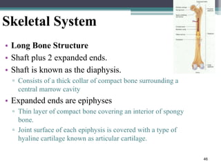
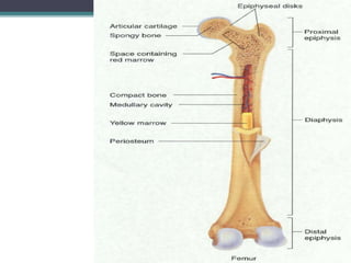
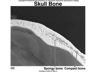
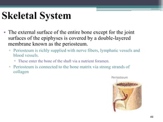
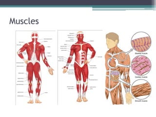
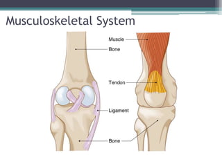
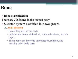
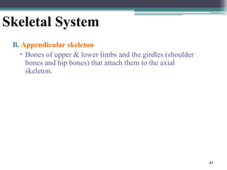
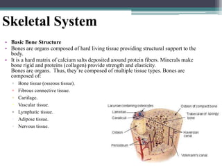
The musculoskeletal system allows for movement of the body and is comprised of bones, muscles, cartilage, tendons and ligaments. The skeletal system provides structure and protection to the body through bones and bone marrow. Bones provide support, protect organs, allow for movement through muscles attaching to them, and store minerals. There are over 200 bones in the human body that make up the axial skeleton (skull, spine, ribcage) and appendicular skeleton (limbs and their attachments).
The document discusses the gross and microscopic anatomy of long bones. Grossly, long bones are composed of a diaphysis or shaft made of compact bone covered by periosteum. The epiphyses at the ends are made of thin compact bone enclosing spongy bone. Articular cartilage provides a smooth, slippery joint surface. Microscopically, osteocytes are found in lacunae surrounding central haversian canals, communicating through canaliculi and perforating canals.
This document summarizes key aspects of skeletal physiology:
- It describes the four types of bones and the typical structures of long bones, including the diaphysis, epiphyses, articular cartilage, periosteum, medullary cavity, and endosteum.
- It also discusses the major bone cells (osteoblasts, osteoclasts, osteocytes), the Haversian system, bone matrix composition, and methods of measuring bone mineral density.
- Additional sections cover bone formation through intramembranous and endochondral ossification, fracture repair, cartilage types, bone growth, joint classification, and homeostasis functions of bones.
The document discusses different types of joints in the human body. It begins by explaining that joints allow movement between bones and come in many varieties. It then covers three main types of joints - fibrous joints, cartilaginous joints, and synovial joints. Synovial joints are the most mobile and complex. They are further classified into six categories based on their structure and motion: gliding, hinge, pivot, condylar, saddle, and ball and socket. Each joint type allows for specific ranges of motion.
Macroscopic & Microscopic Structure of Skeletal SystemSado Anatomist
The document discusses the skeletal system at both the macroscopic and microscopic levels. It describes the components of the skeletal system as bone, cartilage, tendons, and ligaments. It then explains the main functions of bones and the skeletal system, which include support, protection, assistance in movement, mineral homeostasis, blood cell production, and triglyceride storage. The document proceeds to classify and describe the different types of bones based on their shapes and locations. Finally, it examines the detailed microscopic structure of long bones and bone tissue.
The Indian Dental Academy is the Leader in continuing dental education , training dentists in all aspects of dentistry and
offering a wide range of dental certified courses in different formats.for more details please visit
www.indiandentalacademy.com
Bones provide support, protection, movement, mineral storage, and blood cell formation. With aging, bones become thinner and more porous due to increased bone absorption. This bone loss leads to loss of height and increased curvature of the spine and joints, contributing to osteoporosis.
general anatomy and development of bonesTaimurKhan87
Bones can be classified based on location in the skeletal system or shape. There are two types of bone tissue: compact bone forming the outer walls and spongy bone in the interior. Bones form through two processes - intramembranous ossification where connective tissue is replaced with bone, and endochondral ossification where cartilage is replaced with bone. Blood supply to long bones comes from nutrient arteries in the diaphysis and metaphyses and periosteal vessels on the surface.
This document discusses the skeletal system and different types of joints. It begins by defining a joint as the connection between two or more bones, and notes their functions of binding parts of the skeleton together and enabling movement. It then describes three classifications of joints: functional (based on movement), structural (based on connecting material), and synovial joints (which have a fluid-filled cavity). Several examples of each type of joint are provided, along with diagrams of their structures.
This document provides an overview of bone anatomy and physiology. It discusses the following key points in 3 sentences or less:
Bone functions as the framework of the body, allows for movement, and protects organs. Bone is made up of cells within a mineralized matrix and makes up 30-40% of body weight. The document outlines the objectives, embryology, gross anatomy including markings, microscopic anatomy, composition, classification, formation, growth, remodeling, blood supply, and roles of PTH, calcitriol, and vitamin D in bone.
There are two main types of bone tissue: compact bone and spongy bone. Compact bone looks solid and is densely packed, consisting of concentric cylindrical layers surrounding central canals. Spongy bone has a porous, sponge-like appearance due to trabeculae that form bone spicules and columns, leaving spaces between them. Both tissues have different microscopic structures that give them their distinct macroscopic appearances. Bone tissue serves important functions as the main structural support of the body, protecting internal organs, providing attachment sites for tendons and muscles, housing bone marrow, and storing minerals.
Cartilage is a type of connective tissue found in joints that provides structure and elasticity. There are three main types of cartilage - hyaline, elastic, and fibrocartilage. Joints allow bones to articulate and move. The two main types are diarthrodial synovial joints, which have a joint cavity and allow free movement, and synarthrodial joints, which are solid joints with little to no movement. Common synovial joints include hinge joints, pivot joints, condyloid joints, saddle joints, and ball and socket joints. Cartilage, synovial fluid, menisci, and other structures work together to provide support, lubrication, and smooth movement between bones.
There are two types of bone tissue: compact bone which is dense and forms the outer layer, and spongy bone which is less dense and found in low stress areas. Bones are composed of osteocytes embedded in bone matrix, as well as osteoblasts which build bone, osteoclasts which resorb bone, and osteogenic cells which develop into osteoblasts. A long bone has a central shaft made of compact bone surrounding marrow, expanded ends made of inner spongy and outer compact bone lined with marrow, and growth plates between the shaft and ends.
The document discusses bones and cartilages. It describes the structure and types of cartilage, including elastic, fibrocartilage, and hyaline cartilage. It then covers the gross structure of bones, including compact and cancellous bone. Bone cells like osteoblasts, osteoclasts and osteocytes are also discussed. The functions of bone include mechanical support, mineral storage, and endocrine functions. Bone formation occurs through endochondral and intramembranous ossification. The document also outlines different types of bones such as long, short, flat, and irregular bones.
This document provides an overview of bone histology. It defines bone as a mineralized connective tissue composed of bone matrix and three cell types: osteoblasts, osteocytes, and osteoclasts. It describes the microscopic structure of compact and spongy bone, including osteons, central canals, lamellae, and trabeculae. It explains the functions of osteoblasts in bone formation, osteoclasts in bone resorption, and osteocytes in bone maintenance. Finally, it discusses the periosteum and endosteum, which cover the external and internal bone surfaces and provide nutrition and new osteoblasts.
This document discusses the different types of joints in the human body. It begins by defining a joint and classifying joints by function and structure. The three main types by function are synarthrosis (immovable), amphiarthrosis (slightly movable), and diarthrosis (freely movable). The three main types by structure are fibrous, cartilaginous, and synovial joints. It then provides details on the different types of synovial joints and examples of various joints in the body like the shoulder, elbow, hip, knee, ankle joints. It concludes with a brief description of bursae and fontanels.
This document provides an overview of bone anatomy and physiology. It defines bone, describes its various functions, and classifications including by position, shape, development, and structure. The document discusses the composition of bone, including its organic and inorganic components. It describes the anatomy of bone including its blood and nerve supply. Finally, it provides details on the histology of bone, the different cell types involved in bone formation and resorption, and the processes of ossification and bone remodeling throughout life.
The skeletal system has several important functions:
1. It provides structure and support for the body, protects internal organs, and allows for movement through muscle attachment points.
2. Bones store minerals like calcium and aid in mineral homeostasis. Certain bones also produce blood cells.
3. The skeletal system is composed of bones, cartilage, ligaments, and tendons. Bones are living tissues with osteogenic cells, osteoblasts, osteocytes, and osteoclasts. They have an extracellular matrix containing collagen and minerals like hydroxyapatite.
The document discusses skeletal physiology, including the four types of bones, structures of long bones, constituents of bone tissue, cells found in bones, functions of bones, and bone development. It also covers cartilage structure and types, bone and cartilage growth, bone fracture repair, and joints. Key points include that long bones have a diaphysis and epiphyses, bones provide support, protection, movement, mineral storage, and blood cell formation. Cartilage includes hyaline, elastic, and fibrocartilage. Bones and cartilage both grow through cell activity but cartilage also grows through the deposition of new matrix.
Long bones have several key structures: cartilage supports the ends, epiphyseal plates allow for growth, the epiphysis are the top and bottom sections, the diaphysis is the long shaft, the periosteum is the outer layer, the medullary cavity stores blood cells and fat, compact bone is hard bone, and cancellous bone has a spongy or honeycomb appearance.
The document discusses the structure and functions of bones in the human skeletal system. It notes that bone is living tissue that is constantly being recreated. Bones provide structure and protection to the body, allow for movement through attachment of muscles and acting as levers, and store minerals and produce blood cells. The five categories of bone are defined based on shape: long bones, short bones, flat bones, irregular bones, and sesamoid bones.
General Osteology by Dr. Rahul Singh ThakurDrRahul Thakur
The document provides an overview of general osteology. It discusses that the skeleton is composed of around 206 bones and is divided into the axial and appendicular skeleton. The axial skeleton contains 80 bones including those of the skull, vertebral column, ribs, and sternum. The appendicular skeleton contains 126 bones including the shoulder girdle, upper limbs, pelvic girdle, and lower limbs. Bone has several functions like providing a rigid framework, serving as levers for muscles, protecting organs, storing minerals, and containing marrow. Bones are classified based on their shape, development, region, and microstructure.
The document summarizes bone structure and markings. It discusses:
1) Bones are classified as either axial (skull, spine, ribs) or appendicular (limbs).
2) Bones are also classified by shape as long, short, flat, or irregular. Long bones make up the limbs, short bones are cube-shaped, and flat bones are thin and curved.
3) Bones have various projections, depressions, and openings that serve as sites for muscle attachment, joints, and conduits for blood vessels.
4) Long bones consist of a hollow shaft and expanded ends, with an outer layer of compact bone and inner spongy bone containing marrow.
The skeletal system is composed of bones and associated tissues that provide structure, support, protection, movement, and mineral storage. Bones are living organs composed of cells and an extracellular matrix. There are two main types of bones - compact bone, which forms the dense outer layer, and spongy bone, which forms the inner layer. The skeletal system develops through two main processes - intramembranous ossification and endochondral ossification.
Fibrous joints are held together by connective tissue with no cavity present, and are either slightly mobile or immobile. Synovial joints contain synovial fluid and are freely movable, consisting of hyaline cartilage covering bone ends, a synovial membrane surrounding the joint cavity, and a fibrous capsule made of ligaments. Synovial joints allow for gliding, hinge, pivot, condyloid, saddle, and ball-and-socket movements. Long bones have a shaft and two expanded ends, short bones are any shape, flat bones resemble shallow plates, and irregular bones have completely irregular shapes.
The skeletal system is composed of 206 bones that serve important biological and mechanical functions. The axial skeleton includes 80 bones that form the axis of the body and protect organs like the brain, while the appendicular skeleton has 126 bones that make up the limbs and their attachments. Bones are living organs composed of compact and spongy tissues, cells like osteoblasts and osteoclasts, and minerals including calcium that provide structure and strength. The skeleton supports the body, protects organs, allows for movement through leverage, and stores minerals and produces blood cells in the bone marrow. Bones are classified by their shapes including tubular, flat, irregular, and sesamoid.
This document provides an overview of long bone anatomy and classification of joints. It describes the main parts of long bones including the diaphysis, epiphyses, metaphysis and their structures. It also classifies bones based on shape and classifies joints based on both structure and function, describing the characteristics of synovial, cartilaginous and fibrous joints. Key joint types include ball-and-socket, hinge, pivot, gliding and saddle joints.
1. The document discusses the functions and components of bones in the human body. It describes how bones provide structure, protection, movement, mineral storage, blood cell production, and triglyceride storage.
2. The main components of bones are described including the skull, vertebral column, ribs, sternum, and bones of the upper and lower limbs.
3. The types of bones - long, short, flat, irregular, and sesamoid - are defined along with examples of each type.
The musculoskeletal system is comprised of muscles, bones, cartilage, ligaments, and joints. It allows for movement via skeletal muscles attaching to bones. Bones provide support, protection, movement, storage of minerals, blood cell production, and heat generation. The musculoskeletal system includes long bones with a diaphysis and two epiphyses, short bones, flat bones, irregular bones, and sesamoid bones. Joints can be fibrous, cartilaginous, or synovial, each varying in mobility. The musculoskeletal system works with tendons, ligaments, cartilage and joints to provide structure and movement to the body.
Bone fractures occur when there is a break in the continuity of the bone. Fractures can be caused by trauma or certain medical conditions like osteoporosis. The types of fractures include closed (simple) fractures where the bone ends do not penetrate the skin, and open (compound) fractures where the bone ends do penetrate the skin. Fractures heal through a process involving blood clot formation, new blood vessel growth, collagen deposition, and ultimately bone mineralization. Proper healing depends on factors like immobilization and prevention of infection. Complications can include delayed healing, non-union, or malunion where the bone heals in an abnormal position.
This document discusses hematopoiesis, the formation of blood cells. It presents two main theories: the monoplastic theory which states all blood cells originate from one stem cell, and the polyplastic theory which suggests different blood cell types originate from different stem cells. The stages of blood cell development are described starting from the embryonic stage through the hepatic and medullary stages where development occurs in the bone marrow. The processes of erythropoiesis and granulopoiesis, leading to the production and maturation of red blood cells and white blood cells, are outlined in detail through their characteristic cell types.
Gave a talk at StartCon about the future of Growth. I touch on viral marketing / referral marketing, fake news and social media, and marketplaces. Finally, the slides go through future technology platforms and how things might evolve there.
The Six Highest Performing B2B Blog Post FormatsBarry Feldman
If your B2B blogging goals include earning social media shares and backlinks to boost your search rankings, this infographic lists the size best approaches.
1) The document discusses the opportunity for technology to improve organizational efficiency and transition economies into a "smart and clean world."
2) It argues that aggregate efficiency has stalled at around 22% for 30 years due to limitations of the Second Industrial Revolution, but that digitizing transport, energy, and communication through technologies like blockchain can help manage resources and increase efficiency.
3) Technologies like precision agriculture, cloud computing, robotics, and autonomous vehicles may allow for "dematerialization" and do more with fewer physical resources through effects like reduced waste and need for transportation/logistics infrastructure.
32 Ways a Digital Marketing Consultant Can Help Grow Your BusinessBarry Feldman
How can a digital marketing consultant help your business? In this resource we'll count the ways. 24 additional marketing resources are bundled for free.
The skeletal and muscular systems are closely integrated because they work together to enable movement. The skeletal system has 206 bones divided into the axial skeleton (bones of the head, trunk and vertebral column) and appendicular skeleton (bones of the limbs and girdles). Bones are classified by shape and composition. Joints connect bones and allow various ranges of motion. The skeletal system provides structure, protection, movement, mineral storage and blood cell formation.
1. Unit - 7- Skeleton anatomy by Thiru muruganthiru murugan
The Skeletal System
By Thiru murugan. M
Unit – 7: Anatomy - The Musculoskeletal system:
The Skeletal system
Anatomical positions
Bones: types, structure, growth and ossification
Axial and appendicular skeleton
Joints: classification, major joints and structure
Application and implications in nursing
The Muscular system:
Types and structure of muscles
Muscle groups: muscles of the head, neck, thorax, abdomen, pelvis, upper limb and lower limbs
Principal muscles: deltoid, biceps, triceps, respiratory, abdominal, pelvic floor muscles, gluteal muscles and vastus lateralis
Major muscles involved in nursing procedures
Skeletal system:
The human skeletal system consists of all of the bones, cartilage, tendons, and ligaments in the body
It Provide framework of the body.
Altogether, the skeleton makes up about 20% of a person's body weight. An adult's skeleton contains 206 bones.
It providing support and protection for the internal organs
The skeletal system also provides attachment points for muscles to allow movements at the joints.
Components of skeletal system:
Cartilage: This smooth and flexible substance covers the tips of your bones where they meet. It enables bones to move without friction (rubbing against each other).
Functions of Cartilage:
Model for bone growth in embryo & fetus
Provides a smooth cushion between adjacent bones
Provides firm flexible support (nose, ears, ribs & trachea)
Excellent shock absorber
Ligaments: Bands of strong connective tissue called ligaments hold bones together.
Functions of Ligaments:
Attach bones to bones
Provide stability
Tendons: Tendons are bands of tissue that connect the ends of a muscle to your bone.
Functions of Tendons:
Attach muscles to bones
Anchors muscle to bone for movement
Joints: A joint is where two or more bones in the body come together.
Anatomical position:
Anatomical position, or standard anatomical position, refers to the positioning of the body when it is standing upright and facing forward with each arm hanging on either side of the body, and the palms facing forward. The legs are parallel, with feet flat on the floor and facing forward.
Bones – types, structure, growth and ossification:
Bones:
Bone are specialized forms of strong connective tissue that forms the skeleton of the body.
It is composed of calcium phosphate and calcium carbonate.
It also serves as a storage area for calcium, playing a large role in calcium balance in the blood
The smallest bone in the human body is called the stirrup or stapes bone, located deep inside the ear & The longest bone in the human is called the femur.
Classification or types of bones:
Bones are divided into 5 types.
Long Bone
Short Bone
Flat Bone
Irregular Bone
Sesamoid Bone
1. Long Bone:
A long bone is one that is cylindrical in shape, being longer than it is wide.
Shape of a bone, not its size.
Long bones are found in: Arms (humerus, ulna, radius) & fingers (metacarpals, phalanges) and also Legs (femur, tibia, fibula),
The document discusses the musculoskeletal system, including bones, joints, muscles, tendons, and ligaments. It describes how bones are composed of compact and spongy bone and how ossification occurs over 20 years. There are 206 bones that are classified as long, flat, or irregular. Bones provide structure, protection, and sites for muscle attachment. Joints, such as hinges, gliding joints, ball-and-socket joints, and pivots, connect bones and allow movement. Joints are held together by ligaments and cartilage while tendons connect muscles to bones. Muscles contract and relax to flex and extend limbs through tendons at joints.
The skeletal system consists of bones, cartilage, joints and ligaments. Bones provide structure, protect organs and allow movement. There are two types of ossification - intramembranous which forms flat bones, and endochondral which forms most other bones through cartilage models. Bones are living tissues that continually remodel through the actions of osteoblasts which build bone and osteoclasts which break it down. Nutrition, exercise, hormones and aging all impact bone remodeling and strength.
The skeletal system consists of bones, cartilage, joints and ligaments that work together to support the body, protect organs and allow for movement. There are two types of ossification that can form bone - intramembranous, where bone develops in connective tissue, and endochondral, where bone replaces cartilage. Bones are constantly remodeled through the actions of osteoblasts which build bone and osteoclasts which break it down. Factors like nutrition, hormones, exercise and aging can impact bone remodeling and strength over time.
The skeletal system is composed of 206 bones and provides support, protection, movement, blood cell production, calcium storage, and endocrine regulation. It consists of two divisions: the axial skeleton which includes the skull, vertebral column, rib cage, and hyoid bone, and provides support and protection for internal organs; and the appendicular skeleton of 126 bones including the pectoral girdle, upper and lower limbs, and pelvic girdle. Cartilage, found at joints, provides flexibility and cushioning between bones. The three types are hyaline, fibro, and elastic cartilage located in various parts of the body.
The human skeleton is composed of 206 bones that provide structure, protect internal organs, allow for movement, and store minerals. There are 80 axial bones including the skull and vertebrae, and 126 appendicular bones such as the arms, legs, shoulders, and hips. Bones are living tissue composed of compact, cancellous, and subchondral bone tissues. The skeleton works with muscles to facilitate movement and provides anchors for muscles. Bones also serve important metabolic functions like mineral storage and blood cell production.
Bone is a complex living tissue that provides structure, protection, and support. There are several types of bone tissue - cortical bone is dense and hard, forming the outer shell, while cancellous bone is spongy and light. Bones also contain bone marrow, which produces blood cells. Bones are made of an organic collagen matrix and inorganic hydroxyapatite crystals. They contain various bone cells that maintain the balance between bone formation and resorption. Bones come in different shapes suited to their functions, including long bones in the arms and legs, flat bones in the skull, and irregularly shaped bones.
1 GNM anatomy Unit -13 - Skeletal system.pptxthiru murugan
By:M. Thiru murugan
Unit – 13:
Formation and growth of bones
Tendons, ligaments and cartilages
Classification of bones, joints
Joint movement
Axial and appendicular skeleton
Skeletal system:
The human skeletal system consists of all of the bones, cartilage, tendons, and ligaments in the body & It Provide framework of the body
Altogether, the skeleton makes up about 20 percent of a person's body weight. An adult's skeleton contains 206 bones
Formation and growth of bones:
Ossification, or osteogenesis, is the process of bone formation.
In fetal life the bone growth occurs in 2 process: intramembranous and endochondral ossification
Intramembranous ossification is the process of bone development from fibrous membranes. It is involved in the formation of the flat bones of the skull, the mandible, and the clavicles.
Endochondral ossification is the process of bone development from hyaline cartilage. All of the bones of the body (except for the flat bones) are formed through endochondral ossification
Stages of bone growth:
Cartilage “model” of bone forms. This model continues to grow as ossification takes place.
Ossification begins at a primary ossification center in the middle of bone.
Ossification then starts to occur at secondary ossification centers at the ends of bone.
The medullary cavity forms. This cavity will contain red bone marrow.
Areas of ossification meet at epiphyseal plates, and articular cartilage forms. Bone growth ends.
Ossification centers:
Bone forms from cartilage, ossification begins with a middle point in the cartilage called the primary ossification center & secondary ossification centers (ends part of the bones) form after birth.
Skeletal maturity:
Throughout childhood, the cartilage remaining in the skeleton keeps growing, and allows for bones to grow in size.
Once all of the cartilage has been replaced by bone, and fusion has taken place at the epiphyseal plates, bones can no longer keep growing in length.
At this point, skeletal maturity has been reached. It generally takes place by age 18 to 25.
Tendons, ligaments and cartilages:
A tendon is a band of tissue that connects muscle to bone.
Functions of Tendons:
Attach muscles to bones
Anchors muscle to bone for movement
Ligament:
A ligament is an elastic band of tissue that connects bone to bone and provides stability to the joint.
Functions of Ligaments:
Attach bones to bones
Provide stability
Cartilage:
Cartilage is a soft, gel-like padding between bones that protects joints and facilitates movement.
Functions of Cartilage:
Model for bone growth in embryo & fetus
Provides a smooth cushion between adjacent bones
Provides firm flexible support (nose, ears, ribs & trachea)
Excellent shock absorber
Classification of bones:
Bones:
Bone are specialized forms of connective tissue that forms the skeleton of the body. It is composed chiefly of calcium phosphate and calcium carbonate. It also serves as a storage area for calcium, playing a large role
The musculoskeletal system comprises the bones, muscles, cartilage, tendons and ligaments in the body. It provides form, support and movement. The muscular system includes voluntary and involuntary muscles that enable movement and maintain posture. The skeletal system comprises bones and joints, including 206 bones that make up the framework of the body. Bones are classified as flat, irregular, long or short. Joints include fibrous, cartilaginous and synovial joints like the ball and socket hip joint that allow movement. Together the musculoskeletal system provides structure, movement and protection to the body.
The skeletal system is composed of bones and associated tissues that perform several essential functions:
1. Support - Bones provide structural support for the body and protection for internal organs.
2. Movement - Skeletal muscles use bones as levers to enable movement of the body.
3. Mineral storage - Bones store minerals like calcium and phosphorus.
There are over 200 bones in the human body that are classified as long, short, flat, or irregular. Bones are living tissues composed of cells like osteoblasts, osteocytes, and osteoclasts embedded in an organic bone matrix and inorganic minerals. Compact bone forms the dense outer layer while spongy bone composes the inner layer. Long bones have
The document describes bone tissue and the process of bone formation. It discusses the six main functions of the skeletal system, the different types of bones based on shape and location, and classifies bone tissue as a connective tissue. It describes the cellular composition of bone tissue and the functions of osteoblasts, osteocytes, and osteoclasts. It also summarizes the processes of intramembranous ossification and endochondral ossification, which are the two methods by which bones initially form and grow.
This document summarizes the histological features of musculoskeletal tissues, including bone, cartilage, skeletal muscle, tendons, ligaments, and peripheral nerves. It describes the main cell types found in bone (osteoblasts, osteocytes, osteoclasts) and their functions. It also discusses the microscopic structure of cortical and cancellous bone, cartilage, skeletal muscle, and peripheral nerves. Key components are highlighted, such as osteons in cortical bone, trabeculae in cancellous bone, the extracellular matrix of cartilage, and the sarcomere structure of muscle fibers.
Bones are rigid organs that form the endoskeleton and have several important functions, including movement, support, protection and storage of minerals. There are two types of ossification that form bones - intramembranous and endochondral ossification. Intramembranous ossification forms some flat bones directly in fibrous membranes, while endochondral ossification first forms cartilage templates that are later replaced by bone. Long bones develop primarily through endochondral ossification, beginning as cartilage that is later invaded by blood vessels and replaced with spongy bone. Growth plates allow bones to lengthen, and remodeling allows bones to increase in thickness. Calcium homeostasis and bone health rely on adequate vitamin D, which facilitates intestinal
The skeletal system has three main functions: providing structure and shape to the body, protecting vital organs, and allowing for bodily movement. It is made up of 206 bones that form the axial skeleton (skull, vertebrae, ribs, sternum) and appendicular skeleton (limbs and girdles). Bones are living tissues composed of compact bone, spongy bone, bone marrow, and various bone cells. They provide structure through their interaction with muscles, tendons, and ligaments at joints like the ball-and-socket hip. The skeletal system also plays roles in blood cell production and mineral storage.
Osteology, derived from the from Greek ὀστέον (ostéon) 'bones', and λόγος (logos) 'study', is the scientific study of bones, practised by osteologists. A subdiscipline of anatomy, anthropology, and paleontology, osteology is the detailed study of the structure of bones, skeletal elements, teeth, microbone morphology, function, disease, pathology, the process of ossification (from cartilaginous molds), and the resistance and hardness of bones (biophysics).[1]
Osteologists frequently work in the public and private sector as consultants for museums, scientists for research laboratories, scientists for medical investigations and/or for companies producing osteological reproductions in an academic context.
Osteology and osteologists should not be confused with osteopathy and its practitioners, osteopaths.
1. The document provides an introduction to osteology, the study of bones. It defines osteology and discusses the classification, structure, and cellular makeup of bones.
2. Bones are classified based on their position in the body as either part of the axial or appendicular skeleton, and by their shape as long, short, flat, irregular, or sesamoid bones.
3. The structures of long bones, which make up the limbs, include a diaphysis, or shaft, and two epiphyses at the ends. Short, flat, and irregular bones differ in their shapes.
4. Bones are made up of compact cortical bone on the outside and spongy cancellous bone
2. Musculoskeletal System
• The musculoskeletal system is the organ system
that gives us the ability to move (locomotion).
The primary functions of this system include:
3. Skeletal System
Functions:
1. Support
The bones of the legs, pelvic, and vertebral column
support the weight of the erect body.
The mandible (jawbone) supports the teeth.
Other bones support various organs and tissues.
2. Protection
The bones of the skull protect the brain.
Ribs and sternum (breast bone) protect the lungs and
heart.
Vertebrae protect the spinal cord.
4. Skeletal System
3. Movement
Skeletal muscles use the bones as levers to move the
body.
4. Reservoir for minerals and adipose tissue
99% of the body’s calcium is stored in bone.
85% of the body’s phosphorous is stored in bone.
5. Hematopoiesis
All blood cells are made in the marrow of certain bones.
5. 6. Triglyceride Storage
Yellow bone marrow
Triglycerides stored in adipose cells
Serves as a potential chemical energy reserve
• 7. Heat generation
6. • The musculoskeletal system is comprised up of
the following tissues:
▫ Muscles
▫ Bones
▫ Cartilage, Ligaments, Tendons
▫ Joints
8. Anatomy and Physiology
• Muscles
▫ Skeletal, smooth, and cardiac
• Joints
▫ The junction between two or more bones
• Tendons
▫ Cord-like structures that attach muscles to the
periosteum of the bone
▫ Attachments of the muscle
Origin and insertion
9. Anatomy and Physiology
• Ligaments
▫ Fibrous tissue connecting two adjacent, freely
movable bones
• Cartilage
▫ Firm, dense type of connective tissue
• Bursae
▫ Small sac filled with synovial fluid
▫ Reduces friction between areas
15. Copyright 2010, John
Wiley & Sons, Inc.
Joints
• Points where bones meet
• Classifications
▫ Structurally: by their anatomy
Fibrous, cartilaginous, or synovial
▫ Functionally: by the degree of movement they
permit
Immovable, slightly movable, and freely movable
16. Structural Classification
• Based on what is between bones:
▫ Space (or not)
▫ Type of connective tissue present
• Types
▫ Fibrous joints - no cavity, just dense irregular
connective tissue
▫ Cartilaginous joints - no cavity, bones held
together by cartilage
▫ Synovial joints - have synovial cavity, dense
irregular tissue of articular capsule, and often
ligaments
17. Functional Classification
• Based on degree of movement they permit
• Types
▫ Synarthrosis: immovable
Pelvis, sutures, teeth
▫ Amphiarthrosis: slightly movable
Epiphyseal plate, tibia-fibula, vertebrae, pelvic
symphysis
▫ Diarthrosis: freely movable
Most joints of the body
All diarthrotic joints are synovial
18. Fibrous Joints
• Suture (synarthrosis)
▫ Joined by thin layer of dense fibrous connective
tissue
▫ Example: between bones of skull
• Syndesmosis
▫ Greater distance between bones and greater amount
of dense irregular connective tissue
▫ Examples
Distal tibia to distal fibula (amphiarthrosis)
Gomphosis (synarthrosis): tooth root in socket
(alveolar process) of mandible or maxilla
19. Fibrous Joints
• Interosseous membrane (amphiarthrosis)
▫ Has greater amount of dense irregular connective
tissue
▫ Examples: extensive membranes between shafts
of some long bones
Radius-ulna
Tibia-fibula
23. Cartilaginous Joints
• Synchondrosis (synarthrosis)
▫ Cartilage connects two areas of bone
▫ Example
Epiphyseal (growth) plate connecting epiphysis and
diaphysis of long bone (synarthrosis)
• Symphysis (amphiarthrosos)
▫ Cartilage connects two bones, but a broad disc of
fibrocartilage is present also
▫ Examples: pubic symphysis and intervertebral discs
26. ▫ Synovial
Most complex joints
Allow a large degree of relative motion between
articulating bones
Surrounded by a fibrous capsule (synovial capsule)
Hip, knee, elbow, ankle, etc.
27. Musculoskeletal System
Synovial fluid:
The joint capsule secretes whitish fluid that
works like oil in a machine, lubricating the joint
surfaces to reduce friction to promote smooth
operation and extend joint life.
32. Joints (Factors Affecting Contact and Range
for Motion at Synovial Joints)
• Range of motion (ROM)
▫ Refers to the range, measured in degrees of a circle,
through which the bones of a joint can be moved
• Factors contribute to keeping the articular
surfaces in contact and affect range of motion:
▫ Structure or shape of the articulating bones
Shape of bones determines how closely they fit together
▫ Strength and tension of the joint ligaments
Ligaments are tense when the joint is in certain positions
Tense ligaments restrict the range of motion
33. Joints (Factors Affecting Contact and Range for
Motion at Synovial Joints)
▫ Arrangement and tension of the muscles
Muscle tension reinforces the restraint placed on a joint by its
ligaments , and thus restricts movement
▫ Contact of soft parts
The point at which one body surface contacts another may limit
mobility
Movement be restricted by the presence of adipose tissue
▫ Hormones
Flexibility may also be affected by hormones
Relaxin increases the flexibility of the pubic symphysis and
loosens the ligaments between the sacrum and hip bone toward
the end of pregnancy
▫ Disuse
Movement may be restricted if a joint has not been used for an
extended period
35. Cartilage: Connective tissue
•Chondroblasts lay down the
matrix,
•become chondrocytes when
surrounded by extra cellular
matrix
•Chondrocytes do not mitose
•Poor healing
•On aging, cartilage becomes
calcified
•Less flexible
•Not bone
40. 40
Bone
• Bone classification
There are 206 bones in the human body.
• Skeleton system classified into two groups:
A. Axial skeleton
Forms long axis of the body.
Includes the bones of the skull, vertebral column, and rib
cage.
These bones are involved in protection, support, and
carrying other body parts.
41. 41
Skeletal System
B. Appendicular skeleton
Bones of upper & lower limbs and the girdles (shoulder
bones and hip bones) that attach them to the axial
skeleton.
42. 42
Skeletal System
• Basic Bone Structure
• Bones are organs composed of hard living tissue providing structural support to the
body.
• It is a hard matrix of calcium salts deposited around protein fibers. Minerals make
bone rigid and proteins (collagen) provide strength and elasticity.
Bones are organs. Thus, they’re composed of multiple tissue types. Bones are
composed of:
▫ Bone tissue (osseous tissue).
▫ Fibrous connective tissue.
▫ Cartilage.
▫ Vascular tissue.
▫ Lymphatic tissue.
▫ Adipose tissue.
▫ Nervous tissue.
43. Types of Osseous Tissue
1. Compact Bone
1. Dense, found in the walls, or cortex
2. Spongy or Cancellous Bone
1. Network of struts and thin plates (trabeculae)
3. Marrow
1. Red and Yellow
44. 44
Skeletal System
• Bone Matrix:
▫ Consists of organic and inorganic components.
▫ 1/3 organic and 2/3 inorganic by weight
▫ Organic component consists of several materials that
are secreted by the osteoblasts:
45. 45
Skeletal System
• Inorganic component of bone matrix
▫ Consists mainly of two components :
1. Calcium phosphate.
2. Calcium hydroxide.
▫ These 2 salts interact to form a compound called
hydroxyapatite.
▫ Bone also contains smaller amounts of magnesium,
▫ fluoride, and sodium.
▫ These minerals give bone its characteristic hardness and the
ability to resist compression.
46. 46
Skeletal System
• Long Bone Structure
• Shaft plus 2 expanded ends.
• Shaft is known as the diaphysis.
▫ Consists of a thick collar of compact bone surrounding a
central marrow cavity
• Expanded ends are epiphyses
▫ Thin layer of compact bone covering an interior of spongy
bone.
▫ Joint surface of each epiphysis is covered with a type of
hyaline cartilage known as articular cartilage.
49. 49
Skeletal System
• The external surface of the entire bone except for the joint
surfaces of the epiphyses is covered by a double-layered
membrane known as the periosteum.
▫ Periosteum is richly supplied with nerve fibers, lymphatic vessels and
blood vessels.
These enter the bone of the shaft via a nutrient foramen.
▫ Periosteum is connected to the bone matrix via strong strands of
collagen
50. 50
Skeletal System
• Internal bone surfaces are covered with a delicate
connective tissue membrane known as the
endosteum.
▫ Covers the spongy bone in the marrow cavities and
lines the canals that pass through compact bone.
▫ Contains both osteoblasts and osteoclasts.
54. 1. Compact Bone
• Osteon = functional unit of
bone
• The mineral matrix is
hydroxyapatite
• Osteoblasts lay down the
matrix in layers (lamellae)
▫ Become osteocytes when
surrounded by EC matrix
▫ Lacunae
• Osteoclasts break
down bone
55.
56. 2. Spongy Bone
• No osteons
• Lacy network of
struts called
trabeculae reinforce
the bone
• Covered by
endosteum
57. 3. Marrow
• Red Marrow
▫ Active
▫ Blood Precursors
• Yellow Marrow
▫ Inactive
▫ Mostly fat
58. 58
Skeletal System
• Bone marrow is a general term for the soft tissue
occupying the medullary cavity of a long bone,
the spaces amid the trabeculae of spongy bone,
and the larger haversian canals.
Red bone marrow looks like blood but with a thicker
consistency.
It consists of a delicate mesh of reticular tissue saturated
with immature red blood cells and scattered adipocytes.
60. 60
Skeletal System
• In a child, the medullary cavity of nearly every
bone is filled with red bone marrow.
• In young to middle-aged adults, the long bones
are filled with fatty yellow bone marrow.
▫ Yellow marrow no longer produces blood, although in
the event of severe or chronic anemia, it can transform
back into red marrow
• In adults, red marrow is limited to the axial
skeleton, pelvic girdle, and proximal heads of the
humerus and the femur.
61. 61
Skeletal System
• Bone tissue is a type of connective tissue consist of
cells and a big amount of extra-cellular matrix.
• Bone cells:
• Osteogenic cells
▫ Undergo cell division; the resulting cells develop
into osteoblasts
62. Osteoblasts
Bone-building cells.
Synthesize and secrete collagen fibers and other organic components of
bone matrix.
Initiate the process of calcification.
Found in both the periosteum and the endosteum
63. 63
The blue arrows indicate the
osteoblasts. The yellow arrows indicate
the bone matrix they’ve just secreted.
64. 64
Skeletal System
Osteocytes
Mature bone cells.
Osteoblasts that have become trapped by the secretion of
matrix.
No longer secrete matrix.
Responsible for maintaining the bone tissue.
65.
66.
67. 67
Skeletal System
Osteoclasts
▫ Huge cells derived from the fusion of as many as 50
monocytes (a type of white blood cell).
▫ Cells that digest bone matrix – this process is called
bone resorption and is part of normal bone growth,
development, maintenance, and repair.
▫ Concentrated in the endosteum.
68.
69. Bone Development
• Osteogenesis (ossification): is the process of bone tissue formation.
• In embryos this leads to the formation of the bony skeleton.
• In children and young adults, ossification occurs as part of bone
growth.
• In adults, it occurs as part of bone re-modeling and bone repair.
70.
71.
72. Formation of the Bony Skeleton
• Before week 8, the human embryonic skeleton is made of
fibrous membranes and hyaline cartilage.
• After week 8, bone tissue begins to replace the fibrous
membranes and hyaline cartilage.
▫ The development of bone from a fibrous membrane is called intra-
membranous ossification.
▫ Plates of Bone e.g. skull
▫ The replacement of hyaline cartilage with bone is known as endo-
chondral ossification.
• Long Bones