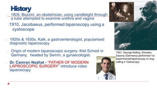
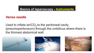
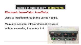
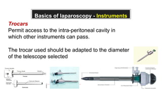
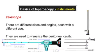
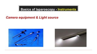
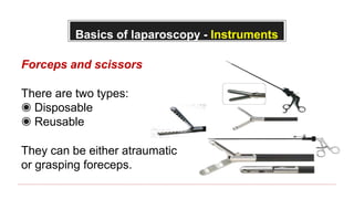
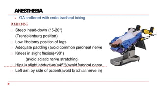
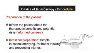
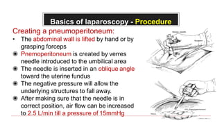
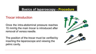
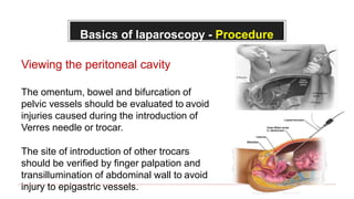
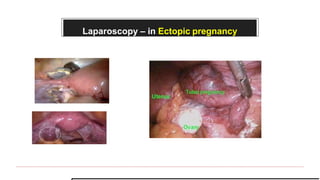
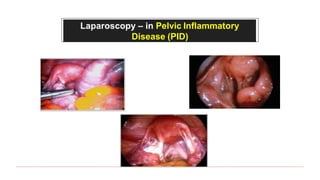
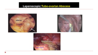
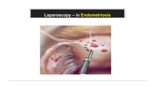
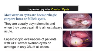
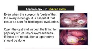
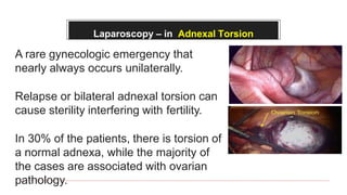
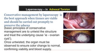
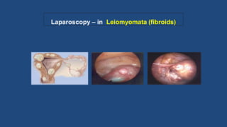
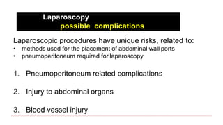
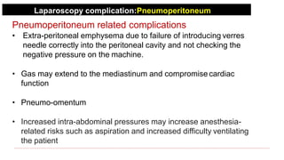
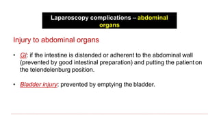
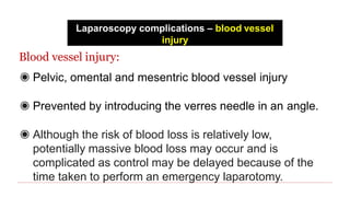
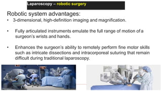
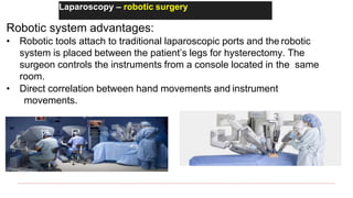
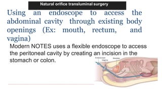
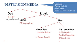
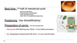
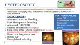
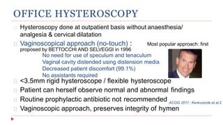
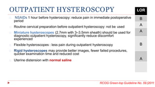
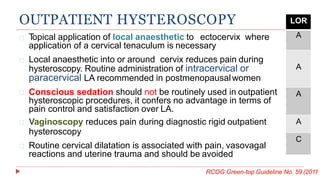
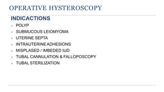
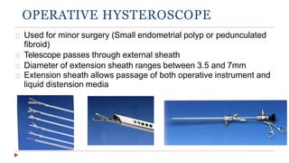
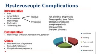
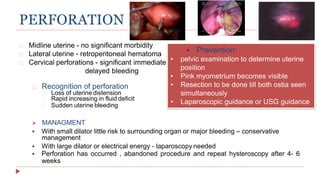
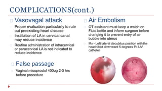
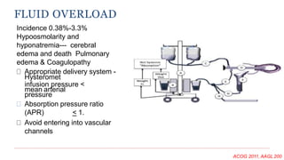
This document provides information about laparoscopy and hysteroscopy procedures. It begins with the basics of laparoscopy, including a definition, brief history, and descriptions of the instruments used. Advantages include reduced postoperative pain and recovery time compared to open surgery. Risks include potential injuries. Hysteroscopy allows direct visualization of the uterine cavity using a small telescope inserted through the cervix. Various devices and distension media options are described. Common indications for both procedures include diagnostic evaluation and treatment of conditions like endometriosis, cysts, and fibroids. Overall the document outlines the key elements of minimally invasive laparoscopic and hysteroscopic surgeries.
This presentation of introduction of laparoscopic surgery made by Dr. R.K. Mishra Director and chief surgeon World Laparoscopy Hospital. Dr. Mishra in this presentation has explained present pas and future of laparoscopic surgery. Laparoscopy is a surgical procedure which uses a special surgical instrument called a laparoscope to look inside the body, or to perform certain operations. World Laparoscopy Hospital is the center of excellence for laparoscopic and da vinci robotic surgery training and considered as one of the best institute in the world. For more detail about laparoscopic surgery please visit: http://www.laparoscopyhospital.com
This presentation will help u know with the history,present and coming up trends in laparoscopy .Also it is an acquaintance presentation regarding laparoscopy.
Laparoscopy in gynaecology presented by drs igbodike emeka philip and dr rotimiigbodikeobgyn
This slide will be helpful if the presentation revolves around laparoscopy in gynaecological practice. Kindly like , clip and share the slide. it is free!
LAPAROSCOPIC HAND INSTRUMENTS, ACCESSORIES AND ERGONOMICSsinghanubhav5
EXTENSIVE COVERAGE OF LAPAROSCOPIC INSTRUMENTS AND THEIR ERGONOMICS TO HELP SURGEONS TO KNOW HOW TO USE THEIR LAP INSTRUMENTS IN MOST APPROPRIATE WAY AND THEIR ERGONOMICS TO BE COMFERTABLE DURING SURGERY AND PATIENTS LIFE ALSO MORE SAFE.
It has not changed the nature of disease
The basic principles of good surgery still apply,including appropriate case selection, excellent exposure,adequate retraction and a high level technical expertise
If a procedure makes no sense with conventional access, it will make no sense with a minimal access approach
The cleaner and gentler the act of operation, the less the patient suffers, the smoother and quicker his convalescence,the more exquisite his healed wound.
We actually do not know what is there stored for us, but we believe that laparoscopy is trending towards advancement and nano and robotic technology is going to replace in future.
3D cameras have come into existence and various newer technologies are being invented.
Slideshow of Laparoscopic Surgery by Prof. R.K. Mishra Prof. R.K. Mishra has the distinction of being first Asian who is honoured as Professor of Minimal Access Surgery by legislated University of Govt. of India. He is is currently the most experienced professor of minimal surgery in the world who has alone as a single faculty trained more than 3000 surgeon and gynaecologists from 108 countries. http://www.laparoscopyhospital.com/drrkmishra.htm
Diagnostic laparoscopy is a minimally invasive surgical
procedure that allows the visual examination of intraabdominal organs in order to detect any pathology. This
procedure allows the direct visual examination of intraabdominal organs including large surface areas of the
liver, gallbladder, spleen, peritoneum, pelvic organs, and
retroperitoneum. Biopsies, aspiration, and cultures can be
obtained, and laparoscopic ultrasound (US) may be used.
Diagnostic laparoscopy is safe and well tolerated and
can be performed in an outpatient or inpatient setting
under general anesthesia (Fig. 1A). There may also be
unique circumstances where office based diagnostic
laparoscopy may be considered under local anesthesia.
These circumstances should include only procedures where
complications and the need for therapeutic procedures
through the same access are extremely unlikely. Manipulation
and biopsy of the viscera is possible through additional ports.
Diagnostic laparoscopy is the most commonly performed
gynecological procedure today. Its greatest advantage is that
it has replaced exploratory laparotomy.
Diagnostic laparoscopy was first introduced in 1901,
when Kelling, performed a peritoneoscopy in a dog and was
called ‘‘celioscopy’’. A Swedish internist named Jacobaeus is
credited with performing the first diagnostic laparoscopy on
human in 1910. He described its application in patients with
ascites and for the early diagnosis of malignant lesions.
In last 10 years, laparoscopy has made a great difference
to the diagnosis of abdominal acute and chronic pain. It
has evolved as an informative and important method of
diagnosing a wide spectrum of both benign and malignant
diseases. Exploratory laparoscopy also allows tissue
biopsy, culture acquisition, and a variety of therapeutic
interventions. Elective diagnostic laparoscopy refers to the
use of the procedure in chronic intra-abdominal disorders.
Emergency diagnostic laparoscopy is performed in patients
presenting with acute abdomen
Basics of laproscopic surgery..
by dr navdeep s kamboj presented at sgrdumsar amritsar.
topics covered--
1 basics of laparoscopy
2 lap cholecystectomy
3 lap appendixcectomy
pneumoperitonem
merits and demerits of laproscopy
ligasure
endoscopy,
laparoscopic instruments
Diagnostic laparoscopy is a minimally invasive surgical procedure that allows the visual examination of intraabdominal organs in order to detect any pathology.
Laparoscopy is an innovative diagnostic and surgical tool in veterinary field. Laparoscopic surgeries revolutionizes the minimally invasive surgical approaches with less surgical trauma and faster recovery.
Most ovarian abnormalities can be managed laparoscopically. Ovarian pathology can occur at any time from fetal life to menopause. First laparoscopic salpingooophorectomy was performed by Semm in 1984.
Minimally invasive/accessed surgery comprises of robotic and non robotic surgery. Non robotic surgery includes laparoscopy, endoscopy, arthroscopy and etc.
DR. NNC LAPAROSCOPY IN PREGNANCY IAGE VARANASI, 17TH MARCH 2024.pptxNiranjan Chavan
Our journey will navigate the evolution of laparoscopy in the context of pregnancy, detailing key milestones, breakthroughs, and advancements in technology and techniques. The presentation highlights how laparoscopy has revolutionized the diagnosis and treatment of conditions such as ectopic pregnancy, ovarian cysts and other gynecological disorders during pregnancy.
The prostate is an exocrine gland of the male mammalian reproductive system
It is a walnut-sized gland that forms part of the male reproductive system and is located in front of the rectum and just below the urinary bladder
Function is to store and secrete a clear, slightly alkaline fluid that constitutes 10-30% of the volume of the seminal fluid that along with the spermatozoa, constitutes semen
A healthy human prostate measures (4cm-vertical, by 3cm-horizontal, 2cm ant-post ).
It surrounds the urethra just below the urinary bladder. It has anterior, median, posterior and two lateral lobes
It’s work is regulated by androgens which are responsible for male sex characteristics
Generalised disease of the prostate due to hormonal derangement which leads to non malignant enlargement of the gland (increase in the number of epithelial cells and stromal tissue)to cause compression of the urethra leading to symptoms (LUTS
- Video recording of this lecture in English language: https://youtu.be/lK81BzxMqdo
- Video recording of this lecture in Arabic language: https://youtu.be/Ve4P0COk9OI
- Link to download the book free: https://nephrotube.blogspot.com/p/nephrotube-nephrology-books.html
- Link to NephroTube website: www.NephroTube.com
- Link to NephroTube social media accounts: https://nephrotube.blogspot.com/p/join-nephrotube-on-social-media.html
Explore natural remedies for syphilis treatment in Singapore. Discover alternative therapies, herbal remedies, and lifestyle changes that may complement conventional treatments. Learn about holistic approaches to managing syphilis symptoms and supporting overall health.
ARTIFICIAL INTELLIGENCE IN HEALTHCARE.pdfAnujkumaranit
Artificial intelligence (AI) refers to the simulation of human intelligence processes by machines, especially computer systems. It encompasses tasks such as learning, reasoning, problem-solving, perception, and language understanding. AI technologies are revolutionizing various fields, from healthcare to finance, by enabling machines to perform tasks that typically require human intelligence.
TEST BANK for Operations Management, 14th Edition by William J. Stevenson, Ve...kevinkariuki227
TEST BANK for Operations Management, 14th Edition by William J. Stevenson, Verified Chapters 1 - 19, Complete Newest Version.pdf
Ethanol (CH3CH2OH), or beverage alcohol, is a two-carbon alcohol
that is rapidly distributed in the body and brain. Ethanol alters many
neurochemical systems and has rewarding and addictive properties. It
is the oldest recreational drug and likely contributes to more morbidity,
mortality, and public health costs than all illicit drugs combined. The
5th edition of the Diagnostic and Statistical Manual of Mental Disorders
(DSM-5) integrates alcohol abuse and alcohol dependence into a single
disorder called alcohol use disorder (AUD), with mild, moderate,
and severe subclassifications (American Psychiatric Association, 2013).
In the DSM-5, all types of substance abuse and dependence have been
combined into a single substance use disorder (SUD) on a continuum
from mild to severe. A diagnosis of AUD requires that at least two of
the 11 DSM-5 behaviors be present within a 12-month period (mild
AUD: 2–3 criteria; moderate AUD: 4–5 criteria; severe AUD: 6–11 criteria).
The four main behavioral effects of AUD are impaired control over
drinking, negative social consequences, risky use, and altered physiological
effects (tolerance, withdrawal). This chapter presents an overview
of the prevalence and harmful consequences of AUD in the U.S.,
the systemic nature of the disease, neurocircuitry and stages of AUD,
comorbidities, fetal alcohol spectrum disorders, genetic risk factors, and
pharmacotherapies for AUD.
Factory Supply Best Quality Pmk Oil CAS 28578–16–7 PMK Powder in Stockrebeccabio
Factory Supply Best Quality Pmk Oil CAS 28578–16–7 PMK Powder in Stock
Telegram: bmksupplier
signal: +85264872720
threema: TUD4A6YC
You can contact me on Telegram or Threema
Communicate promptly and reply
Free of customs clearance, Double Clearance 100% pass delivery to USA, Canada, Spain, Germany, Netherland, Poland, Italy, Sweden, UK, Czech Republic, Australia, Mexico, Russia, Ukraine, Kazakhstan.Door to door service
Hot Selling Organic intermediates
MANAGEMENT OF ATRIOVENTRICULAR CONDUCTION BLOCK.pdfJim Jacob Roy
Cardiac conduction defects can occur due to various causes.
Atrioventricular conduction blocks ( AV blocks ) are classified into 3 types.
This document describes the acute management of AV block.
New Directions in Targeted Therapeutic Approaches for Older Adults With Mantl...i3 Health
i3 Health is pleased to make the speaker slides from this activity available for use as a non-accredited self-study or teaching resource.
This slide deck presented by Dr. Kami Maddocks, Professor-Clinical in the Division of Hematology and
Associate Division Director for Ambulatory Operations
The Ohio State University Comprehensive Cancer Center, will provide insight into new directions in targeted therapeutic approaches for older adults with mantle cell lymphoma.
STATEMENT OF NEED
Mantle cell lymphoma (MCL) is a rare, aggressive B-cell non-Hodgkin lymphoma (NHL) accounting for 5% to 7% of all lymphomas. Its prognosis ranges from indolent disease that does not require treatment for years to very aggressive disease, which is associated with poor survival (Silkenstedt et al, 2021). Typically, MCL is diagnosed at advanced stage and in older patients who cannot tolerate intensive therapy (NCCN, 2022). Although recent advances have slightly increased remission rates, recurrence and relapse remain very common, leading to a median overall survival between 3 and 6 years (LLS, 2021). Though there are several effective options, progress is still needed towards establishing an accepted frontline approach for MCL (Castellino et al, 2022). Treatment selection and management of MCL are complicated by the heterogeneity of prognosis, advanced age and comorbidities of patients, and lack of an established standard approach for treatment, making it vital that clinicians be familiar with the latest research and advances in this area. In this activity chaired by Michael Wang, MD, Professor in the Department of Lymphoma & Myeloma at MD Anderson Cancer Center, expert faculty will discuss prognostic factors informing treatment, the promising results of recent trials in new therapeutic approaches, and the implications of treatment resistance in therapeutic selection for MCL.
Target Audience
Hematology/oncology fellows, attending faculty, and other health care professionals involved in the treatment of patients with mantle cell lymphoma (MCL).
Learning Objectives
1.) Identify clinical and biological prognostic factors that can guide treatment decision making for older adults with MCL
2.) Evaluate emerging data on targeted therapeutic approaches for treatment-naive and relapsed/refractory MCL and their applicability to older adults
3.) Assess mechanisms of resistance to targeted therapies for MCL and their implications for treatment selection
3. Laparoscopy
A surgical procedure in which a
fibre-optic instrument is inserted
through the abdominal wall to view
the organs in the abdomen or
permit small-scale surgery with
help of pneumoperitoneum
4. Laparoscopic surgery
Laparoscopic surgery, also called minimally invasive
surgery (MIS), bandaid surgery, or keyhole surgery, is
a modern surgical technique in which operations are
performed far from their location through small
incisions (usually 0.5–1.5 cm)
5. History
1805, Bozzini, an obstetrician, using candlelight through
a tube attempted to examine urethra and vagina
1910, Jacobaeus, performed laparoscopy using a
cystoscope
1920s & 1930s, Kalk, a gastroenterologist, popularised
diagnostic laparoscopy
Origin of modern laparoscopic surgery- Kiel School in
Germany, headed by Semm, a gynaecologist.
Dr. Camran Nezhat - “FATHER OF MODERN
LAPROSCOPIC SURGERY” introduce video
laparoscopy
1901: George Kelling, Dresden,
Saxony (Germany) performed 1st
experimental laparoscopy on dog,
calling it ‘Celioscopy’
6. ADVANTAGES
Reduced postoperative morbidity - pain, chest & wound
complication
Accelerated recovery
Lesser adhesion formation
Better cosmesis
Reduced contact with body fluids & disease
transmission
Reduced incidence of ventral hernia-
11% in midline vs 4.7% in transverse scar vs 0.7% after laparoscopy.
MAGNIFICATION PRECISION DOCUMENT-
ATION
7. DISADVANTAGES
Expensive equipment
Learning curve
Limitation of intact organ retrieval(tumours)
Trocar related injuries to vessels and viscera
Counter-intuitive motion
Diathermy burns
Hemostasis more difficult
Williams Gynecology 2nd Edition A guide to laparoscopicInsufflation related postoperative pain
surgery
9. Basics of laparoscopy - Instruments
Verres needle
Used to inflate air/CO2 to the peritoneal cavity
(pneumoperitoneum) through the umbilicus where there is
the thinnest abdominal wall.
10. Basics of laparoscopy - Instruments
Electronic laparoflator: Insufflator
Used to insufflate through the verres needle.
Maintains constant intra-abdominal pressure
without exceeding the safety limit.
11. Basics of laparoscopy - Instruments
Trocars
Permit access to the intra-peritoneal cavity in
which other instruments can pass.
The trocar used should be adapted to the diameter
of the telescope selected
12. Basics of laparoscopy - Instruments
Telescope
There are different sizes and angles, each with a
different use.
They are used to visualize the peritoneal cavity.
14. Basics of laparoscopy - Instruments
Forceps and scissors
There are two types:
◉ Disposable
◉ Reusable
They can be either atraumatic
or grasping foreceps.
15. ANESTHESIA
POSITIONING
Steep, head-down (15-20°)
(Trendelenburg position)
Low lithotomy position of legs
Adequate padding (avoid common peroneal nerve
Knees in slight flexion(<90°)
(avoid sciatic nerve stretching)
Hips in slight abduction(<45°)(avoid femoral nerve
Left arm by side of patient(avoid brachial nerve inj
bowel displaced
injury)
GA preffered with endo tracheal tubing
KK Roy et al., Arch Gynecol Obstet (2014) 289:337–340
16. Basics of laparoscopy - Procedure
Preparation of the patient:
◉ Inform the patient about the
therapeutic benefits and potential
risks (informed consent).
◉ Intestinal preparation: Simple
intestinal emptying, for better viewing
and preventing injuries.
17. Basics of laparoscopy - Procedure
Creating a pneumoperitoneum:
• The abdominal wall is lifted by hand or by
grasping forceps
◉ Pnemoperitoneum is created by verres
needle introduced to the umbilical area
◉ The needle is inserted in an oblique angle
toward the uterine fundus
◉ The negative pressure will allow the
underlying structures to fall away.
◉ After making sure that the needle is in
correct position, air flow can be increased
to 2.5 L/min till a pressure of 15mmHg
18. Basics of laparoscopy - Procedure
Trocar introduction
Once the intra-abdominal pressure reaches
15 mmHg the main trocar is introduced after
removal of veress needle.
The position of the trocar must be verified by
inserting the laparoscope and viewing the
pelvic cavity.
19. Basics of laparoscopy - Procedure
Viewing the peritoneal cavity
The omentum, bowel and bifurcation of
pelvic vessels should be evaluated to avoid
injuries caused during the introduction of
Verres needle or trocar.
The site of introduction of other trocars
should be verified by finger palpation and
transillumination of abdominal wall to avoid
injury to epigastric vessels.
20. Basics of laparoscopy - Procedure
During the operative procedure
• 1 or 2 bottles of Ringer’s lactate are used to wash the peritoneal
cavity after laparoscopy.
• Leave 500/1000 ml of ringer’s lactate to reduce the incidence of
post operative pain.
After the procedure
• CO2 gas must be evacuated completely to reducepost-operative
pain
25. Laparoscopy – in Ovarian Cysts
Most ovarian cysts are haemorrhagic
corpora lutea or follicle cysts.
They are usually asymptomatic and
when they cause pain it is almost always
acute.
Laparoscopic evaluations of patients
with CPP reveal ovarian cysts on
average in only 3% of all cases.
26. Laparoscopy – in Ovarian Cysts
Even when the surgeon is ‘certain’ that
the ovary is benign, it is essential that
tissue be sent for histological evaluation.
Open the cyst and inspect the lining for
papillary structures or excrescences.
If these are noted, then a laparotomy
should be done
27. Laparoscopy – in Adnexal Torsion
A rare gynecologic emergency that
nearly always occurs unilaterally.
Relapse or bilateral adnexal torsion can
cause sterility interfering with fertility.
In 30% of the patients, there is torsion of
a normal adnexa, while the majority of
the cases are associated with ovarian
pathology.
28. Laparoscopy – in Adnexal Torsion
Conservative management by laparoscopy is
the best approach when tissues are viable
and should be carried out promptly to
preserve the adnexa
(basic principles of conservative
management are to untwist the structure
and treat the underlying cause ‘ie - ovarian
cyst’).
Once untwisted, the organ must be
observed to ensure color change to normal,
confirming viability and blood supply.
30. Laparoscopy – in Oncologic procedures
For second-look procedures following surgical and chemo
treatment of malignancy.
Laparoscopy has also been used for staging, including
peritoneal washes with biopsy, partial omentectomy, and
pelvic and periaortic lymphadenectomy
Laparoscopically assisted radical vaginal hysterectomy have
also been used by some gynecologic oncologists.
31. Laparoscopy – Tubal sterilization
Tubal sterilization can be done using:
◉ Bipolar coagulation
◉ Clips (filshie clips) and rings
Before the procedure, inform patient
about:
• Chance of irreversibility
• Failure rate 1/200
• Bleeding may occur and we may shift to laparatomy.
32. Laparoscopy – risk factors
Patient related risk factors
◉ Obesity
◉ Age
◉ Previous abdominal surgery
Anesthetic risk factors
◉ Time since last oral intake
◉ Heart disease
◉ Pulmonary disease
33. LPatient related risk factors
Obesity
◉ Laparoscopy becomes more difficult and potentially
more risky.
◉ Placement of laparoscopic instruments becomes much
more difficult
◉ Bleeding from abdominal wall vessels may be more
common because these vessels become difficult to
locate.
◉ Restricted operative field secondary to retroperitoneal fat
deposits in the pelvic sidewalls and increased bowel
excursion into the operative field
34. Laparoscopy – Patient related
risk factors
Age
◉ Older patients are at increased risk of having
concomitant disease processes that affect their
perioperative morbidity and mortality
35. Patient related risk factors
Previous abdominal surgery
◉ Risk of adhesions of omentum and/or bowel to the
anterior abdominal wall after previous abdominal surgery
is greater than 20%.
◉ As laparoscopy requires the insertion of sharp
instruments into the abdominal cavity, a reasonable
assumption is that previous surgery would increase the
risk of bowel injury
36. pre-op work-up
1. Complete blood cell count
2. Pregnancy test
3. Urinalysis
4. ECG
5. Other:
• In patients with known health problems, other laboratory tests, such as liver
function tests or electrolyte evaluations, may be indicated.
• A thorough preoperative medical evaluation, including appropriate
laboratory studies
6. Imaging studies:
• Chest radiography
• Intravenous pyelograph or kidney ultrasoundarium enema
37. Laparoscopy
possible complications
Laparoscopic procedures have unique risks, related to:
• methods used for the placement of abdominal wall ports
• pneumoperitoneum required for laparoscopy
1. Pneumoperitoneum related complications
2. Injury to abdominal organs
3. Blood vessel injury
38. Laparoscopy complication:Pneumoperitoneum
Pneumoperitoneum related complications
• Extra-peritoneal emphysema due to failure of introducing verres
needle correctly into the peritoneal cavity and not checking the
negative pressure on the machine.
• Gas may extend to the mediastinum and compromise cardiac
function
• Pneumo-omentum
• Increased intra-abdominal pressures may increase anesthesia-
related risks such as aspiration and increased difficulty ventilating
the patient
39. Laparoscopy complications – abdominal
organs
Injury to abdominal organs
• GI: if the intestine is distended or adherent to the abdominal wall
(prevented by good intestinal preparation) and putting the patient on
the telendelenburg position.
• Bladder injury: prevented by emptying the bladder.
40. Laparoscopy complications – blood vessel
injury
Blood vessel injury:
◉ Pelvic, omental and mesentric blood vessel injury
◉ Prevented by introducing the verres needle in an angle.
◉ Although the risk of blood loss is relatively low,
potentially massive blood loss may occur and is
complicated as control may be delayed because of the
time taken to perform an emergency laparotomy.
41. recent advances
3 innovations that have been introduced
in the field of laparoscopy:
◉ Robotic surgery
◉ Natural orifice transluminal surgery (NOTES)
◉ Single incision laparoscopic surgery (SILS).
Of these 3 developing technologies, robotic surgery is
having the largest impact on clinical care.
42. Laparoscopy – robotic surgery
Robotic system advantages:
• 3-dimensional, high-definition imaging and magnification.
• Fully articulated instruments emulate the full range of motion of a
surgeon’s wrists and hands.
• Enhances the surgeon’s ability to remotely perform fine motor skills
such as intricate dissections and intracorporeal suturing that remain
difficult during traditional laparoscopy.
43. Laparoscopy – robotic surgery
Robotic system advantages:
• Robotic tools attach to traditional laparoscopic ports and the robotic
system is placed between the patient’s legs for hysterectomy. The
surgeon controls the instruments from a console located in the same
room.
• Direct correlation between hand movements and instrument
movements.
44. Natural orifice transluminal surgery
Using an endoscope to access the
abdominal cavity through existing body
openings (Ex: mouth, rectum, and
vagina)
Modern NOTES uses a flexible endoscope to access
the peritoneal cavity by creating an incision in the
stomach or colon.
45. Single incision laparoscopic surgery
Single incision laparoscopic surgery
(SILS) refers to performing laparoscopy
through a single incision.
46. Advantages vs disadvantages
◉ Minimizes the number
of incisions
◉ In turn results in
decreased pain,
improved cosmetics
◉ Reduces the risks
associated with a
secondary port
placement.
Advantages Disadvantages
visibility, depth
perception,
maneuverability,
reach, and the
ability to create
counter-traction
are all limited.
47. Laparoscopy – risk vs benefit
Laparoscopy is a hybrid surgical approach that shares
characteristics of both minor and major surgery.
To patients, laparoscopic procedures often seem
to be minor surgery because of the small incisions,
relatively small amount of postoperative pain, and
short convalescent period.
Its an intra-abdominal procedure - therefore, it
shares all intraoperative and postoperative risks
of laparotomy, including infection and injury to
adjacent intra-abdominal structures
49. HISTORY
First attemt for visualisation of abdominal organ-
Bozzini 1806 – illumination of urethra by candle
Pantaleoni 1869 introduced hysteroscopy for diagnosis of intrauterine
ds.
Rubin 1925 : used cystourethroscope to look into uterus. Used water to
distend Uterus and to wash lense, used carbondioxide
1990s : Hysteroscopic surgical procedures
became popular , demonstrated
equivalent or better results than traditional
laparoscopic surgery of uterus
50. Hysteroscopy involves passing a small diameter telescope
either flexible or rigid through the cervix to directly inspect the
uterine cavity. Excellent images can be obtained.
Flexible hysteroscope: may be used in outpatient settings
Rigid instrument: employ circulating fluids therefore can be
used to visualize uterine cavity even if the women is bleeding.
52. TYPES
Rigid hysteroscope
4-mm scope offers the sharpest and
clearest view
Narrow, 3.5 mm - minimal dilation of
the cervix.
Ideal for office hysteroscopy
Flexible hysteroscope
• Can deflect over a range of 120-160°
• New equipment replaces image fiber
bundle with a video chip, eliminating
unwanted ground glass artifact (Moire
effect)
• Directed biopsies,transcervical
tubocornual recanalization, chorionic
villus sampling, IUD retrieval
53. LiquidGas
CO2
LMW
Electrolyte
- Normal Saline
Non electrolyte
1.5% Glycine
Sorbitol/Mannitol
5%dextrose
,
- Ringer lactate
32% dextran
Nontoxic
Transmits light
Good view
Does not mix with blood
Not used Commonly
DISTENSION MEDIA
HMW
54. Best time- 1st half of menstrual cycle
Isthmus hypotonic
Endometrium proliferative
Less cervical mucus
Less risk of unexpected pregnancy
Positioning : low dorsolithotomy
Preparation of cervix : for cervical stenosis
Misoprostol 200-400mcg sl/pv 30min - 6 hrs before procedure
Inj. Vasopressin Intracervical 0.05 U/mL, 4 cc at 4 and 8 o'clock
55. HYSTEROSCOPY
INDICATIONS :
Abnormal uterine bleeding
Post Menopausal bleeding
Abnormal HSG/USG
Uterine abnormalities (septae)
Suspected intra- uterine pathology (polyps,
myomas,adhesions,foreignnbodies)
Recurrent Pregnancy loss
Before IVF
Unexplained infertility
o Hysteroscopy is considered the gold standard for diagnosis of intrauterine lesions
o Transvaginal sonography / HSG and are most commonly used for UTERINE CAVITY
ABNORMALITY
CONTRAINDICATIONS
• Acute PID
• Active herpes infection
• Pregnency
• Medically Unstable
patient
Endometrial polyp
Cystic endometrial
changes
56. OFFICE HYSTEROSCOPY
Hysteroscopy done at outpatient basis without anaesthesia/
analgesia & cervical dilatation
Vaginoscopical approach (no-touch) : Most popular approach: first
Routine prophylactic antibiotic not recommended
Vaginoscopic approach, preserves integrity of hymen
proposed by BETTOCCHI AND SELVEGGI in 1996
No need for use of speculum and tenaculum
Vaginal cavity distended using distension media
Decreased patient discomfort (99.1%)
No assistants required
<3.5mm rigid hysteroscope / flexible hysteroscope
Patient can herself observe normal and abnormal findings
ACOG 2011 ; Kerkvoorde et al 2
57. OUTPATIENT HYSTEROSCOPY
NSAIDs 1 hour before hysteroscopy: reduce pain in immediate postoperative
period
Routine cervical preparation before outpatient hysteroscopy: not be used
Miniature hysteroscopes (2.7mm with 3–3.5mm sheath) should be used for
diagnostic outpatient hysteroscopy, significantly reduce discomfort
experienced
Flexible hysteroscopes : less pain during outpatient hysteroscopy
Rigid hysteroscopes may provide better images, fewer failed procedures,
quicker examination time and reduced cost
Uterine distension with normal saline
RCOG Green-top Guideline No. 59 (2011
LOR
B
A
A
B
A
of efficacy en
58. Topical application of local anaesthetic to ectocervix where
application of a cervical tenaculum is necessary
OUTPATIENT HYSTEROSCOPY LOR
A
A
A
A
C
Local anaesthetic into or around cervix reduces pain during
hysteroscopy. Routine administration of intracervical or
paracervical LA recommended in postmenopausalwomen
Conscious sedation should not be routinely used in outpatient
hysteroscopic procedures, it confers no advantage in terms of
pain control and satisfaction over LA.
Vaginoscopy reduces pain during diagnostic rigid outpatient
hysteroscopy
Routine cervical dilatation is associated with pain, vasovagal
reactions and uterine trauma and should be avoided
RCOG Green-top Guideline No. 59 (2011
60. OPERATIVE HYSTEROSCOPE
Used for minor surgery (Small endometrial polyp or pedunculated
fibroid)
Telescope passes through external sheath
Diameter of extension sheath ranges between 3.5 and 7mm
Extension sheath allows passage of both operative instrument and
liquid distension media
62. PERFORATION
or
Midline uterine - no significant morbidity
Lateral uterine - retroperitoneal hematoma
Cervical perforations - significant immediate
delayed bleeding
Recognition of perforation
Loss of uterine distension
Rapid increasing in fluid deficit
Sudden uterine bleeding
MANAGMENT
With small dilator little risk to surrounding organ or major bleeding – conservative
management
With large dilator or electrical energy - laparoscopy needed
Perforation has occurred , abandoned procedure and repeat hysteroscopy after 4- 6
weeks
Prevention:
• pelvic examination to determine uterine
position
• Pink myometrium becomes visible
• Resection to be done till both ostia seen
simultaneously
• Laparoscopic guidance or USG guidance
63. COMPLICATIONS(cont.)
Vasovagal attack
Proper evaluation particularly to rule
out preexisting heart disease
Instillation of LA in cervical canal
may reduce incidence
Routine administration of intracervical
or paracervical LA is not indicated to
reduce incidence
Air Embolism
OT assistant must keep a watch on
Fluid bottle and inform surgeon before
changing it to prevent entry of air
bubble into uterus
Mx - Left lateral decubitus position withthe
head tilted downward 5 degrees f/b IJV
catheter
False passage
Vaginal misoprostol 400ug 2-3 hrs
before procedure
64. FLUID OVERLOAD
Incidence 0.38%-3.3%
Hypoosmolarity and
hyponatremia--- cerebral
edema and death Pulmonary
edema & Coagulopathy
Appropriate delivery system -
Hysteromet
infusion pressure <
mean arterial
pressure
Absorption pressure ratio
(APR) < 1.
Avoid entering into vascular
channels
ACOG 2011, AAGL200
65. TAKE HOME MESSAGE
Laparoscopy has grown rapidly & become technique ofchoice
Basic knowledge of instruments & energy sources is necessary beforeany surgery
Primary incision for laparoscopy should be vertical from base of umbilicus
Manometer test : most reliable test for veressentry
Direct trocar entry : less minor complication & failedentry
For failed entry/ scarred abdomen – open technique / palmer pointentry
Non-midline incision ≥ 7mm & midline ≥10 mm requires deep sheathclosure
Harmonic : poor maintenance of residual tip temperature & minimal thermalspread
Ligasure : adequate maintenance of residual tip temperature but more lateral thermalspread
Whenever possible use bipolar energy sources over mono-polar in lowest possiblevoltage
66. TAKE HOME MESSAGE
Hysteroscopy done in 1st half of menstrualcycle
For outpatient hysteroscopy vaginoscopic approach is preferred
Miniature hysteroscopes (2.7mm with 3–3.5mm sheath) - significantly reduce patientdiscomfort
Local Anaesthesia
Intra cervical - reduces vasovagal symptoms, decreases thepain
Para cervical - significantly decreases the pain, not reduces vasovagalsymptoms
Distension media
Normal saline 0.9% – diagnostic, operative hysteroscopy with instrument / with bipolar
Glycine 1.5% - monopolar energy