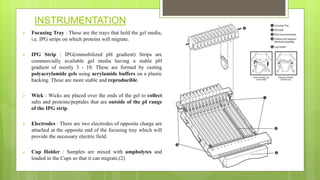
 SUBMITTED BY:- MOHAMMAD JAVED
 M.PHARM 1ST SEM
 PHARMACEUTICAL CHEMISTRY
 SUBJECT:- MODERN PHARMACEUTICAL ANALYTICAL
TECHNIQUE
 TOPIC:- ISO-ELECTRIC FOCUSING.
CONTENTS
1. Introduction
2. Graphical representation
3. Principle
4. Instrumentation
5. Factor affecting seperation
6. Advantage and disadvantage
7. Summary
8. Reference
INTRODUCTION
• It was first discovered by H.Sevennson, Sweden.(1)
• We use this technique as protein separation on the basis of Iso-elelectric point( p.I).
• It is also called electro-focusing.
• It is a technique for separating different molecule in their iso-electric point. It is a type of
zone electrophoresis performed by protein.(2)
• Iso-electric point(pI):- The pH at which net electric charge on protein becomes zero.
• Below p.I- positive charge(acidic)
• Above p.I- negative charge(basic)
• Protein moves towards the electrode with opposite charge.

 Graphical representation of iso-electric point of Alanine
PRINCIPLE
• It is performed in pH gradient.
• The proteins are amphoteric molecules with acidic and basic buffering group.
• In acidic environment the basic group becomes positively charged while in Basic
environment the acidic group becomes negatively charged.
• Proteins are positively charged in solution at pH value below pI,migrate towards
cathode.
• Proteins are negatively charged in solution at pH value above pI, migrate towards
anode.
• At pH=pI the protein have no charge and stop.(3)
INSTRUMENTATION
 Focusing Tray : These are the trays that hold the gel media,
i.e. IPG strips on which proteins will migrate.
 IPG Strip : IPG(immobilized pH gradient) Strips are
commercially available gel media having a stable pH
gradient of mostly 3 - 10. These are formed by casting
polyacrylamide gels using acrylamide buffers on a plastic
backing. These are more stable and reproducible.
 Wick : Wicks are placed over the ends of the gel to collect
salts and proteins/peptides that are outside of the pI range
of the IPG strip.
 Electrodes : There are two electrodes of opposite charge are
attached at the opposite end of the focusing tray which will
provide the necessary electric field.
 Cup Holder : Samples are mixed with ampholytes and
loaded in the Cups so that it can migrate.(2)
FACTOR AFFECTING SEPERATION
i. pH
ii. Intensity of electric field
iii. Temperature
iv. Starting and find voltage
v. Time
vi. Mode of ramping
ADVANTAGE AND DISADVANTAGE
 Advantages:
 Proteins that as by little 0.001 pH unit can be separated.
 I.E.F is a powerful analytical tool for separation of protein.
 Disadvantage:-
 Inconsistency
 Limited stability of solution
 Inadequate purity of application as a standard.
SUMMARY
• Food and agricultural industries.
• In enzymology, immunology, membrane bio chemistry.
• Forensic and human genetic lab.
• I.E.F is the 1st step in 2D gel electrophoresis and then furthur separated by SDS-
PAGE.
• It is used to separate different protein/peptide molecule on the basis of the pI and
charge value.
REFERENCE
1. Rilbe, H. (1973, June). HISTORICAL AND THEORETICAL ASPECTS OF
ISOELECTRIC FOCUSING. Annals of the New York Academy of Sciences,
209(1 Isoelectric F), 11–22.
2. Mahadik k, Sathiyanarayan L:, Instrumental Method of analysis: Nirali
publication, August 2020; Page no,: 13.4.3
3. Pergande, M., & Cologna, S. (2017, January 25). Isoelectric Point
Separations of Peptides and Proteins. Proteomes, 5(4), 4.
https://doi.org/10.3390/proteomes5010004

ISO ELECTRIC FOCUSING(ANALYSIS).pptx

  • 1.
     SUBMITTED BY:-MOHAMMAD JAVED  M.PHARM 1ST SEM  PHARMACEUTICAL CHEMISTRY  SUBJECT:- MODERN PHARMACEUTICAL ANALYTICAL TECHNIQUE  TOPIC:- ISO-ELECTRIC FOCUSING.
  • 2.
    CONTENTS 1. Introduction 2. Graphicalrepresentation 3. Principle 4. Instrumentation 5. Factor affecting seperation 6. Advantage and disadvantage 7. Summary 8. Reference
  • 3.
    INTRODUCTION • It wasfirst discovered by H.Sevennson, Sweden.(1) • We use this technique as protein separation on the basis of Iso-elelectric point( p.I). • It is also called electro-focusing. • It is a technique for separating different molecule in their iso-electric point. It is a type of zone electrophoresis performed by protein.(2) • Iso-electric point(pI):- The pH at which net electric charge on protein becomes zero. • Below p.I- positive charge(acidic) • Above p.I- negative charge(basic) • Protein moves towards the electrode with opposite charge.
  • 4.
      Graphical representationof iso-electric point of Alanine
  • 5.
    PRINCIPLE • It isperformed in pH gradient. • The proteins are amphoteric molecules with acidic and basic buffering group. • In acidic environment the basic group becomes positively charged while in Basic environment the acidic group becomes negatively charged. • Proteins are positively charged in solution at pH value below pI,migrate towards cathode. • Proteins are negatively charged in solution at pH value above pI, migrate towards anode. • At pH=pI the protein have no charge and stop.(3)
  • 7.
    INSTRUMENTATION  Focusing Tray: These are the trays that hold the gel media, i.e. IPG strips on which proteins will migrate.  IPG Strip : IPG(immobilized pH gradient) Strips are commercially available gel media having a stable pH gradient of mostly 3 - 10. These are formed by casting polyacrylamide gels using acrylamide buffers on a plastic backing. These are more stable and reproducible.  Wick : Wicks are placed over the ends of the gel to collect salts and proteins/peptides that are outside of the pI range of the IPG strip.  Electrodes : There are two electrodes of opposite charge are attached at the opposite end of the focusing tray which will provide the necessary electric field.  Cup Holder : Samples are mixed with ampholytes and loaded in the Cups so that it can migrate.(2)
  • 8.
    FACTOR AFFECTING SEPERATION i.pH ii. Intensity of electric field iii. Temperature iv. Starting and find voltage v. Time vi. Mode of ramping
  • 9.
    ADVANTAGE AND DISADVANTAGE Advantages:  Proteins that as by little 0.001 pH unit can be separated.  I.E.F is a powerful analytical tool for separation of protein.  Disadvantage:-  Inconsistency  Limited stability of solution  Inadequate purity of application as a standard.
  • 10.
    SUMMARY • Food andagricultural industries. • In enzymology, immunology, membrane bio chemistry. • Forensic and human genetic lab. • I.E.F is the 1st step in 2D gel electrophoresis and then furthur separated by SDS- PAGE. • It is used to separate different protein/peptide molecule on the basis of the pI and charge value.
  • 11.
    REFERENCE 1. Rilbe, H.(1973, June). HISTORICAL AND THEORETICAL ASPECTS OF ISOELECTRIC FOCUSING. Annals of the New York Academy of Sciences, 209(1 Isoelectric F), 11–22. 2. Mahadik k, Sathiyanarayan L:, Instrumental Method of analysis: Nirali publication, August 2020; Page no,: 13.4.3 3. Pergande, M., & Cologna, S. (2017, January 25). Isoelectric Point Separations of Peptides and Proteins. Proteomes, 5(4), 4. https://doi.org/10.3390/proteomes5010004