This document discusses ischemia and risk factors for atherosclerosis. It defines ischemia as an imbalance between myocardial oxygen supply and demand. Major risk factors include high blood pressure, high cholesterol, smoking, obesity, and diabetes. The stages of atherosclerosis and strategies for prevention and treatment are discussed, including lifestyle changes, medications like ACE inhibitors, and diagnostic tests. Angina symptoms and types are summarized.
The prolong complications of coronary artery disease such as angina pectoris, myocardial infarction, cardiac heart failure, its management and surgical mgt.
The prolong complications of coronary artery disease such as angina pectoris, myocardial infarction, cardiac heart failure, its management and surgical mgt.
A brief description for 2nd year MBBS students about IHD- MI,Unstable Angina by Dr Sabu Augustine. content from other presentations (ppts)and text books
Angina pectoris is a clinical syndrome usually characterized by episodes of pain or pressure in the anterior chest . The cause is usually insufficient coronary blood flow which results in a decreased oxygen supply to meet an increased myocardial demand for oxygen in response to physical exertion or emotional stress.
Lung Cancer: Artificial Intelligence, Synergetics, Complex System Analysis, S...Oleg Kshivets
RESULTS: Overall life span (LS) was 2252.1±1742.5 days and cumulative 5-year survival (5YS) reached 73.2%, 10 years – 64.8%, 20 years – 42.5%. 513 LCP lived more than 5 years (LS=3124.6±1525.6 days), 148 LCP – more than 10 years (LS=5054.4±1504.1 days).199 LCP died because of LC (LS=562.7±374.5 days). 5YS of LCP after bi/lobectomies was significantly superior in comparison with LCP after pneumonectomies (78.1% vs.63.7%, P=0.00001 by log-rank test). AT significantly improved 5YS (66.3% vs. 34.8%) (P=0.00000 by log-rank test) only for LCP with N1-2. Cox modeling displayed that 5YS of LCP significantly depended on: phase transition (PT) early-invasive LC in terms of synergetics, PT N0—N12, cell ratio factors (ratio between cancer cells- CC and blood cells subpopulations), G1-3, histology, glucose, AT, blood cell circuit, prothrombin index, heparin tolerance, recalcification time (P=0.000-0.038). Neural networks, genetic algorithm selection and bootstrap simulation revealed relationships between 5YS and PT early-invasive LC (rank=1), PT N0—N12 (rank=2), thrombocytes/CC (3), erythrocytes/CC (4), eosinophils/CC (5), healthy cells/CC (6), lymphocytes/CC (7), segmented neutrophils/CC (8), stick neutrophils/CC (9), monocytes/CC (10); leucocytes/CC (11). Correct prediction of 5YS was 100% by neural networks computing (area under ROC curve=1.0; error=0.0).
CONCLUSIONS: 5YS of LCP after radical procedures significantly depended on: 1) PT early-invasive cancer; 2) PT N0--N12; 3) cell ratio factors; 4) blood cell circuit; 5) biochemical factors; 6) hemostasis system; 7) AT; 8) LC characteristics; 9) LC cell dynamics; 10) surgery type: lobectomy/pneumonectomy; 11) anthropometric data. Optimal diagnosis and treatment strategies for LC are: 1) screening and early detection of LC; 2) availability of experienced thoracic surgeons because of complexity of radical procedures; 3) aggressive en block surgery and adequate lymph node dissection for completeness; 4) precise prediction; 5) adjuvant chemoimmunoradiotherapy for LCP with unfavorable prognosis.
- Video recording of this lecture in English language: https://youtu.be/lK81BzxMqdo
- Video recording of this lecture in Arabic language: https://youtu.be/Ve4P0COk9OI
- Link to download the book free: https://nephrotube.blogspot.com/p/nephrotube-nephrology-books.html
- Link to NephroTube website: www.NephroTube.com
- Link to NephroTube social media accounts: https://nephrotube.blogspot.com/p/join-nephrotube-on-social-media.html
Anti ulcer drugs and their Advance pharmacology ||
Anti-ulcer drugs are medications used to prevent and treat ulcers in the stomach and upper part of the small intestine (duodenal ulcers). These ulcers are often caused by an imbalance between stomach acid and the mucosal lining, which protects the stomach lining.
||Scope: Overview of various classes of anti-ulcer drugs, their mechanisms of action, indications, side effects, and clinical considerations.
Title: Sense of Smell
Presenter: Dr. Faiza, Assistant Professor of Physiology
Qualifications:
MBBS (Best Graduate, AIMC Lahore)
FCPS Physiology
ICMT, CHPE, DHPE (STMU)
MPH (GC University, Faisalabad)
MBA (Virtual University of Pakistan)
Learning Objectives:
Describe the primary categories of smells and the concept of odor blindness.
Explain the structure and location of the olfactory membrane and mucosa, including the types and roles of cells involved in olfaction.
Describe the pathway and mechanisms of olfactory signal transmission from the olfactory receptors to the brain.
Illustrate the biochemical cascade triggered by odorant binding to olfactory receptors, including the role of G-proteins and second messengers in generating an action potential.
Identify different types of olfactory disorders such as anosmia, hyposmia, hyperosmia, and dysosmia, including their potential causes.
Key Topics:
Olfactory Genes:
3% of the human genome accounts for olfactory genes.
400 genes for odorant receptors.
Olfactory Membrane:
Located in the superior part of the nasal cavity.
Medially: Folds downward along the superior septum.
Laterally: Folds over the superior turbinate and upper surface of the middle turbinate.
Total surface area: 5-10 square centimeters.
Olfactory Mucosa:
Olfactory Cells: Bipolar nerve cells derived from the CNS (100 million), with 4-25 olfactory cilia per cell.
Sustentacular Cells: Produce mucus and maintain ionic and molecular environment.
Basal Cells: Replace worn-out olfactory cells with an average lifespan of 1-2 months.
Bowman’s Gland: Secretes mucus.
Stimulation of Olfactory Cells:
Odorant dissolves in mucus and attaches to receptors on olfactory cilia.
Involves a cascade effect through G-proteins and second messengers, leading to depolarization and action potential generation in the olfactory nerve.
Quality of a Good Odorant:
Small (3-20 Carbon atoms), volatile, water-soluble, and lipid-soluble.
Facilitated by odorant-binding proteins in mucus.
Membrane Potential and Action Potential:
Resting membrane potential: -55mV.
Action potential frequency in the olfactory nerve increases with odorant strength.
Adaptation Towards the Sense of Smell:
Rapid adaptation within the first second, with further slow adaptation.
Psychological adaptation greater than receptor adaptation, involving feedback inhibition from the central nervous system.
Primary Sensations of Smell:
Camphoraceous, Musky, Floral, Pepperminty, Ethereal, Pungent, Putrid.
Odor Detection Threshold:
Examples: Hydrogen sulfide (0.0005 ppm), Methyl-mercaptan (0.002 ppm).
Some toxic substances are odorless at lethal concentrations.
Characteristics of Smell:
Odor blindness for single substances due to lack of appropriate receptor protein.
Behavioral and emotional influences of smell.
Transmission of Olfactory Signals:
From olfactory cells to glomeruli in the olfactory bulb, involving lateral inhibition.
Primitive, less old, and new olfactory systems with different path
Ozempic: Preoperative Management of Patients on GLP-1 Receptor Agonists Saeid Safari
Preoperative Management of Patients on GLP-1 Receptor Agonists like Ozempic and Semiglutide
ASA GUIDELINE
NYSORA Guideline
2 Case Reports of Gastric Ultrasound
micro teaching on communication m.sc nursing.pdfAnurag Sharma
Microteaching is a unique model of practice teaching. It is a viable instrument for the. desired change in the teaching behavior or the behavior potential which, in specified types of real. classroom situations, tends to facilitate the achievement of specified types of objectives.
TEST BANK for Operations Management, 14th Edition by William J. Stevenson, Ve...kevinkariuki227
TEST BANK for Operations Management, 14th Edition by William J. Stevenson, Verified Chapters 1 - 19, Complete Newest Version.pdf
TEST BANK for Operations Management, 14th Edition by William J. Stevenson, Verified Chapters 1 - 19, Complete Newest Version.pdf
Pulmonary Thromboembolism - etilogy, types, medical- Surgical and nursing man...VarunMahajani
Disruption of blood supply to lung alveoli due to blockage of one or more pulmonary blood vessels is called as Pulmonary thromboembolism. In this presentation we will discuss its causes, types and its management in depth.
Explore natural remedies for syphilis treatment in Singapore. Discover alternative therapies, herbal remedies, and lifestyle changes that may complement conventional treatments. Learn about holistic approaches to managing syphilis symptoms and supporting overall health.
MANAGEMENT OF ATRIOVENTRICULAR CONDUCTION BLOCK.pdfJim Jacob Roy
Cardiac conduction defects can occur due to various causes.
Atrioventricular conduction blocks ( AV blocks ) are classified into 3 types.
This document describes the acute management of AV block.
6. PREVENTION OF ATHEROMATOUS DISEASE
ACE-Is improve endothelial function and prolong life
Regular exercise increases circulating HDL.
Antioxidants (e.g. vitamin C and vitamin E) improve endothelial
function.
7.
8. LIPOPROTEIN TRANSPORT
Lipids and cholesterol are transported in the bloodstream as
complexes of lipid and protein known as lipoproteins. These
consist of a central core of hydrophobic lipid encased in a
hydrophilic coat of polar phospholipid, free cholesterol
and apoprotein.
There are four main classes of lipoprotein
1. HDL particles (contain apoA1 and apoA2), diameter 7-20 nm
2. LDL particles (contain apoB-100), diameter 20-30 nm
3. VLDL particles (contain apoB-100), diameter 30-80 nm
4. chylomicrons (contain apoB-48), diameter 100-1000 nm.
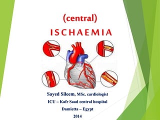
9. Ischaemic heart disease(IHD) is defined as acute or
chronic form of cardiac disability arising from
imbalances between the myocardial supply and
demand for oxygenated blood.
Since, narrowing or obstruction of the coronary
arterial system is the most common cause of
myocardial anoxia, the alternate term coronary
artery disease(CAD) is used synonymously with IHD.
10. SIGNS AND SYMPTOMS
Ischaemic heart disease may be present with any of the following
problems:
Angina pectoris
Acute chest pain: ACS {unstable angina or MI}.("heart attack",
severe chest pain unrelieved by rest associated with evidence
of acute heart damage)
Heart failure (dyspnoea or limb oedema).
Heart burn.
11. Angina occurs when the oxygen supply to the myocardium is
insufficient for its needs.
The pain has a characteristic distribution in the
chest,
arm and
neck,
It is brought on by
1. exertion,
2. cold or
3. excitement.
12. TYPES OF ANGINA stable=unstable=varient=MI
Predictable
Occurs on exercise, emotion or eating.
Caused by increase demand of the heart and by a fixed narrowing
of coronary vessels, almost always by atheroma.
Blood flow fails to increase during increased demand despite the
local factors mediated vasodilation and so ischemic pain.
So, the diastolic pressure increases and this causes a endocrinal
crunch and thus causing Ischaematic pain in this region.
Relieved by taking rest and reducing the myocardial workload.
13. TYPES OF ANGINA stable=unstable=varient=MI
This is characterized by pain that occurs with less and less
exertion, culminating in pain at rest.
The pathology is similar to that involved in myocardial
infarction, namely platelet-fibrin thrombus associated with a
ruptured atheromatous plaque, but without complete occlusion
of the vessel.
14. TYPES OF ANGINA stable=unstable=varient=MI
Uncommon
Occurs at rest generally during sleep
Caused by Large Coronary artery spasm
Therapy is with Coronary artery vasodilators.(e.g. organic
nitrates, calcium antagonists).
15. TYPES OF ANGINA stable=unstable=varient=MI
Myocardial infarction occurs when a coronary artery has been
blocked by thrombus.
This may be fatal and is a common cause of death, usually as a
result of mechanical failure of the ventricle or from arrhythmia.
Cardiac myocytes rely on aerobic metabolism. If the supply of
oxygen remains below a critical value, a sequence of events
leading to cell death (by necrosis or apoptosis) detected
clinically by an elevation of circulating troponin.
16. Effects of myocardial ischaemia: This leads to cell death by one of two
pathways: Necrosis or apoptosis
17. Pathophysiology of myocardial ischemia
Myocardial oxigen supply is decreased
Narrowed coronary arteries (sclerosis, thrombus,
spasmus, coronary embolism, vasculitis)
Hypotension
Severe anemia
Methemoglobinemia, increased carboxyhemoglobin
Myocardial oxigen demand is increased
Left ventricle hypertrophy
Fever
Hyperthyroidism
Tachycardy
18. The clinical manifestations of ischemic heart disease
Ischemic heart disease without clinical symptoms.
Sudden death can be the presenting manifestation.
Cardiomegaly and heart failure that may have caused
no symptoms prior the development of heart failure –
ischemic cardiomyopathy.
Angina pectoris. Stable angina pectoris.
Unstable angina/Non ST-elevation myocardial
infarction (NSTEMI)/STEMI = acut coronary
syndromes
19. The typical clinical features of angina pectoris
The typical location of pain is retrosternal.
When the patient is asked to localize the sensation, he or
she will typically place their hand over the sternum,
somtetimes with a clenched fist, to indicate the
squezzing. The pain can not be localized with one finger.
Usually described as heaviness, pressure, squezzing, or
choking.
Usually associates with gradual intensification of
symptoms over a period of minutes.
It lasts typically 2-5 min.
It can radiate to either shoulder and to both arms
(especially the ulnar surfaces of the forearm and hand.
It can also arise in or radiate to the back, interscapular
region, root of neck, jaw, teeth, and epigastrium. Rarely
localized below the umbilicus or above the mandible.
Exertional angina is typically relieved by rest and
nitroglycerin.
20. Associated symptoms and signs of angina pectoris
Associated symptoms
Dyspnoe
Fatique, faintness
Nausea, vomiting
Sweating
Sense of impending doom (mostly in case of
myocardial infarction)
Physical signs
Third and fourth heart sounds
Apical systolic murmur due to mitral regurgitation
(impaired papillary muscle function)
Pulmonary congestion
21. Summary of the characteristics of angina pectoris
Typical angina pectoris:
Retrosternal chest pain (discomfort)
Complaints occur after exertion or emotional stress
The pain is relieved by rest and nitroglycerin
Atypical angina pectoris: especially in women and
diabetics, angina may be atypical in location and not
strictly related to provocing factors)
Pseudoangina: Only one or no one out of three
characteristics.
23. Diagnostic tests in patients with chest discomfort
2.
If the patient’s history or examination is consistent
with pulmonary embolism
D-dimer, CT-angiography or a lung scan, echocardiography combined with
lower extremity venous ultrasound
With aortic dissection
Chest CT scan with contrast, MRI, or transesophageal echocardiography
No evidence of life-threatening conditions, the clinician should
then focus on serious chronic conditions with the potential to
cause major complications, the most common of which is stable
angina
- exercise electrocradiography, stress echocardiography or
stress perfusion imaging
- Pericarditis (, blood pressure pattern, echocardiography)
If not, could the discomfort be due to an acute condition that
warrants specific therapy?
- Pneumonia – Chest X-ray
- Herpes zoster – physical examination
If not, another treatable chronic condition
24. Serum cardiac markers
Released into the circulation from necrotic heart muscle
CK (creatine kinase) rises 4-8 hrs after onset of MI
and normalize by 48-72 hrs
not specific for myocardial necrosis
MB isoenzyme of CK is more specific
Cardiac specific troponins: more sensitive and
specific than CK and CKMB for identification
of myocardial necrosis
Myoglobin- first serum marker to rise after MI, but
lacks specificity.
25. Diagnostic Decision Tree
for Coronary Artery Disease
NEGATIVE
Nuclear Medicine NEGATIVE
rule out CAD
POSITIVE
NON DIAGNOSTIC
POSITIVE
NON DIAGNOSTIC
~ 4.4 m
procedures/y
Stress ECG
Ultrasound
$ 400.-
rule out CAD
medication
Stress Perfusion Imaging
medication
~ 2.2 m
procedures/y
X-Ray Coronary
Angiography or IVUS
$ 300.-
$ 700.-
>$ 3,000.-
~ 13 m
procedures/y
Radiation
Invasive
26. MR Coronary Angiography after 0.125 mmol/kg
Injection of B-22956/1 [Res. : 0.7 0.7 0.7 mm3]
1 min
postcontrast
33 min
postcontrast
17 min
postcontrast
RCA RCA
RCA
RCA
LAD LAD
30. The main therapeutic drugs include drugs to
improve cardiac function by maintaining
oxygenation and reducing cardiac work as well as
treating pain and preventing further thrombosis.
Combinations of thrombolytic, antiplatelet and
antithrombotic (a heparin preparation) drugs to
open the blocked artery and prevent re-occlusion.
31. Antiplatelet treatment. Clopidogrel 300mg if undergoing PCI- Glycoprotein IIb/IIIa inhibitors
(abciximab) if undergoing PCI
Throbolytic therapy then, Heparin eg enoxaparin 1mg/kg 12 hourly
ACEIs and ARBs also reduce cardiac work and improve survival, treat hypertension and may
lower the risk of recurrent myocardial infarction
Aspirin : 160-325 mg chewable aspirin leads to rapid buccal absorption, inhibition of COX in
platelets and reduction of TXA2
βB reduce cardiac work and thereby the metabolic needs of the heart, and are used as soon as
the patient is stable. Atenolol 5mg over 5 mins repeated after 10-15 mins
CCB
Cholesterol lowering medications, such as statins, are useful to decrease the amount of LDL.
Nitroglycerin 0.4mg sublingual +- IV
Oxygen if there is arterial hypoxia.
Opioids (given with an antiemetic) to prevent pain
Tight glycaemic control
Optimise potassium and magnesium
33. PCI Procedural refinements: Stents
Expandable metal mesh tubes that buttresses the dilated segment, limit
restenosis.
Drug eluting stents: further reduce cellular proliferation in response to the
injury of dilatation.
35. STEMI
ASA, beta blockers, antithrombin therapy
<12 hrs >12 hrs
Eligible for
Lytic therapy
Lytic C/I Not a candidate
For reperfusion
Persistent
symptoms
Thrombolysis Primary PCI no yes
Other medical therapy Consider reperfusion
(ACEI, nitrates, beta blockers, antiplatelets, antithrombin,statins)
36. TThhee FFoouurr DDss
TTiimee ooff OOnnsseett
ED Time Point 1:
DOOR
ED Time Point 2:
DATA
ED Time Point 3:
DECISION
ED Time Point 4:
DRUG
Time Interval III
Decision to drug
Time Interval II
ECG to decision to treat
Time Interval I
Door to ECG
NHAAP Recommendations. U.S. Department of Health NIH Publication: 1997:97-3787.
37. Unstable angina/NSTEMI
Aspirin, antithrombin - nitrates - GP IIb-IIIa
antagonist- Betablockers (or CCB)
Assess clinical status
High risk/unstable Stable
(Recurrent ischemia, LV dysfunction
Widespread EKG changes, positive
enzyme markers)
Stress test
yes
Cardiac catheterization Severe ischemia
no
Revascularization (PCI/CABG) Medical therapy
42. Mode of action
Activate plasminogen to form plasmin which
degrades fibrin breaking up thrombi
Streptokinase, alteplase, reteplase, tenecteplase
Streptokinase – antibodies within 4 ds
Alteplase, reteplase followed by heparin for 48 hs
43. Indications Contraindications
Acute ST elevation
myocardial infarction
Acute pulmonary
embolism
Acute ischaemic stroke
within 3 hours
Recent haemorrhage
trauma or surgery
Recent dental extraction
Coagulation defects;
bleeding disorders
Aortic dissection
History of cerebrovascular
disease
Active peptic ulceration
Severe menorrhagia
Severe hypertension
Active cavitating lung
disease
Acute pancreatitis
Severe liver disease
Oesophageal varices
Previous reaction to
streptokinase
Relative contraindications
Venepuncture (non-compressible
site)
Recent invasive procedure
External chest compressions
Pregnancy
Abdominal aortic aneurysm
Diabetic retinopathy
Anticoagulant therapy
44. Side effects
Nausea and vomiting
Bleeding
Reperfusion arrhythmias
Hypotension
Back pain
Allergic reactions (esp streptokinase)
46. Differencial diagnosis of
chest discomfort
Acute myocardial infarction
The duration of the pain often more than 30 min
Often more severe than angina
Unrielived by nitroglicerin
May be associated with evidence of heart failure or
arrhythmia
Aortic dissection
Tearing, ripping pain with abrupt onset
Associated with hypertension, and/or connective tissue
disorder
Depending on the location of dissection:
Loss of peripheral pulse
Pericardial tamponad
Murmur of aortic insufficiency
47. Differencial diagnosis of
chest discomfort
Pericarditis
The duration of the pain is hours to days
Sharp, retrosternal pain that is aggravated by coughing, deep
breath, or changes in body position (relieved by sitting and leaning
forward)
Pulmonary embolism
Abrupt onset of the pain. Location is often lateral
Associated symptoms are dyspnea, tachycardy,and occasionally
hemoptysis
Pneumothorax
Sudden onset of pleuritic chest pain. Location:lateral to side of
pneumothorax
Dyspnea, decreased breath sounds, tympanic percussion sound.
Pneumonia or pleuritis
Localized sharp, knifelike pain
Pain is aggravated by inspiration and coughing
Dyspnea, fever, rales, occasionally pleural rub
48. Differencial diagnosis of
chest discomfort
Esophageal reflux
Deep, burning discomfort that may be exacerbated by
alcohol, aspirin, or some foods
Worsened by postprandial recumbency, relieved by
antacids
Ulcer disease
Symptoms do not associated with exertion
Prolonged burning pain
Typically occurs 60 to 90 min after meals, when
postprandial acid production is no longer neutralized
by food in the stomach
Gallbladder disease
Prolonged colic pain
Occurs an hour or more after meals
49. Differencial diagnosis of
chest discomfort
Neuromusculoskeletal diseases
Cervical disk disease: compression of nerve roots –
dermatomal distribution (pain in dermatomal
distribution can also be caused by intercostal muscle
cramp and herpes zoster)
The pain is aggravated by movement
Costochondral and chondrosternal syndromes
(Tietze’s syndrome)
direct pressure on the costochondral-costosternal junctions
may reproduce the pain.
Psychiatric conditions
Th symptoms are frquently described as visceral
tightness or aching that last more than 30 min.
50. Pathophysiology of acut
coronary syndrome
UA/NSTEMI: Caused by a reduction in oxygen supply
and/or by an increase in myocardial oxygen demand
superimposed on an atherosclerotic coronary plaque.
STEMI: coronary blood flow decreases abruptly after a
thrombotic occlusion of a coronary artery previously
affected by atherosclerosis.
51. Diagnosis
Electrocardiogram
Echocardiograms
Stress Tests
Nuclear Imaging
Angiography
It is may be ordered if your doctor
suspects a problem with the heart muscle
or one of the valves that channel blood
through the heart.
52. Diagnosis
Electrocardiogram
Echocardiograms
Stress Tests
Nuclear Imaging
Angiography
They are used to show how the heart
reacts to physical exertion. Exercise stress
tests are usually performed on a treadmill
or exercise bicycle.
53. Diagnosis
Electrocardiogram
Echocardiograms
Stress Tests
Nuclear Imaging
Angiography
involves the use of small amounts of
short-lived radioactive material, which is
injected into the bloodstream.
A special camera (live-motion x-ray)
detects the radioactivity of these
materials, and the images displayed show
how your heart pumps blood.
This is useful in identifying any areas of
abnormal motion or for assessing the
blood supply to the heart muscle.
54. Diagnosis
Electrocardiogram
Echocardiograms
Stress Tests
Nuclear Imaging
Angiography
Is the most accurate means by which to
examine the coronary arteries.
It requires a surgical procedure called
cardiac catheterization. During the
procedure, catheters (small thin plastic
tubes) are placed in the artery of the leg
or arm, and directed using an x-ray
machine to the opening of each of the
coronary arteries
55. Ten important treatment elements of stable angina include:
AA aspirin and anti-anginals
BB beta-blockers and blood pressure control
CC cholesterol and cigarettes
DD diet and diabetes
EE education and exercise
56.
57. 1. ORGANIC NITRATES
It is used in the treatment of angina pectoris are simple nitric
and nitrous acid esters of glycerol. They differ in their
volatility, e.g. isosorbide dinitrate and isosorbide mononitrate
are solids at room temperature, nitroglycerin is only
moderately volatile, and amyl nitrite is extremely volatile.
These compounds cause a rapid reduction in myocardial
oxygen demand, followed by rapid relief of symptoms.
They are effective in stable and unstable angina as well as in
variant angina pectoris.
58. Mechanism of action
Nitrates decrease coronary vasoconstriction or spasm and increase
perfusion of the myocardium by relaxing coronary arteries.
In addition, they relax veins, decreasing preload and myocardial oxygen
consumption.
Organic nitrates, such as nitroglycerin, which is also known as glyceryl
trinitrate, are thought to relax vascular smooth muscle by their intracellular
conversion to nitrite ions, and then to nitric oxide, which in turn activates
guanylate cyclase and increases the cells' cyclic guanosine monophosphate
(GMP). Elevated cGMP ultimately leads to dephosphorylation of the
myosin light chain, resulting in vascular smooth muscle relaxation.
59. Pharmacokinetics
The time to onset of action varies from 1 minute for nitroglycerin to more
than 1 hour for isosorbide mononitrate .
Significant first-pass metabolism of nitroglycerin occurs in the liver.
Therefore, it is common to take the drug either sublingually or via a
transdermal patch, thereby avoiding this route of elimination.
Isosorbide mononitrate owes its improved bioavailability and long
duration of action to its stability against hepatic breakdown.
Oral isosorbide dinitrate undergoes denitration to two mononitrates, both of
which possess antianginal activity.
60. Adverse effects
The most common adverse effect of nitroglycerin, as well as of
the other nitrates, is headache.
High doses of organic nitrates can also cause postural
hypotension, facial flushing, and tachycardia. Sildenafil
potentiates the action of the nitrates.
To preclude the dangerous hypotension that may occur, this
combination is contraindicated.
61. 2. β BLOCKERS
The β blockers decrease the oxygen demands of the myocardium by lowering
both the rate and the force of contraction of the heart.
They suppress the activation of the heart by blocking β receptors, and they
reduce the work of the heart by decreasing heart rate, contractility, cardiac
output, and blood pressure. With β blockers, the demand for oxygen by the
myocardium is reduced both during exertion and at rest.
Propranolol is the prototype for this class of compounds, but it is not cardio
selective.
Thus, other β blockers, such as metoprolol or atenolol, are preferred.
Agents with intrinsic sympathomimetic activity (for example, pindolol) are
less effective and should be avoided in angina.
62. 3. CALCIUM-CHANNEL BLOCKERS
Calcium channels
Three types of Ca2+ channel
(a ) Voltage sensitive channel : Activated when membrane potential drops
to around -40 m V or lower.
(b) Receptor operated channel : Activated by Adr and other agonists-independent
of membrane depolarization
(NA contracts even depolarized aortic smooth me bypromoting influx of
Ca2+ through this channel and releasing Ca2+ from sarcoplasmic reticulum).
(c) Leak channel : Small amounts of Ca2+ leak into resting cell and are
pumped out by Ca2+
63.
64. Only the voltage sensitive L-type channels are blocked by the CCBs.
The three groups of CCBs viz. phenylalkylamines (verapamil),
benzothiazepine (diltiazem) and dihydropyridines (nifedipine) bind to
their own specific binding sites on the α1, subunit; all restricting Ca 2+
entry, though characteristics of channel blockade differ.
Further, different drugs may have differing affinities for various site
specific isoforms of the L-channels.
This may account for the differences in action exhibited by various
CCBs. The vascular smooth muscle has a more depolarized mernbrane
(RMP about -40 mV) than heart.
This may contribute to vascular selectivity of certain CCBs.
67. Clinical uses of calcium channel blockers
Arrhythmias (verapamil):
To slow ventricular rate in rapid atrial fibrillation.
To prevent recurrence of supraventricular tachycardia (SVT).
Hypertension:
Usually a dihydropyridine drug (e.g. amlodipine or slow-release
nifedipine).
To prevent angina (e.g. dihydropyridine or diltiazem)
68. Clinical Application of hs-CRP for
Cardiovascular Risk Prediction
1 mg/L 3 mg/L 10 mg/L
Low
Risk
Moderate
Risk
High
Risk
>100 mg/L
Acute Phase Response
Ignore Value, Repeat Test in 3 weeks
Ridker PM. Circulation 2003;107:363-9