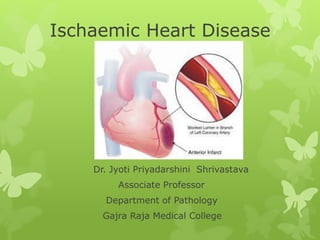
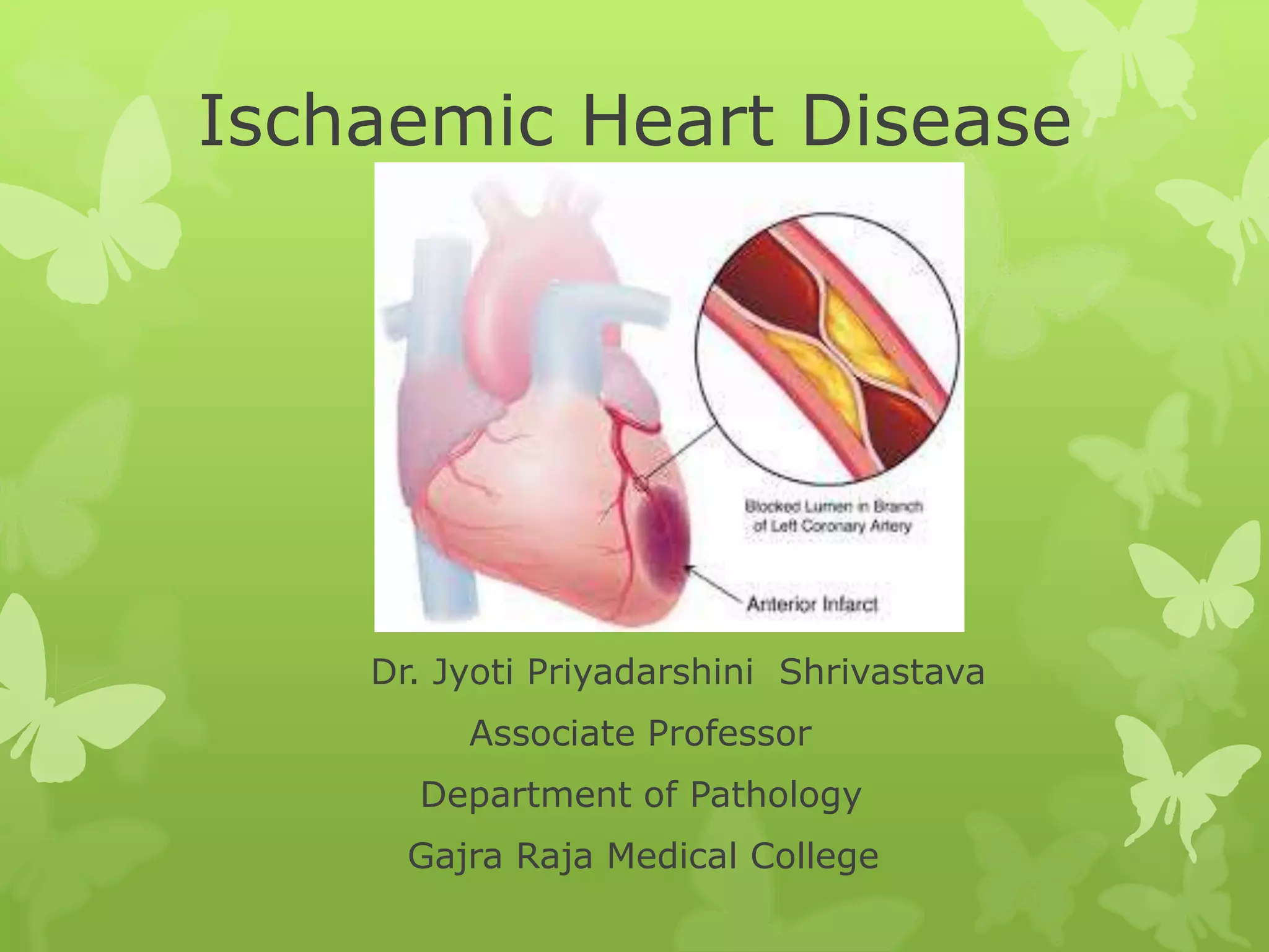
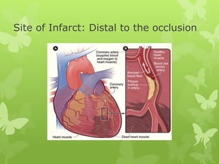
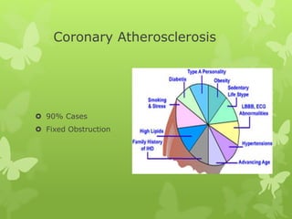
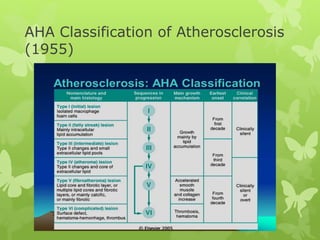
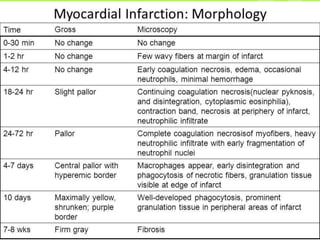
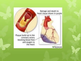
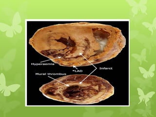
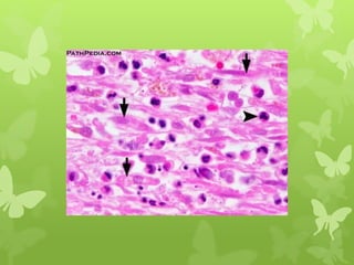
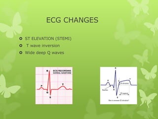
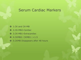
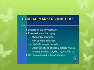
Ischaemic Heart Disease
Dr. Jyoti Priyadarshini Shrivastava
Associate Professor
Department of Pathology
Gajra Raja Medical College
Blood Supply of Heart
 Rt.and Lt.Coronary Arteries-Arise from root of AORTA
 Supply blood during Diastole
 Veins: Cardiac Vein and Coronary Sinus
 Myocardium-Max.Supply
Site of Infarct: Distal to the occlusion
TERMS
Ischaemia: Ischemia is a restriction in blood
supply to tissues, causing a shortage of oxygen that is
needed for cellular metabolism (to keep tissue alive).
Infarction: Tissue death (necrosis) due to inadequate
blood supply to the affected area. Latin infarctus,
"stuffed into.
Angina: Angina is pressure, squeezing, burning, or
tightness in the chest. The pain or discomfort usually
starts behind the breastbone & can occur in the arms,
shoulders, neck, jaw, throat, or back. The pain may
feel like indigestion.
DEFINITION OF IHD
Ischaemic Heart Disease (IHD) is defined as
acute or chronic form of cardiac disability due
to imbalance between myocardial supply and
demand of oxygenated blood.
Synonym : CAD
2020: commonest leading cause of Death
IHD
1.Asymptomatic
2.Angina Pectoris
3.Acute Myocardial Infarction
4.Chronic Ischemic Heart
Disease
5.Sudden Cardiac Death
ETIOPATHOGENESIS
Etiology
1.Coronary Atherosclerosis
2.Superadded changes in Coronary
Atherosclerosis
Non-Atherosclerotic Causes
Coronary Atherosclerosis
 90% Cases
 Fixed Obstruction
Specific lesions in Atherosclerosis
 1.Distribution:
a.Ant.Descending branch
b.Rt.Coronary
c.Circumflex a.
 2. Location :
Area of involvement-3-4 cm. from coronary Ostia
Bifurcation of Arteries
Atherosclerotic plaques are seen throughout the coronaries.
Superadded changes in Coronary Atherosclerosis
Acute changes in chr.atheromatous
plaque(spasm)
Haemorrhage
Fissure
Ulcer------thrombosis,embolization
Coronary Artery Thrombosis-Transmural MI
Local platelet aggregation & coronary artery
spasm
AHA Classification of Atherosclerosis
(1955)
Non Atherosclerotic Causes
 <10% cases
Vasospasm
Stenosis
Arteritis
Thrombotic diseases
Embolism
Trauma
Aneurysms
Compression
Acute Coronary Syndrome
Absolute Medical Emergency
 Triad:Umbrella term
1. Acute Myocardial Infarction
2. Unstable Angina
3. Sudden Cardiac Death
ANGINA PECTORIS
 Clinical syndrome
 Transient Myocardial Ischemia
 Symptom:
1.Paroxysmal Pain in the substernum/precordial region
2.Aggravated by Increased Demand
3.Relievedby rest.
Site:Left arm
Neck
Jaw
Right Arm
Age/sex: 50 yrs./Males
Clinical Patterns of Angina
 3 types:
1.Stable or Typical Angina
2.Prinzmetal’s Variant Angina
3.Unstable or Crescendo Angina
Stable or Typical Angina
Perfusion less on demand
Relieved on rest
Chr.sclerosing coronary AS
ST depressed---Poor perfusion of
Subendocardium
No enzyme elevation
No Irreversible Myocardial injury
Prinzmetals Variant Angina
 Pain at rest
 Sudden vasospasm
 ST elevation –Transmural Ischemia
 Vaodilator(Nitroglycerine )effective
Crescendo or Unstable Angina
 Pre-Infarction Angina or
 Acute Coronary Insufficiency
Pain frequent
prolonged
rest
D/d- AMI has ST elevation
Causes: Stenosing coronary AS
Complicated plaque
Vasospasm
Infarction is prevented by Collaterals.
Myocardial cell necrosis+
ACUTE MYOCARDIAL INFARCTION
Fatal consequence of CAD
Collaterals are protective
Exercise –Good collateral
AS directly proportional to AMI
 Incidence:10-25%
 Age:Elderly
<40 yrs.-
 Sex: Males
ETIOPATHOGENESIS
 AS in any of the 3 major trunks
 >75% lumen block
 90% cases of MI
1.Myocardial Ischemia
2.Role of Platelets
3.Acute Plaque Rupture
4.Non AS causes
5.Transmural vs subendocardial infarcts
 Transmural:Disrupted Atheromatous plaque
 Subendocardial:Decreasedperfusion of Myocardium
Age of Infarct
 Newly formed Infarcts: Acute,recent or fresh
 Advanced Infarcts : Old,Healed or Organised
Location Of Infarcts
 Left Ventricle
 Rt.Atrial
 LAD Coronary A.-Ant.part LV
(40-50%) Apex
IV Septum
 RCA Stenosis: Post.part of LV
(30-40%) Post. 1/3 of IV Septum
 Lt.Circumflex CA : Lat.wall of LV
(15-20%)
MORPHOLOGIC FEATURES
Grade 0
Grade I
Dense Neutrophilic Infiltrate
6 DAY OLD
Ischemic Necrosis in MI
Infarct Healing
EM Changes
1.perinuclear glycogen lost(5 min)
2.mitochondrial sweeling(20-30
min.)
3.sarcolemmal disruption
4.clump nuclear chromatin
Infarct area under EM
 1.Glycogen Depletion
 2.Loss of K+
 3.Na+ intake
 4.Ca+ Influx
 5.enzyme leakage
Diagnosis of MI
1.Clinical features
2.E.C.G
3.Serum Enzyme Markers
Clinical Features
 Pain
 Indigestion
 Apprehension
 Shock : SBP <80 MM Hg
 Oliguria: <20 ml/hour
 Low grade fever-Leucocytosis
Elevated ESR
 Acute Pulmonary edema
ECG CHANGES
 ST ELEVATION (STEMI)
 T wave inversion
 Wide deep Q waves
Serum Cardiac Markers
 1.CK and CK-MB
 2.CK-MB2-Cardiac
 3.CK-MB1-Extracardiac
 4.CKMB2: CKMB1::>1.5
 5.CKMB Disappears after 48 hours
LDH
 LDH1:LDH2>1
 Rise after 24 hours
 Peak-3-6 days
 Normal in 2 weeks
Cardiac Specific Troponins (cTn)
 Troponins: Cardiac n skeletal muscles
 cTn : specific
 2 types:
cTn T(7-10 days)
cTn I (10-14 days)
 Risewith CKMB-4-6 hours
Myoglobin
 1st marker to rise
 Lacks cardiac specificity
 Excreted within 24 hours
 SGOT(Aspartate Aminitransferase)-8 hours
3-4 days
COMPLICATIONS
 Arrythmias
 Congestive Heart Failure
 Cardiogenic Shock
 Mural Thrombus and Thromboembolism
 Rupture
 Cardiac Aneurysm
 Pericarditis
 Post myocardial Infarction Syndrome
Sudden cardiac Death
 Morning: Hypercoagulability++
 Thromboplastin release by ateromatous plaque.
 Ventricular arrythmia+electrical disturbance
 Within 1 hour----DEATH
References
 Robbins Textbook of Pathology,10th edn.
 Textbook of Pathology,Harsh Mohan ,6th edn.
 Textbook of Pathology,1st edn.,Vinay Kamal
 Wikipedia
THANK YOU

Ischaemic Heart Disease(IHD)

  • 1.
    Ischaemic Heart Disease Dr.Jyoti Priyadarshini Shrivastava Associate Professor Department of Pathology Gajra Raja Medical College
  • 2.
    Blood Supply ofHeart  Rt.and Lt.Coronary Arteries-Arise from root of AORTA  Supply blood during Diastole  Veins: Cardiac Vein and Coronary Sinus  Myocardium-Max.Supply
  • 4.
    Site of Infarct:Distal to the occlusion
  • 5.
    TERMS Ischaemia: Ischemia isa restriction in blood supply to tissues, causing a shortage of oxygen that is needed for cellular metabolism (to keep tissue alive). Infarction: Tissue death (necrosis) due to inadequate blood supply to the affected area. Latin infarctus, "stuffed into. Angina: Angina is pressure, squeezing, burning, or tightness in the chest. The pain or discomfort usually starts behind the breastbone & can occur in the arms, shoulders, neck, jaw, throat, or back. The pain may feel like indigestion.
  • 7.
    DEFINITION OF IHD IschaemicHeart Disease (IHD) is defined as acute or chronic form of cardiac disability due to imbalance between myocardial supply and demand of oxygenated blood. Synonym : CAD 2020: commonest leading cause of Death
  • 8.
    IHD 1.Asymptomatic 2.Angina Pectoris 3.Acute MyocardialInfarction 4.Chronic Ischemic Heart Disease 5.Sudden Cardiac Death
  • 9.
  • 10.
    Etiology 1.Coronary Atherosclerosis 2.Superadded changesin Coronary Atherosclerosis Non-Atherosclerotic Causes
  • 11.
    Coronary Atherosclerosis  90%Cases  Fixed Obstruction
  • 12.
    Specific lesions inAtherosclerosis  1.Distribution: a.Ant.Descending branch b.Rt.Coronary c.Circumflex a.  2. Location : Area of involvement-3-4 cm. from coronary Ostia Bifurcation of Arteries Atherosclerotic plaques are seen throughout the coronaries.
  • 13.
    Superadded changes inCoronary Atherosclerosis Acute changes in chr.atheromatous plaque(spasm) Haemorrhage Fissure Ulcer------thrombosis,embolization Coronary Artery Thrombosis-Transmural MI Local platelet aggregation & coronary artery spasm
  • 14.
    AHA Classification ofAtherosclerosis (1955)
  • 15.
    Non Atherosclerotic Causes <10% cases Vasospasm Stenosis Arteritis Thrombotic diseases Embolism Trauma Aneurysms Compression
  • 16.
    Acute Coronary Syndrome AbsoluteMedical Emergency  Triad:Umbrella term 1. Acute Myocardial Infarction 2. Unstable Angina 3. Sudden Cardiac Death
  • 19.
    ANGINA PECTORIS  Clinicalsyndrome  Transient Myocardial Ischemia  Symptom: 1.Paroxysmal Pain in the substernum/precordial region 2.Aggravated by Increased Demand 3.Relievedby rest. Site:Left arm Neck Jaw Right Arm Age/sex: 50 yrs./Males
  • 21.
    Clinical Patterns ofAngina  3 types: 1.Stable or Typical Angina 2.Prinzmetal’s Variant Angina 3.Unstable or Crescendo Angina
  • 22.
    Stable or TypicalAngina Perfusion less on demand Relieved on rest Chr.sclerosing coronary AS ST depressed---Poor perfusion of Subendocardium No enzyme elevation No Irreversible Myocardial injury
  • 23.
    Prinzmetals Variant Angina Pain at rest  Sudden vasospasm  ST elevation –Transmural Ischemia  Vaodilator(Nitroglycerine )effective
  • 24.
    Crescendo or UnstableAngina  Pre-Infarction Angina or  Acute Coronary Insufficiency Pain frequent prolonged rest D/d- AMI has ST elevation Causes: Stenosing coronary AS Complicated plaque Vasospasm Infarction is prevented by Collaterals. Myocardial cell necrosis+
  • 25.
    ACUTE MYOCARDIAL INFARCTION Fatalconsequence of CAD Collaterals are protective Exercise –Good collateral AS directly proportional to AMI
  • 26.
  • 27.
    ETIOPATHOGENESIS  AS inany of the 3 major trunks  >75% lumen block  90% cases of MI 1.Myocardial Ischemia 2.Role of Platelets 3.Acute Plaque Rupture 4.Non AS causes 5.Transmural vs subendocardial infarcts
  • 30.
     Transmural:Disrupted Atheromatousplaque  Subendocardial:Decreasedperfusion of Myocardium
  • 32.
    Age of Infarct Newly formed Infarcts: Acute,recent or fresh  Advanced Infarcts : Old,Healed or Organised
  • 33.
    Location Of Infarcts Left Ventricle  Rt.Atrial  LAD Coronary A.-Ant.part LV (40-50%) Apex IV Septum  RCA Stenosis: Post.part of LV (30-40%) Post. 1/3 of IV Septum  Lt.Circumflex CA : Lat.wall of LV (15-20%)
  • 34.
  • 38.
  • 39.
  • 40.
  • 42.
  • 43.
  • 44.
  • 45.
    EM Changes 1.perinuclear glycogenlost(5 min) 2.mitochondrial sweeling(20-30 min.) 3.sarcolemmal disruption 4.clump nuclear chromatin
  • 47.
  • 49.
     1.Glycogen Depletion 2.Loss of K+  3.Na+ intake  4.Ca+ Influx  5.enzyme leakage
  • 50.
    Diagnosis of MI 1.Clinicalfeatures 2.E.C.G 3.Serum Enzyme Markers
  • 51.
    Clinical Features  Pain Indigestion  Apprehension  Shock : SBP <80 MM Hg  Oliguria: <20 ml/hour  Low grade fever-Leucocytosis Elevated ESR  Acute Pulmonary edema
  • 52.
    ECG CHANGES  STELEVATION (STEMI)  T wave inversion  Wide deep Q waves
  • 53.
    Serum Cardiac Markers 1.CK and CK-MB  2.CK-MB2-Cardiac  3.CK-MB1-Extracardiac  4.CKMB2: CKMB1::>1.5  5.CKMB Disappears after 48 hours
  • 56.
    LDH  LDH1:LDH2>1  Riseafter 24 hours  Peak-3-6 days  Normal in 2 weeks
  • 57.
    Cardiac Specific Troponins(cTn)  Troponins: Cardiac n skeletal muscles  cTn : specific  2 types: cTn T(7-10 days) cTn I (10-14 days)  Risewith CKMB-4-6 hours
  • 58.
    Myoglobin  1st markerto rise  Lacks cardiac specificity  Excreted within 24 hours  SGOT(Aspartate Aminitransferase)-8 hours 3-4 days
  • 61.
    COMPLICATIONS  Arrythmias  CongestiveHeart Failure  Cardiogenic Shock  Mural Thrombus and Thromboembolism  Rupture  Cardiac Aneurysm  Pericarditis  Post myocardial Infarction Syndrome
  • 62.
    Sudden cardiac Death Morning: Hypercoagulability++  Thromboplastin release by ateromatous plaque.  Ventricular arrythmia+electrical disturbance  Within 1 hour----DEATH
  • 63.
    References  Robbins Textbookof Pathology,10th edn.  Textbook of Pathology,Harsh Mohan ,6th edn.  Textbook of Pathology,1st edn.,Vinay Kamal  Wikipedia
  • 64.

Editor's Notes

  • #43 Centralpallor,necrotic fibre