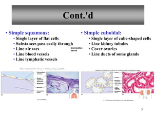
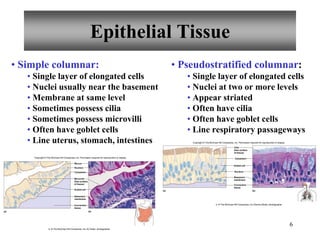
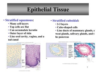
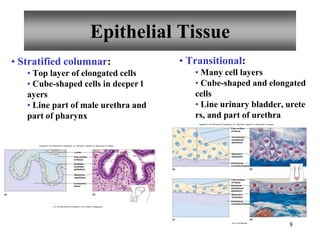
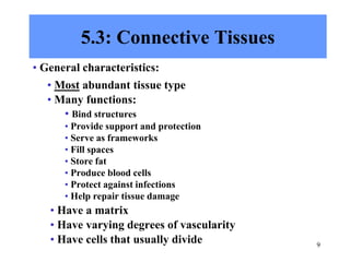
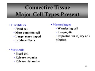
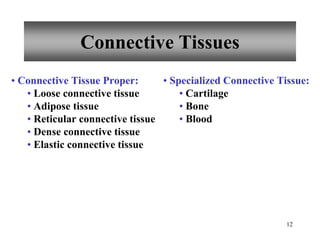
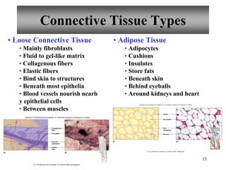
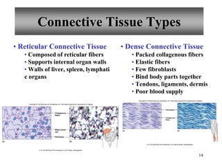
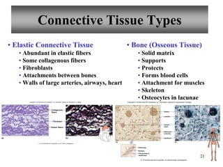
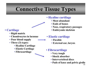
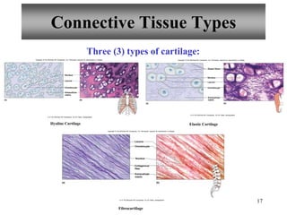
1
Tissues
Copyright © The McGraw-Hill Companies, Inc. Permission required for reproduction or display.
2
5.1: Introduction
• Similar cells with a common function are called tissu
es.
• The study of tissues is called histology.
• There are four (4) primary or major tissue types:
1. Epithelial Tissue
2. Connective Tissue
3. Muscle Tissue
4. Nervous Tissue
3
5.2: Epithelial Tissue
• General characteristics:
• Cover organs and the body
• Line body cavities
• Line hollow organs
• Have a free surface
• Have a basement membrane
• Are avascular
• Cells readily divide
• Cells tightly packed
• Cells often have desmosomes
• Function in protection, secretion, absorption, and excretion
• Classified according to cell shape and number of cell layers
• Classified according to cell shape and number of
cell layers
Classification
4
5
Cont.'d
• Simple squamous:
• Single layer of flat cells
• Substances pass easily through
• Line air sacs
• Line blood vessels
• Line lymphatic vessels
• Simple cuboidal:
• Single layer of cube-shaped cells
• Line kidney tubules
• Cover ovaries
• Line ducts of some glands
(b)
(a)
Free surface
of tissue
Simple
squamous
epithelium
Basement
Nucleus
Connective
tissue
Connective
tissue
Copyright
©
The
McGraw-Hill
Companies,
Inc.
Permission
required
for
reproduction
or
display.
b,d: © Ed Reschke
Nucleus
Basement
membrane
Free surface
of tissue
Simple
cuboidal
epithelium
Connective
tissue
Lumen
(a) (b)
Copyright © The McGraw-Hill Companies, Inc. Permission required for reproduction or display.
b: © The McGraw-Hill Companies, Inc./Al Telser, photographer
6
• Simple columnar:
• Single layer of elongated cells
• Nuclei usually near the basement
• Membrane at same level
• Sometimes possess cilia
• Sometimes possess microvilli
• Often have goblet cells
• Line uterus, stomach, intestines
• Pseudostratified columnar:
• Single layer of elongated cells
• Nuclei at two or more levels
• Appear striated
• Often have cilia
• Often have goblet cells
• Line respiratory passageways
Nucleus
Basement
membrane
Microvilli
(free surface
of tissue)
Connective
tissue
Mucus
Cytoplasm
Goblet cell
(a) (b)
Copyright © The McGraw-Hill Companies, Inc. Permission required for reproduction or display.
b: © The McGraw-Hill Companies, Inc./Al Telser, photographer.
(a) (b)
Cilia
(free surface
of tissue)
Goblet cell
Basement
membrane
Nucleus
Connective
tissue
Cytoplasm
Copyright © The McGraw-Hill Companies, Inc. Permission required for reproduction or display.
b: © The McGraw-Hill Companies, Inc./Dennis Strete, photographer
Epithelial Tissue
7
• Stratified squamous:
• Many cell layers
• Top cells are flat
• Can accumulate keratin
• Outer layer of skin
• Line oral cavity, vagina, and a
nal canal
• Stratified cuboidal:
• 2-3 layers
• Cube-shaped cells
• Line ducts of mammary glands, s
weat glands, salivary glands, and t
he pancreas
Basement
membrane
Layer of
dividing
cells
Connective
tissue
Free surface
of tissue
Squamous
cells
(b)
(a)
Copyright © The McGraw-HillCompanies, Inc. Permission required for reproduction or display.
b: © The McGraw-Hill Companies, Inc./Al Telser, photographer
(a) (b)
Stratified
cuboidal
epithelium
Free surface
of tissue
Lumen
Basement
membrane
Connective
tissue
Nucleus
Copyright © The McGraw-Hill Companies, Inc. Permission required for reproduction or display.
b: © The McGraw-Hill Companies, Inc./Al Telser, photographer.
Epithelial Tissue
8
• Stratified columnar:
• Top layer of elongated cells
• Cube-shaped cells in deeper l
ayers
• Line part of male urethra and
part of pharynx
• Transitional:
• Many cell layers
• Cube-shaped and elongated
cells
• Line urinary bladder, urete
rs, and part of urethra
(b)
(a)
Lumen
Stratified
columnar
epithelium
Connective
tissue
Basement
membrane
Free surface
of tissue
Copyright © The McGraw-Hill Companies, Inc. Permission required for reproduction or display.
b: © The McGraw-Hill Companies, Inc./Al Telser, photographer
Stretched
transitional
epithelium
Basement
membrane
Underlying
connective tissue
Basement
membrane
Underlying
connective tissue
Unstretched
transitional
epithelium
(b)
(a)
(d)
(c)
Free surface
of tissue
Free surface
of tissue
Copyright © The McGraw-Hill Companies, Inc. Permission required for reproduction or display.
b,d: © Ed Reschke
Epithelial Tissue
9
5.3: Connective Tissues
• General characteristics:
• Most abundant tissue type
• Many functions:
• Bind structures
• Provide support and protection
• Serve as frameworks
• Fill spaces
• Store fat
• Produce blood cells
• Protect against infections
• Help repair tissue damage
• Have a matrix
• Have varying degrees of vascularity
• Have cells that usually divide
10
Connective Tissue
Major Cell Types Present
• Fibroblasts
• Fixed cell
• Most common cell
• Large, star-shaped
• Produce fibers
• Macrophages
• Wandering cell
• Phagocytic
• Important in injury or i
nfection
• Mast cells
• Fixed cell
• Release heparin
• Release histamine
11
Connective Tissue
Fiber Types Present
• Collagenous fibers
• Thick
• Composed of collagen
• Great tensile strength
• Abundant in dense CT
• Hold structures together
• Tendons, ligaments
• Elastic fibers
• Bundles of microfibrils e
mbedded in elastin
• Fibers branch
• Elastic
• Vocal cords, air passages
• Reticular fibers
• Very thin collagenous fibers
• Highly branched
• Form supportive networks
12
Connective Tissues
• Connective Tissue Proper:
• Loose connective tissue
• Adipose tissue
• Reticular connective tissue
• Dense connective tissue
• Elastic connective tissue
• Specialized Connective Tissue:
• Cartilage
• Bone
• Blood
13
Connective Tissue Types
• Loose Connective Tissue
• Mainly fibroblasts
• Fluid to gel-like matrix
• Collagenous fibers
• Elastic fibers
• Bind skin to structures
• Beneath most epithelia
• Blood vessels nourish nearb
y epithelial cells
• Between muscles
• Adipose Tissue
• Adipocytes
• Cushions
• Insulates
• Store fats
• Beneath skin
• Behind eyeballs
• Around kidneys and heart
Elastic
fiber
(a) (b)
Collagenous
fiber
Fibroblast
Ground
substance
Copyright © The McGraw-Hill Companies, Inc. Permission required for reproduction or display.
b: © The McGraw-Hill Companies, Inc./Dennis Strete, photographer
Fat droplet
(a) (b)
Nucleus
Cell
membrane
Cytsol
Copyright © The McGraw-Hill Companies, Inc. Permission required for reproduction or display.
b: © The McGraw-Hill Companies, Inc./Dennis Strete, photographer
14
Connective Tissue Types
• Reticular Connective Tissue
• Composed of reticular fibers
• Supports internal organ walls
• Walls of liver, spleen, lymphati
c organs
• Dense Connective Tissue
• Packed collagenous fibers
• Elastic fibers
• Few fibroblasts
• Bind body parts together
• Tendons, ligaments, dermis
• Poor blood supply
Collagenous
fibers
Fibroblast
White blood
cell
(a) (b)
Copyright © The McGraw-Hill Companies, Inc. Permission required for reproduction or display.
b: © The McGraw-Hill Companies, Inc./Al Telser, photographer
Fibroblasts
Collagenous
fibers
(a) (b)
Copyright © The McGraw-Hill Companies, Inc. Permission required for reproduction or display.
b: © The McGraw-Hill Companies, Inc./Dennis Strete, photographer
15
Connective Tissue Types
• Elastic Connective Tissue
• Abundant in elastic fibers
• Some collagenous fibers
• Fibroblasts
• Attachments between bones
• Walls of large arteries, airways, heart
• Bone (Osseous Tissue)
• Solid matrix
• Supports
• Protects
• Forms blood cells
• Attachment for muscles
• Skeleton
• Osteocytes in lacunae
Elastic fibers
Collagenous
fibers
Fibroblast
(a) (b)
Copyright © The McGraw-Hill Companies, Inc. Permission required for reproduction or display.
b: © The McGraw-Hill Companies, Inc./Al Telser, photographer
21
Canaliculi
Osteocyte
in lacuna
Central
canal
Lamella
Nucleus
Osteocyte
Cell process in
canaliculus
(a) (b)
Osteon
Copyright © The McGraw-Hill Companies, Inc. Permission required for reproduction or display
b: © The McGraw-Hill Companies, Inc./Dennis Strete, photographer
16
Connective Tissue Types
• Cartilage
• Rigid matrix
• Chondrocytes in lacunae
• Poor blood supply
• Three (3) types:
• Hyaline Cartilage
• Elastic Cartilage
• Fibrocartilage
• Hyaline cartilage
• Most abundant
• Ends of bones
• Nose, respiratory passages
• Embryonic skeleton
• Elastic cartilage
• Flexible
• External ear, larynx
• Fibrocartilage
• Very tough
• Shock absorber
• Intervertebral discs
• Pads of knee and pelvic girdle
17
Connective Tissue Types
Three (3) types of cartilage:
Hyaline Cartilage Elastic Cartilage
Fibrocartilage
Chondrocyte
Nucleus
Extracellular
matrix
(a) (b)
Lacuna
Copyright © The McGraw-Hill Companies, Inc. Permission required for reproduction or display.
b: © The McGraw-Hill Companies, Inc./Al Telser, photographer
Chondrocyte
Elastic fibers
Nucleus
Extracellular
matrix
(a) (b)
Lacuna
Copyright © The McGraw-Hill Companies, Inc. Permission required for reproduction or display.
b: © The McGraw-Hill Companies, Inc./Al Telser, photographer
Chondrocyte
Nucleus
Collagenous
fiber
Extracellular
matrix
(a) (b)
Lacuna
Copyright © The McGraw-Hill Companies, Inc. Permission required for reproduction or display.
b: © The McGraw-Hill Companies, Inc./Al Telser, photographer
18
Connective Tissue Types
• Blood
• Fluid matrix called plasma
• Red blood cells
• White blood cells
• Platelets
• Transports
• Defends
• Involved in clotting
• Throughout body in blood ve
ssels
• Heart
Red blood
cells
Plasma
(extracellular
matrix of blood)
Platelets
White blood
cell
(a) (b)
Copyright © The McGraw-Hill Companies, Inc. Permission required for reproduction or display.
b: © The McGraw-Hill Companies, Inc./Al Telser, photographer
19
5.5: Muscle Tissues
• General characteristics:
• Muscle cells also called mus
cle fibers
• Contractile
• Three (3) types:
• Skeletal muscle
• Smooth muscle
• Cardiac muscle
• Skeletal muscle
• Attached to bones
• Striated
• Voluntary
• Smooth muscle
• Walls of organs
• Skin
• Walls of blood vessels
• Involuntary
• Non-striated
• Cardiac muscle
• Heart wall
• Involuntary
• Striated
• Intercalated discs
20
5.6: Nervous Tissue
• Found in brain, spinal cord, and
peripheral nerves
• Functional cells are neurons
• Neuroglial cells support and
bind nervous tissue components
• Sensory reception
• Conduction of nerve impulses
Cell
membrane
Neuroglial
cells
Cytoplasm
Cellular
process
Nucleus
(a) (b)
Copyright © The McGraw-Hill Companies, Inc. Permission required for reproduction or display.
b: © Ed Reschke.

Tissues.pptx

  • 1.
    1 Tissues Copyright © TheMcGraw-Hill Companies, Inc. Permission required for reproduction or display.
  • 2.
    2 5.1: Introduction • Similarcells with a common function are called tissu es. • The study of tissues is called histology. • There are four (4) primary or major tissue types: 1. Epithelial Tissue 2. Connective Tissue 3. Muscle Tissue 4. Nervous Tissue
  • 3.
    3 5.2: Epithelial Tissue •General characteristics: • Cover organs and the body • Line body cavities • Line hollow organs • Have a free surface • Have a basement membrane • Are avascular • Cells readily divide • Cells tightly packed • Cells often have desmosomes • Function in protection, secretion, absorption, and excretion • Classified according to cell shape and number of cell layers
  • 4.
    • Classified accordingto cell shape and number of cell layers Classification 4
  • 5.
    5 Cont.'d • Simple squamous: •Single layer of flat cells • Substances pass easily through • Line air sacs • Line blood vessels • Line lymphatic vessels • Simple cuboidal: • Single layer of cube-shaped cells • Line kidney tubules • Cover ovaries • Line ducts of some glands (b) (a) Free surface of tissue Simple squamous epithelium Basement Nucleus Connective tissue Connective tissue Copyright © The McGraw-Hill Companies, Inc. Permission required for reproduction or display. b,d: © Ed Reschke Nucleus Basement membrane Free surface of tissue Simple cuboidal epithelium Connective tissue Lumen (a) (b) Copyright © The McGraw-Hill Companies, Inc. Permission required for reproduction or display. b: © The McGraw-Hill Companies, Inc./Al Telser, photographer
  • 6.
    6 • Simple columnar: •Single layer of elongated cells • Nuclei usually near the basement • Membrane at same level • Sometimes possess cilia • Sometimes possess microvilli • Often have goblet cells • Line uterus, stomach, intestines • Pseudostratified columnar: • Single layer of elongated cells • Nuclei at two or more levels • Appear striated • Often have cilia • Often have goblet cells • Line respiratory passageways Nucleus Basement membrane Microvilli (free surface of tissue) Connective tissue Mucus Cytoplasm Goblet cell (a) (b) Copyright © The McGraw-Hill Companies, Inc. Permission required for reproduction or display. b: © The McGraw-Hill Companies, Inc./Al Telser, photographer. (a) (b) Cilia (free surface of tissue) Goblet cell Basement membrane Nucleus Connective tissue Cytoplasm Copyright © The McGraw-Hill Companies, Inc. Permission required for reproduction or display. b: © The McGraw-Hill Companies, Inc./Dennis Strete, photographer Epithelial Tissue
  • 7.
    7 • Stratified squamous: •Many cell layers • Top cells are flat • Can accumulate keratin • Outer layer of skin • Line oral cavity, vagina, and a nal canal • Stratified cuboidal: • 2-3 layers • Cube-shaped cells • Line ducts of mammary glands, s weat glands, salivary glands, and t he pancreas Basement membrane Layer of dividing cells Connective tissue Free surface of tissue Squamous cells (b) (a) Copyright © The McGraw-HillCompanies, Inc. Permission required for reproduction or display. b: © The McGraw-Hill Companies, Inc./Al Telser, photographer (a) (b) Stratified cuboidal epithelium Free surface of tissue Lumen Basement membrane Connective tissue Nucleus Copyright © The McGraw-Hill Companies, Inc. Permission required for reproduction or display. b: © The McGraw-Hill Companies, Inc./Al Telser, photographer. Epithelial Tissue
  • 8.
    8 • Stratified columnar: •Top layer of elongated cells • Cube-shaped cells in deeper l ayers • Line part of male urethra and part of pharynx • Transitional: • Many cell layers • Cube-shaped and elongated cells • Line urinary bladder, urete rs, and part of urethra (b) (a) Lumen Stratified columnar epithelium Connective tissue Basement membrane Free surface of tissue Copyright © The McGraw-Hill Companies, Inc. Permission required for reproduction or display. b: © The McGraw-Hill Companies, Inc./Al Telser, photographer Stretched transitional epithelium Basement membrane Underlying connective tissue Basement membrane Underlying connective tissue Unstretched transitional epithelium (b) (a) (d) (c) Free surface of tissue Free surface of tissue Copyright © The McGraw-Hill Companies, Inc. Permission required for reproduction or display. b,d: © Ed Reschke Epithelial Tissue
  • 9.
    9 5.3: Connective Tissues •General characteristics: • Most abundant tissue type • Many functions: • Bind structures • Provide support and protection • Serve as frameworks • Fill spaces • Store fat • Produce blood cells • Protect against infections • Help repair tissue damage • Have a matrix • Have varying degrees of vascularity • Have cells that usually divide
  • 10.
    10 Connective Tissue Major CellTypes Present • Fibroblasts • Fixed cell • Most common cell • Large, star-shaped • Produce fibers • Macrophages • Wandering cell • Phagocytic • Important in injury or i nfection • Mast cells • Fixed cell • Release heparin • Release histamine
  • 11.
    11 Connective Tissue Fiber TypesPresent • Collagenous fibers • Thick • Composed of collagen • Great tensile strength • Abundant in dense CT • Hold structures together • Tendons, ligaments • Elastic fibers • Bundles of microfibrils e mbedded in elastin • Fibers branch • Elastic • Vocal cords, air passages • Reticular fibers • Very thin collagenous fibers • Highly branched • Form supportive networks
  • 12.
    12 Connective Tissues • ConnectiveTissue Proper: • Loose connective tissue • Adipose tissue • Reticular connective tissue • Dense connective tissue • Elastic connective tissue • Specialized Connective Tissue: • Cartilage • Bone • Blood
  • 13.
    13 Connective Tissue Types •Loose Connective Tissue • Mainly fibroblasts • Fluid to gel-like matrix • Collagenous fibers • Elastic fibers • Bind skin to structures • Beneath most epithelia • Blood vessels nourish nearb y epithelial cells • Between muscles • Adipose Tissue • Adipocytes • Cushions • Insulates • Store fats • Beneath skin • Behind eyeballs • Around kidneys and heart Elastic fiber (a) (b) Collagenous fiber Fibroblast Ground substance Copyright © The McGraw-Hill Companies, Inc. Permission required for reproduction or display. b: © The McGraw-Hill Companies, Inc./Dennis Strete, photographer Fat droplet (a) (b) Nucleus Cell membrane Cytsol Copyright © The McGraw-Hill Companies, Inc. Permission required for reproduction or display. b: © The McGraw-Hill Companies, Inc./Dennis Strete, photographer
  • 14.
    14 Connective Tissue Types •Reticular Connective Tissue • Composed of reticular fibers • Supports internal organ walls • Walls of liver, spleen, lymphati c organs • Dense Connective Tissue • Packed collagenous fibers • Elastic fibers • Few fibroblasts • Bind body parts together • Tendons, ligaments, dermis • Poor blood supply Collagenous fibers Fibroblast White blood cell (a) (b) Copyright © The McGraw-Hill Companies, Inc. Permission required for reproduction or display. b: © The McGraw-Hill Companies, Inc./Al Telser, photographer Fibroblasts Collagenous fibers (a) (b) Copyright © The McGraw-Hill Companies, Inc. Permission required for reproduction or display. b: © The McGraw-Hill Companies, Inc./Dennis Strete, photographer
  • 15.
    15 Connective Tissue Types •Elastic Connective Tissue • Abundant in elastic fibers • Some collagenous fibers • Fibroblasts • Attachments between bones • Walls of large arteries, airways, heart • Bone (Osseous Tissue) • Solid matrix • Supports • Protects • Forms blood cells • Attachment for muscles • Skeleton • Osteocytes in lacunae Elastic fibers Collagenous fibers Fibroblast (a) (b) Copyright © The McGraw-Hill Companies, Inc. Permission required for reproduction or display. b: © The McGraw-Hill Companies, Inc./Al Telser, photographer 21 Canaliculi Osteocyte in lacuna Central canal Lamella Nucleus Osteocyte Cell process in canaliculus (a) (b) Osteon Copyright © The McGraw-Hill Companies, Inc. Permission required for reproduction or display b: © The McGraw-Hill Companies, Inc./Dennis Strete, photographer
  • 16.
    16 Connective Tissue Types •Cartilage • Rigid matrix • Chondrocytes in lacunae • Poor blood supply • Three (3) types: • Hyaline Cartilage • Elastic Cartilage • Fibrocartilage • Hyaline cartilage • Most abundant • Ends of bones • Nose, respiratory passages • Embryonic skeleton • Elastic cartilage • Flexible • External ear, larynx • Fibrocartilage • Very tough • Shock absorber • Intervertebral discs • Pads of knee and pelvic girdle
  • 17.
    17 Connective Tissue Types Three(3) types of cartilage: Hyaline Cartilage Elastic Cartilage Fibrocartilage Chondrocyte Nucleus Extracellular matrix (a) (b) Lacuna Copyright © The McGraw-Hill Companies, Inc. Permission required for reproduction or display. b: © The McGraw-Hill Companies, Inc./Al Telser, photographer Chondrocyte Elastic fibers Nucleus Extracellular matrix (a) (b) Lacuna Copyright © The McGraw-Hill Companies, Inc. Permission required for reproduction or display. b: © The McGraw-Hill Companies, Inc./Al Telser, photographer Chondrocyte Nucleus Collagenous fiber Extracellular matrix (a) (b) Lacuna Copyright © The McGraw-Hill Companies, Inc. Permission required for reproduction or display. b: © The McGraw-Hill Companies, Inc./Al Telser, photographer
  • 18.
    18 Connective Tissue Types •Blood • Fluid matrix called plasma • Red blood cells • White blood cells • Platelets • Transports • Defends • Involved in clotting • Throughout body in blood ve ssels • Heart Red blood cells Plasma (extracellular matrix of blood) Platelets White blood cell (a) (b) Copyright © The McGraw-Hill Companies, Inc. Permission required for reproduction or display. b: © The McGraw-Hill Companies, Inc./Al Telser, photographer
  • 19.
    19 5.5: Muscle Tissues •General characteristics: • Muscle cells also called mus cle fibers • Contractile • Three (3) types: • Skeletal muscle • Smooth muscle • Cardiac muscle • Skeletal muscle • Attached to bones • Striated • Voluntary • Smooth muscle • Walls of organs • Skin • Walls of blood vessels • Involuntary • Non-striated • Cardiac muscle • Heart wall • Involuntary • Striated • Intercalated discs
  • 20.
    20 5.6: Nervous Tissue •Found in brain, spinal cord, and peripheral nerves • Functional cells are neurons • Neuroglial cells support and bind nervous tissue components • Sensory reception • Conduction of nerve impulses Cell membrane Neuroglial cells Cytoplasm Cellular process Nucleus (a) (b) Copyright © The McGraw-Hill Companies, Inc. Permission required for reproduction or display. b: © Ed Reschke.