Download to read offline

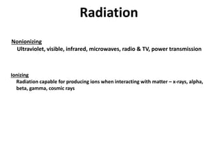
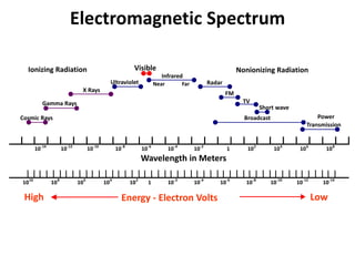
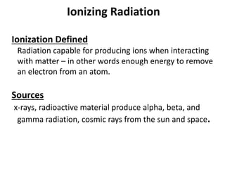
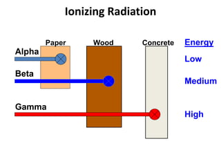


Radiation can be ionizing or non-ionizing. Ionizing radiation like x-rays and gamma rays can damage cells by ionizing them. Non-ionizing radiation like visible light and microwaves do not have enough energy to ionize atoms. The health effects of radiation depend on dose, exposure time, and radiation type. Proper safety protocols aim to keep radiation exposure as low as reasonably achievable.