Downloaded 3,744 times


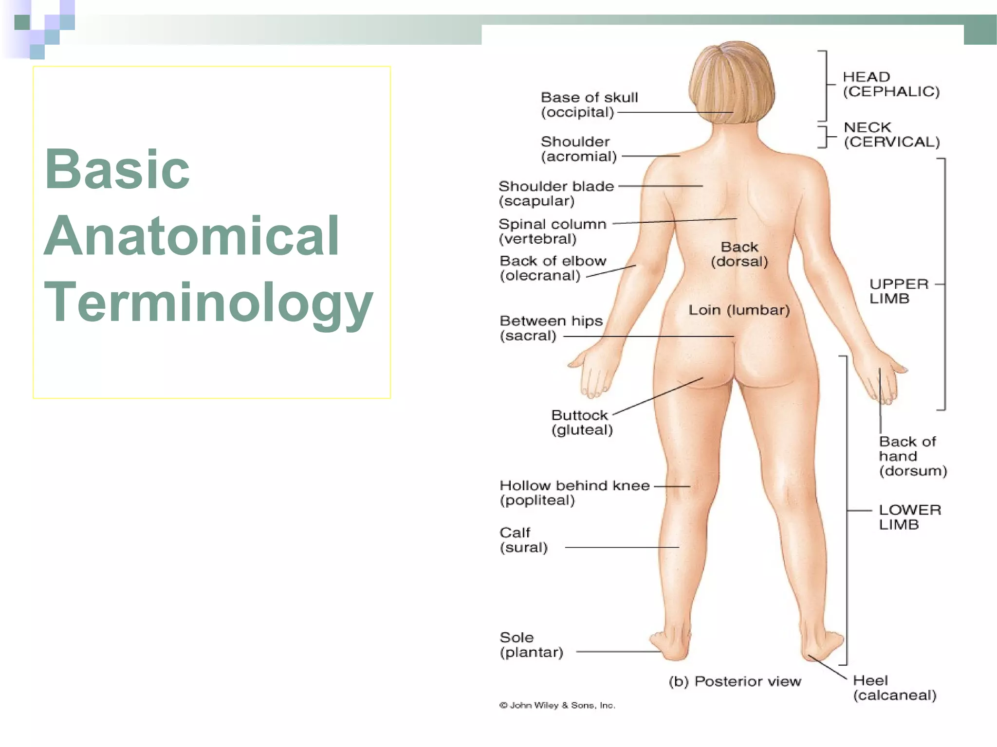
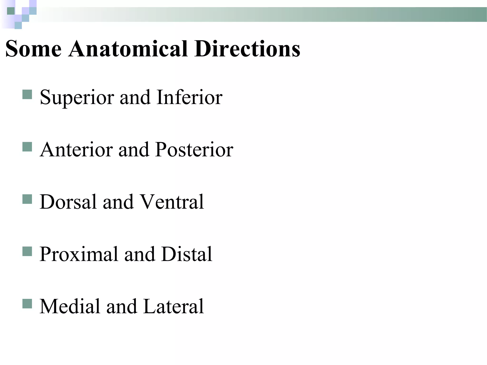


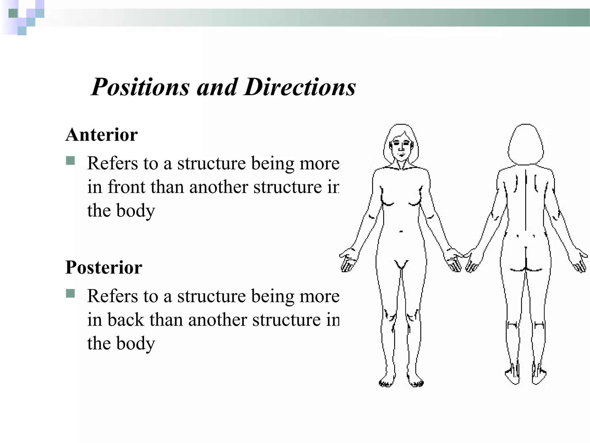


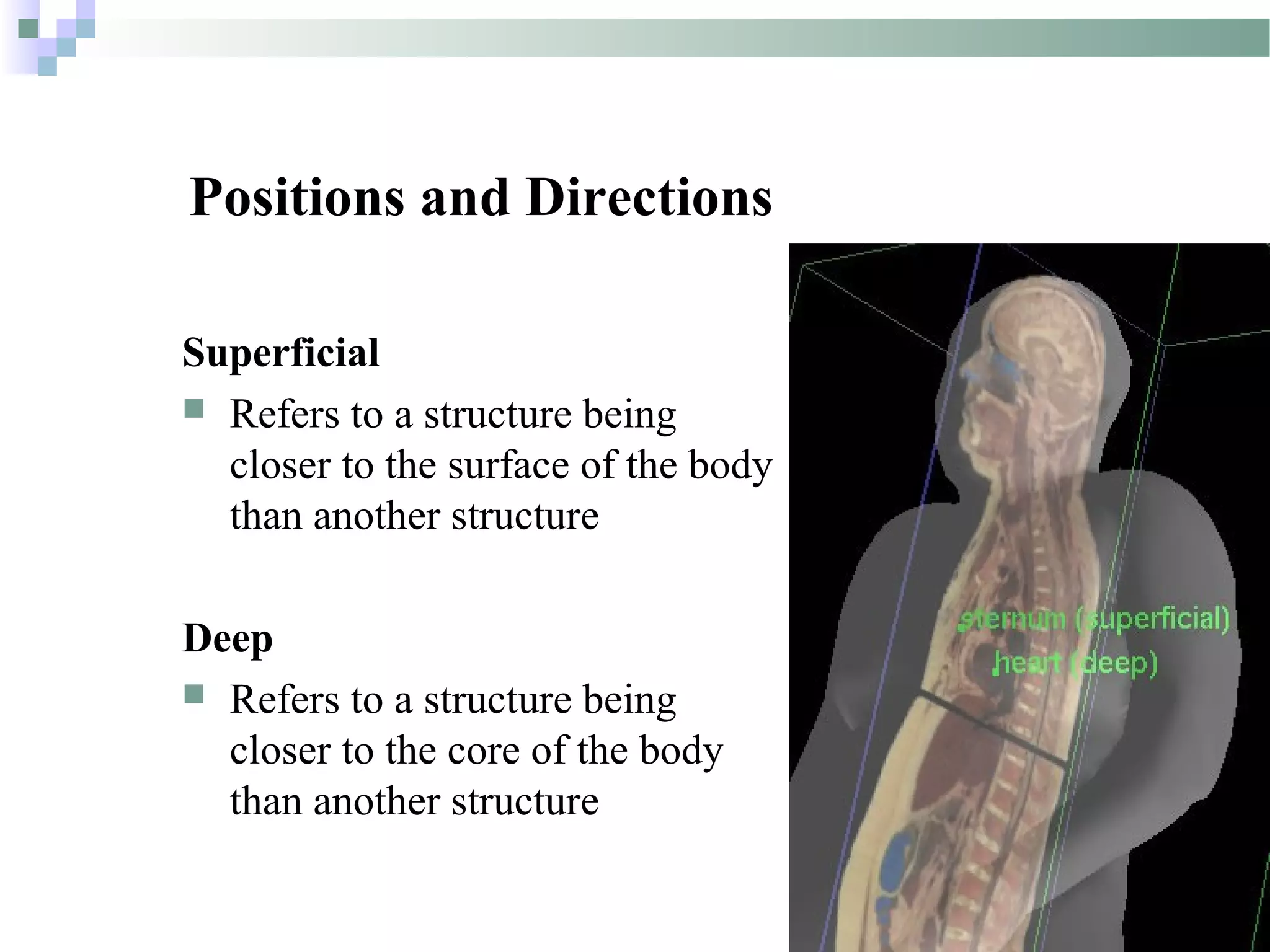
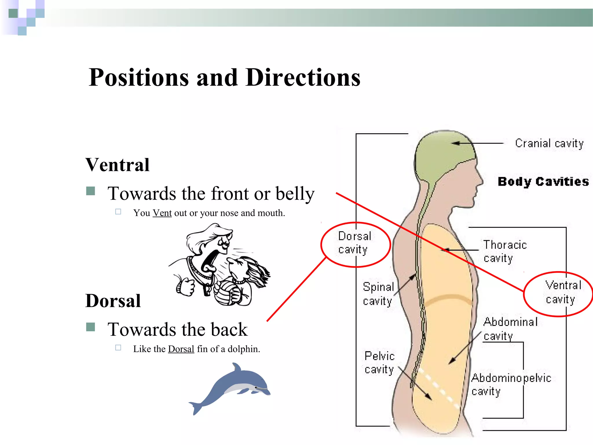
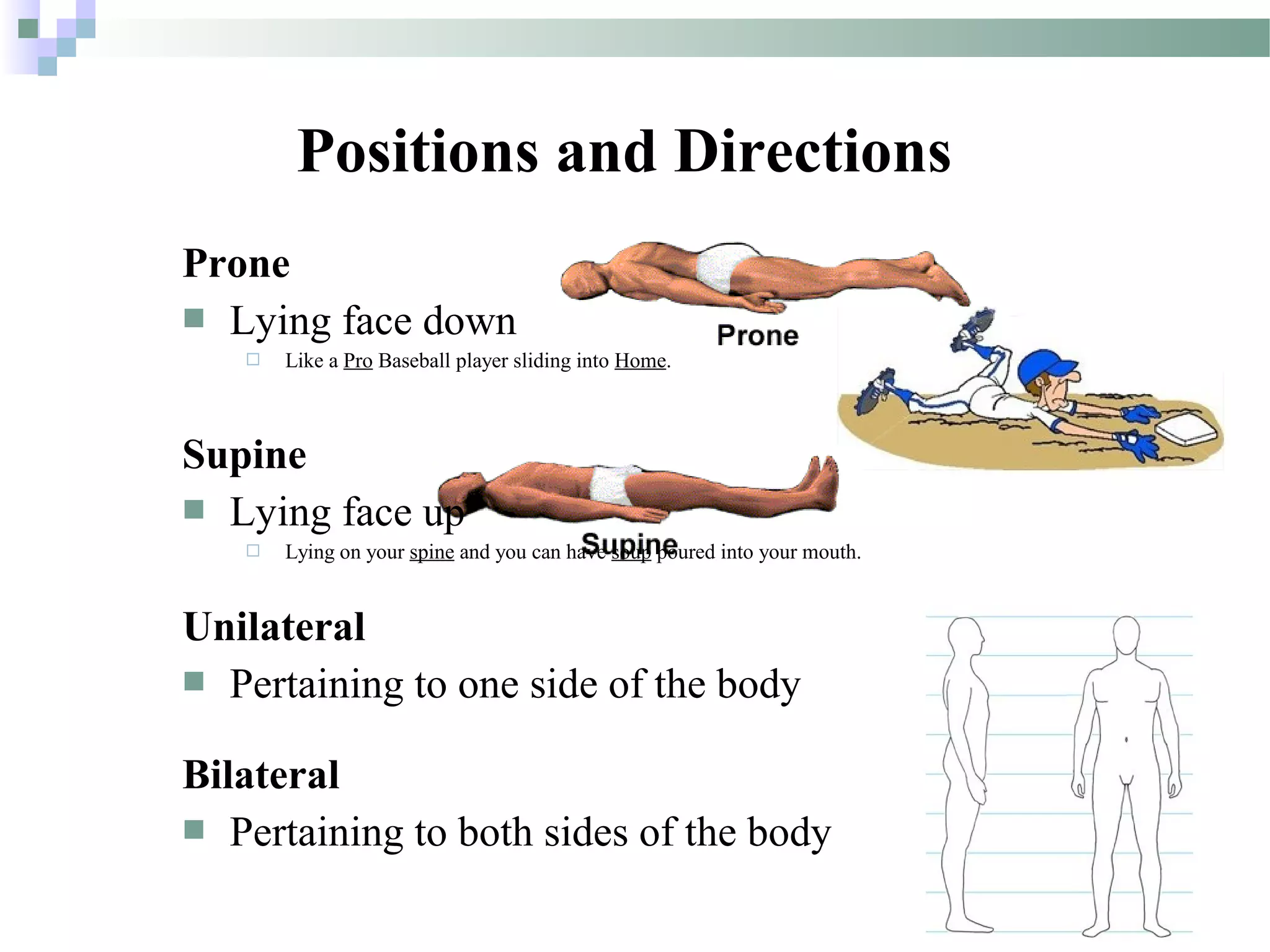


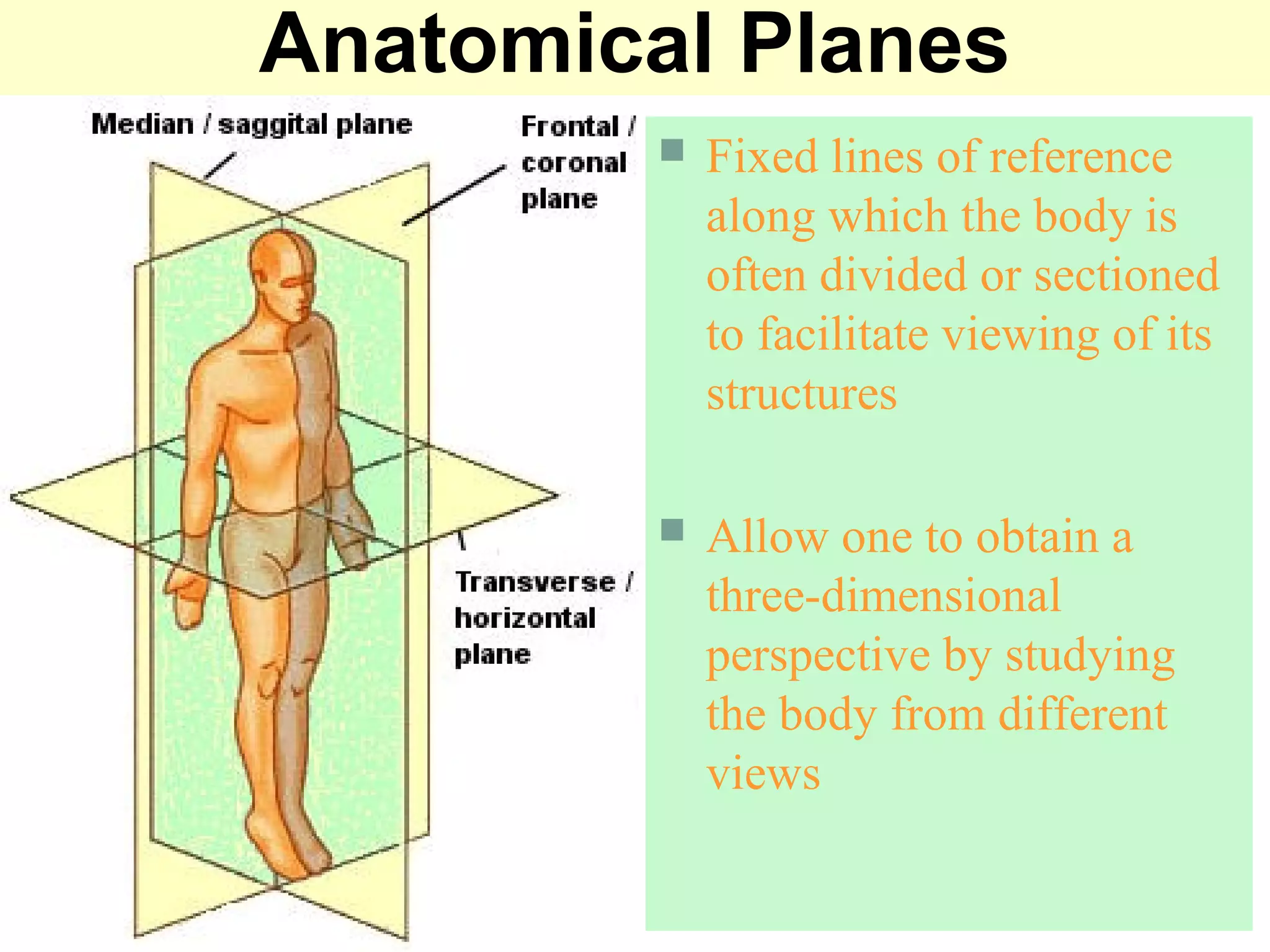
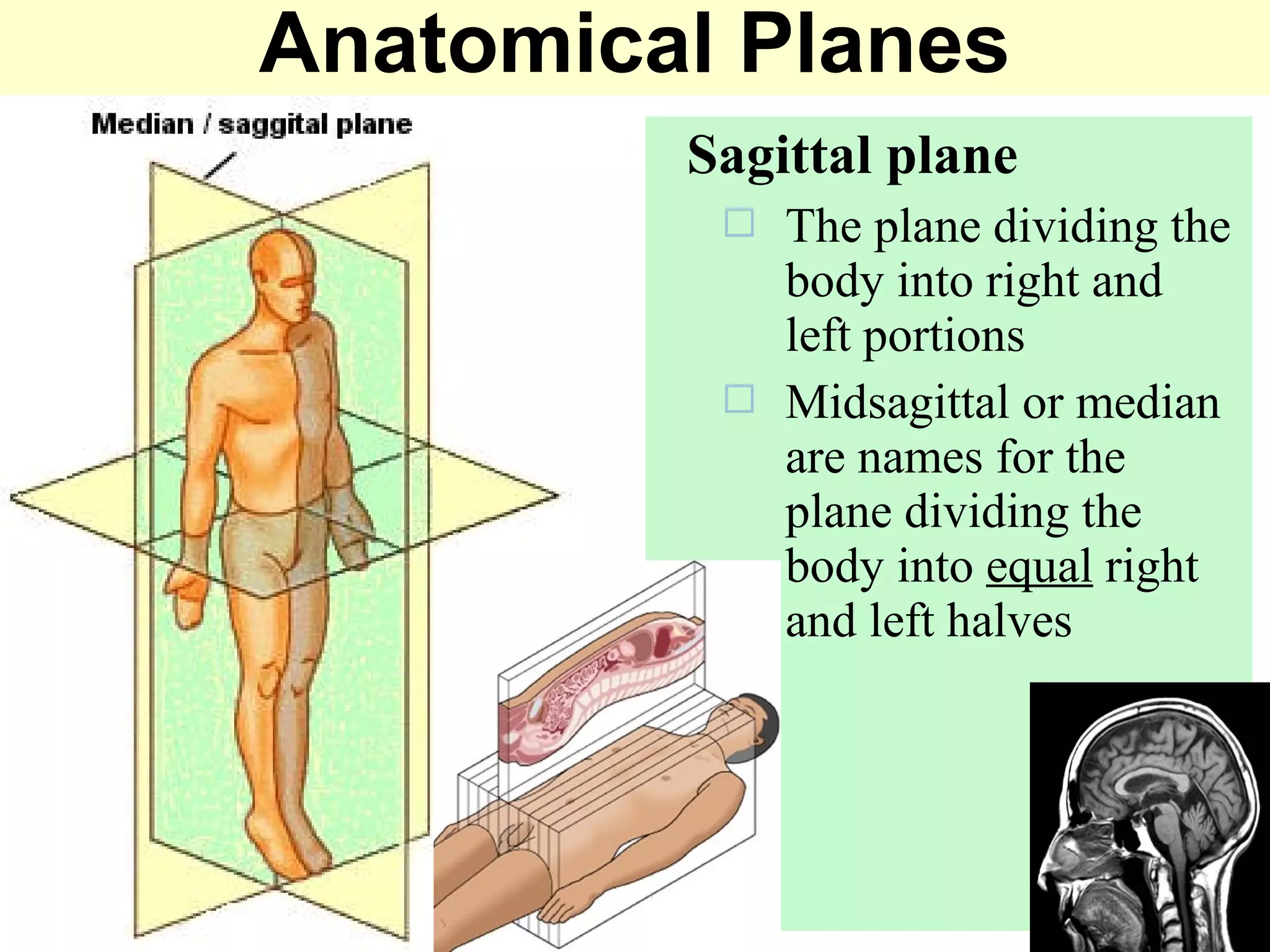

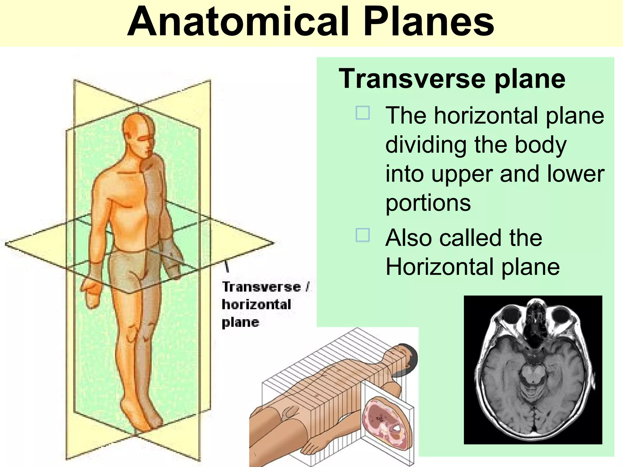


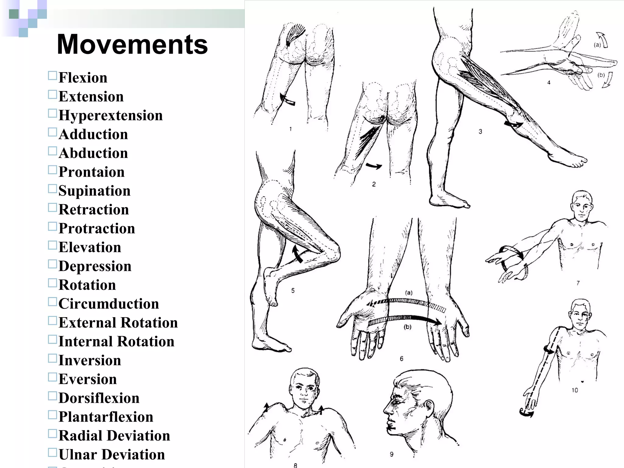
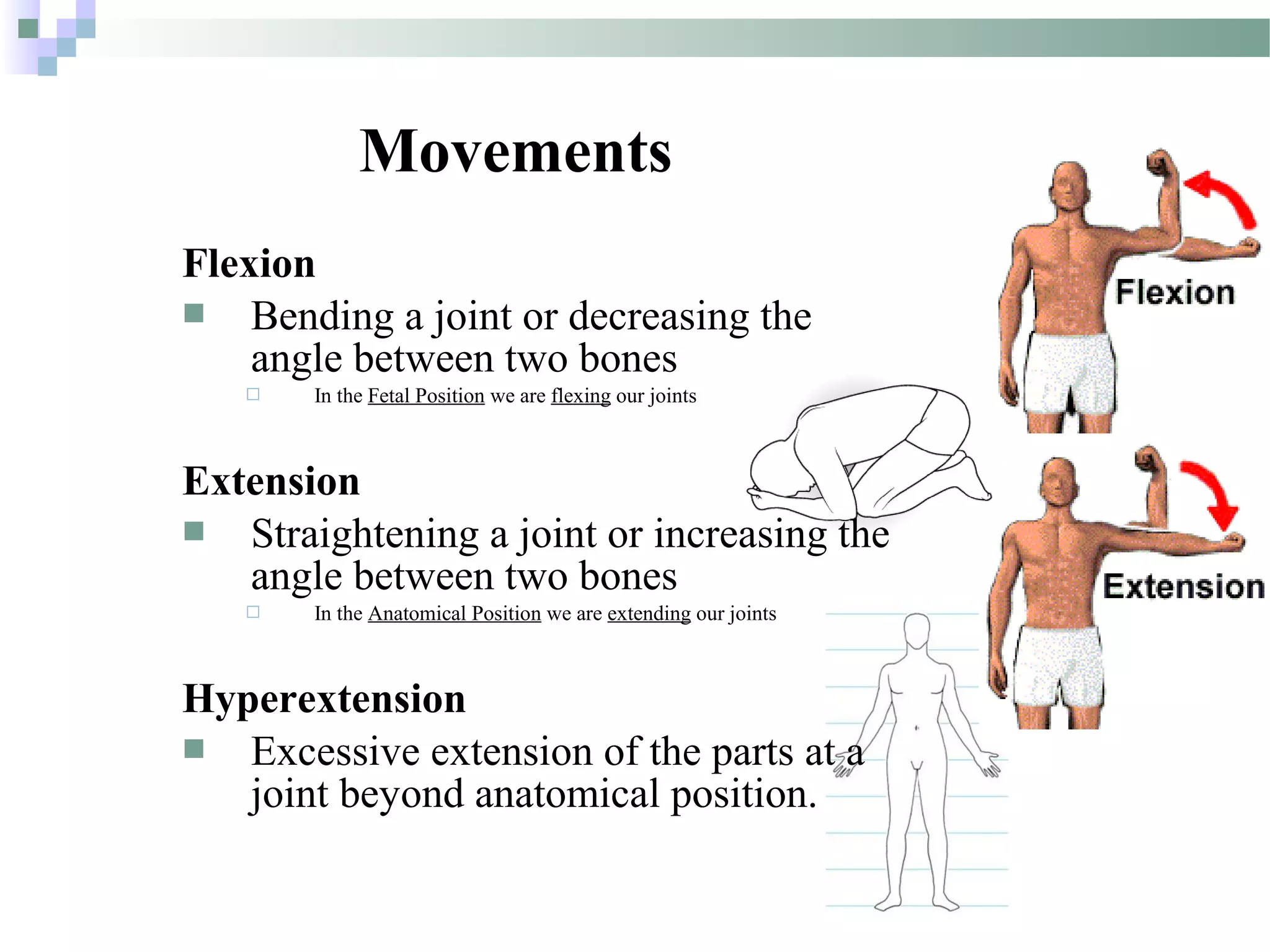

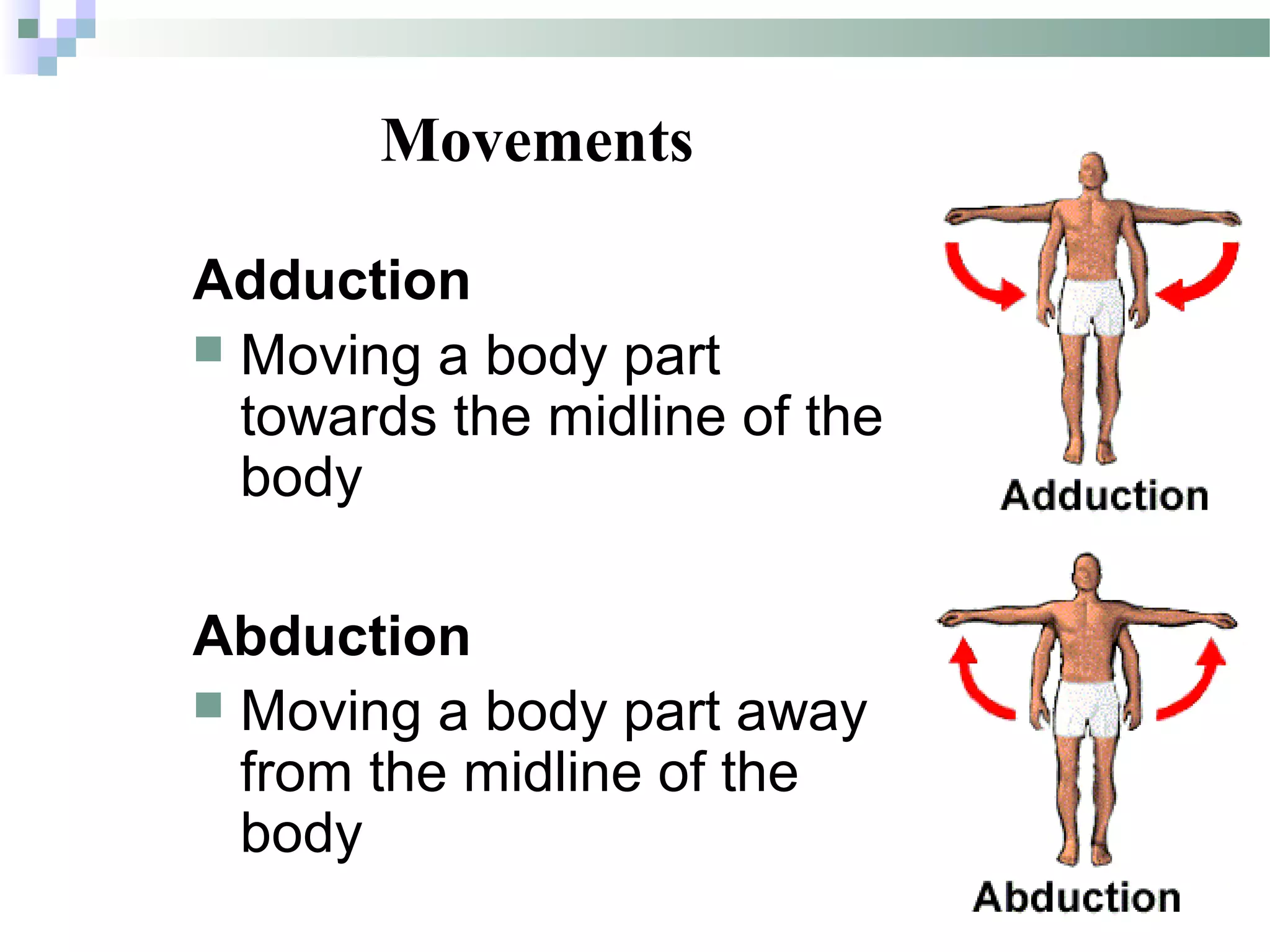
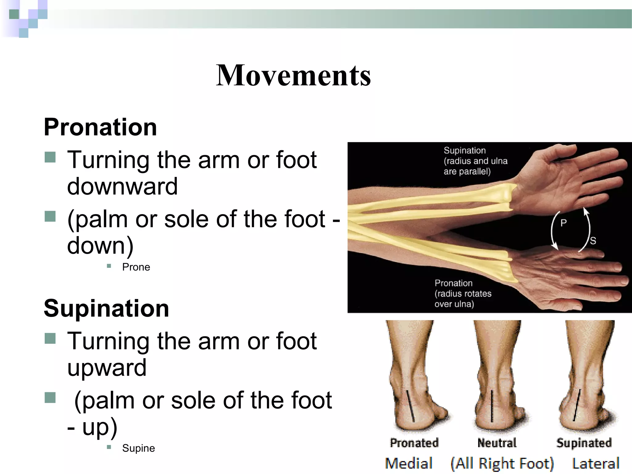
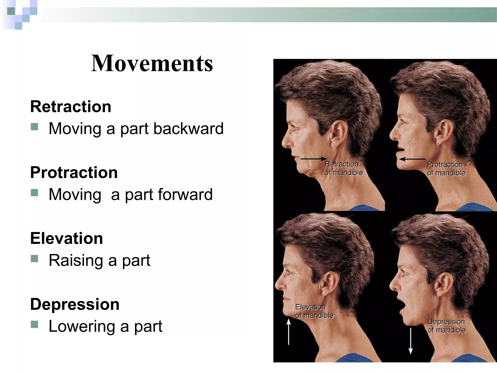
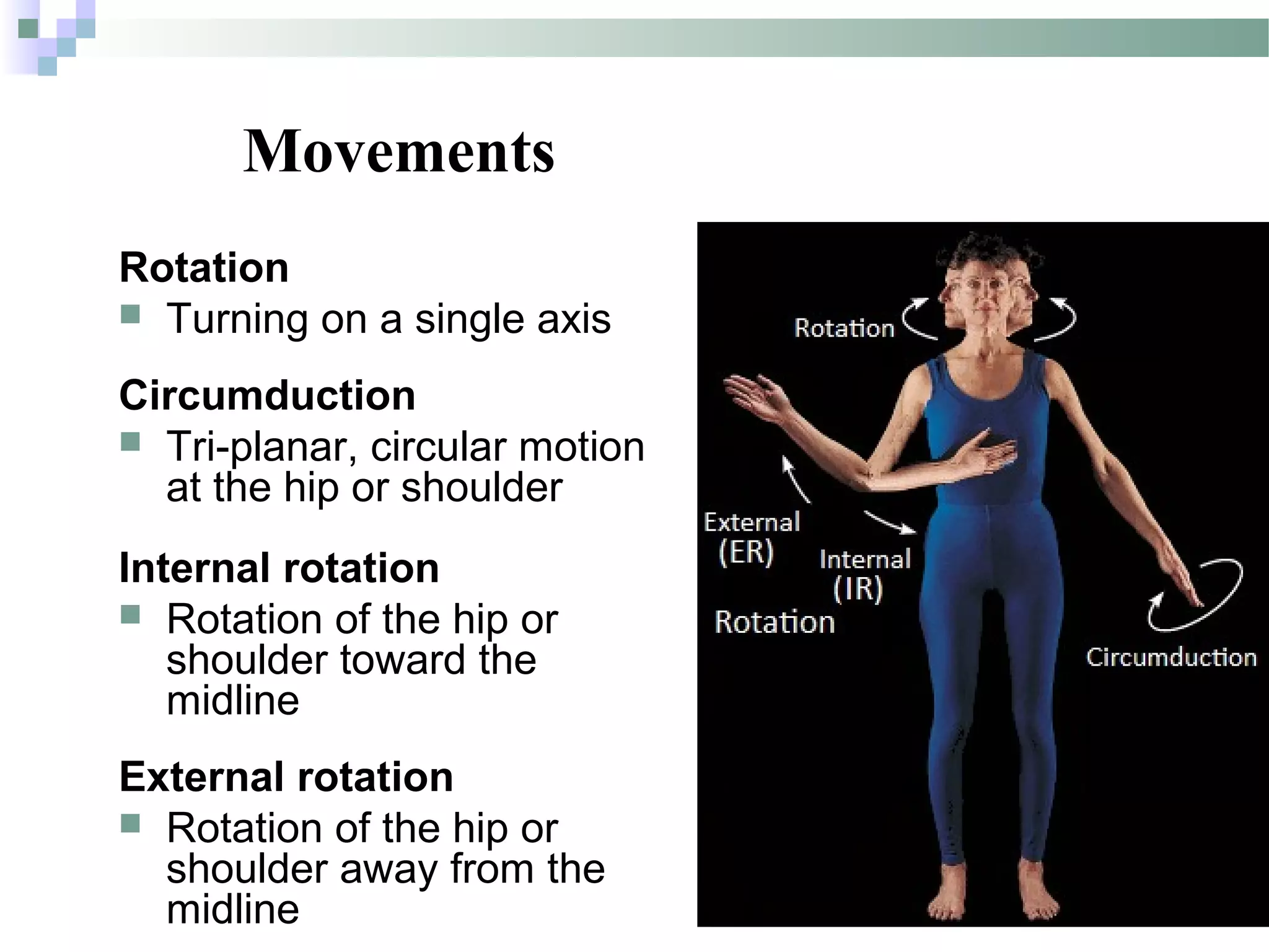


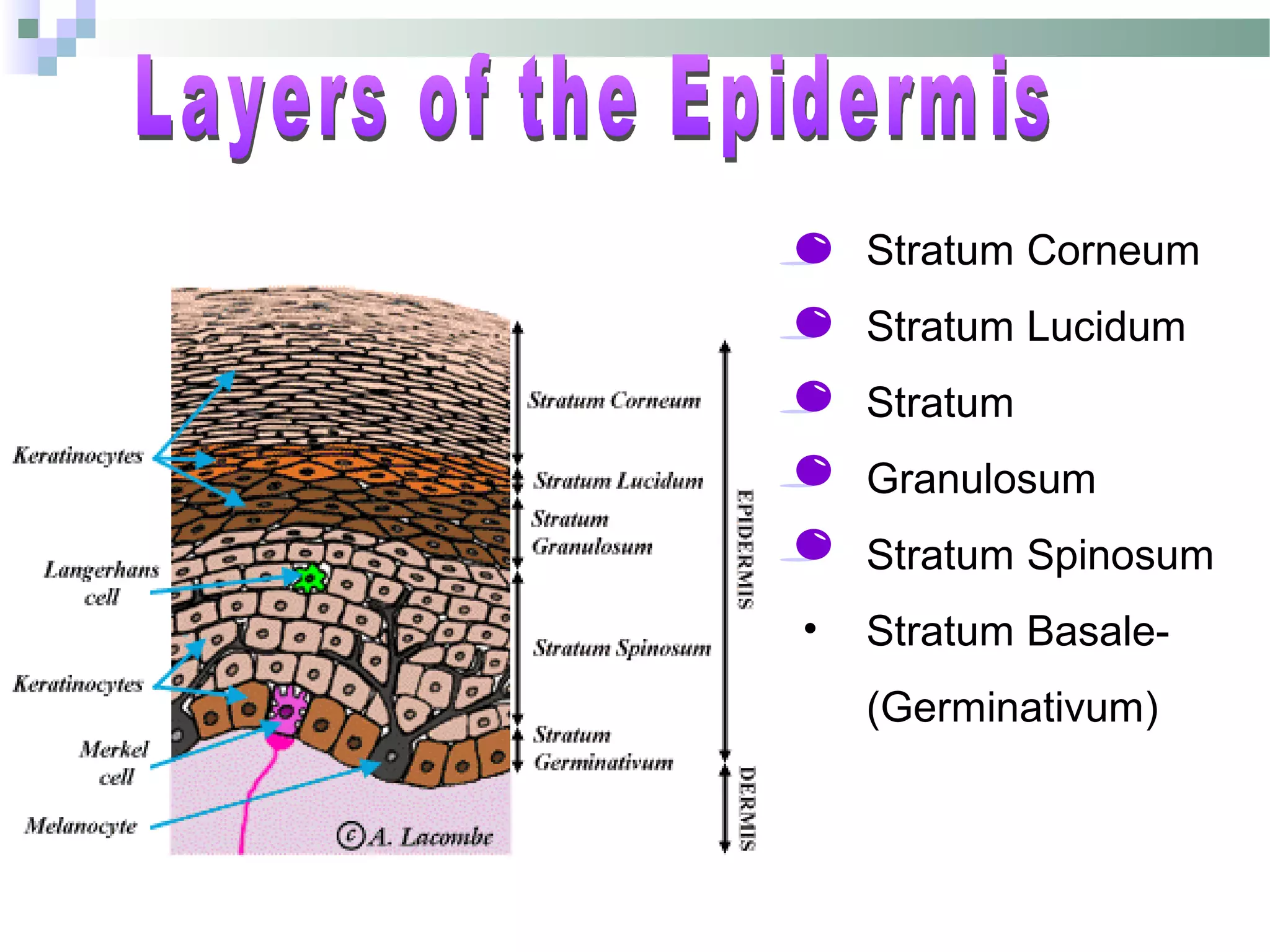


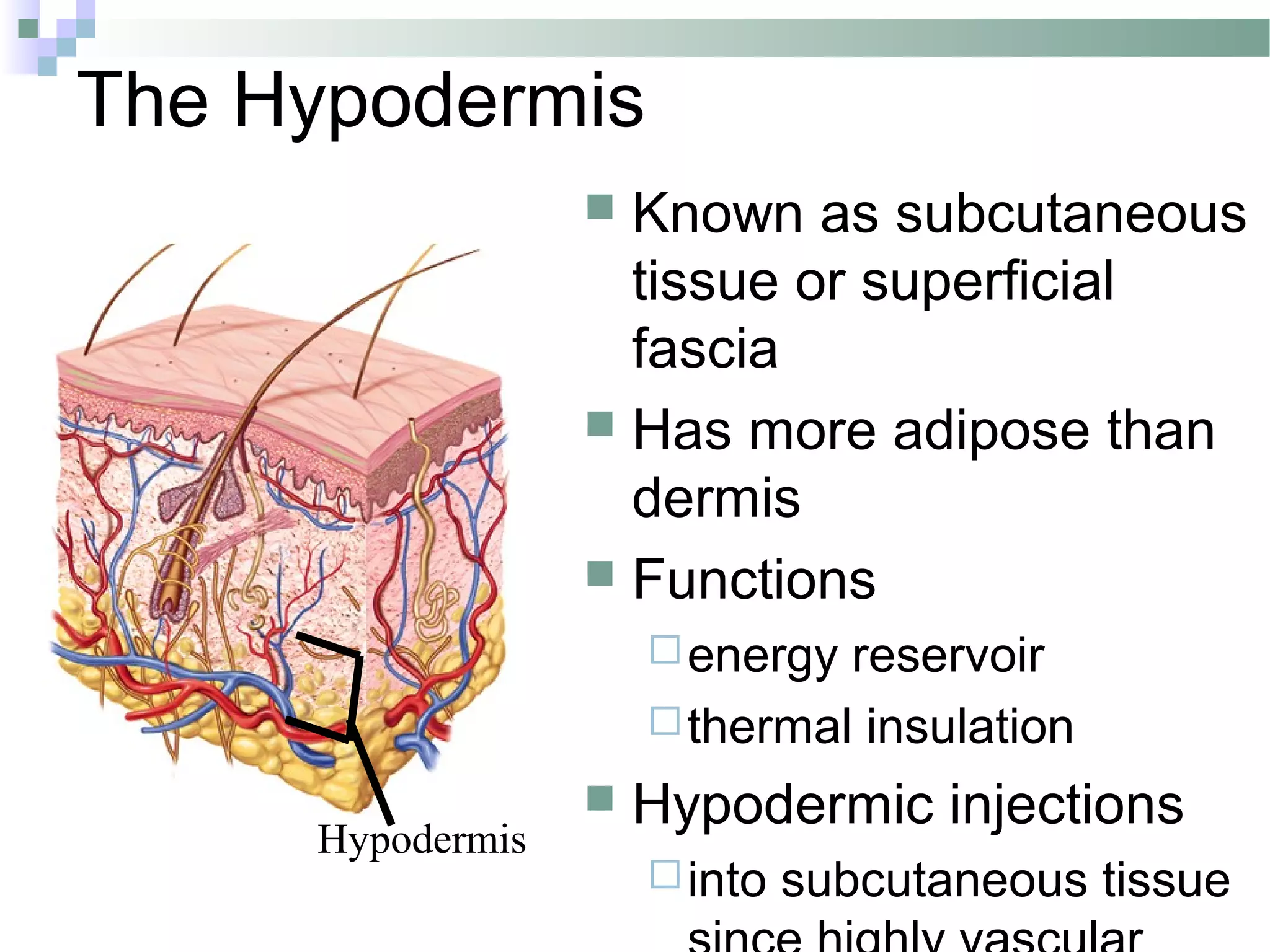


The document provides a comprehensive overview of human anatomy, covering medical terminology, anatomical planes, directions, and movements. It details the structure and function of the integumentary system, including skin layers, hair, nails, and associated glands. Key concepts such as anatomical positions, tissue types, and movements of the body are also explained.