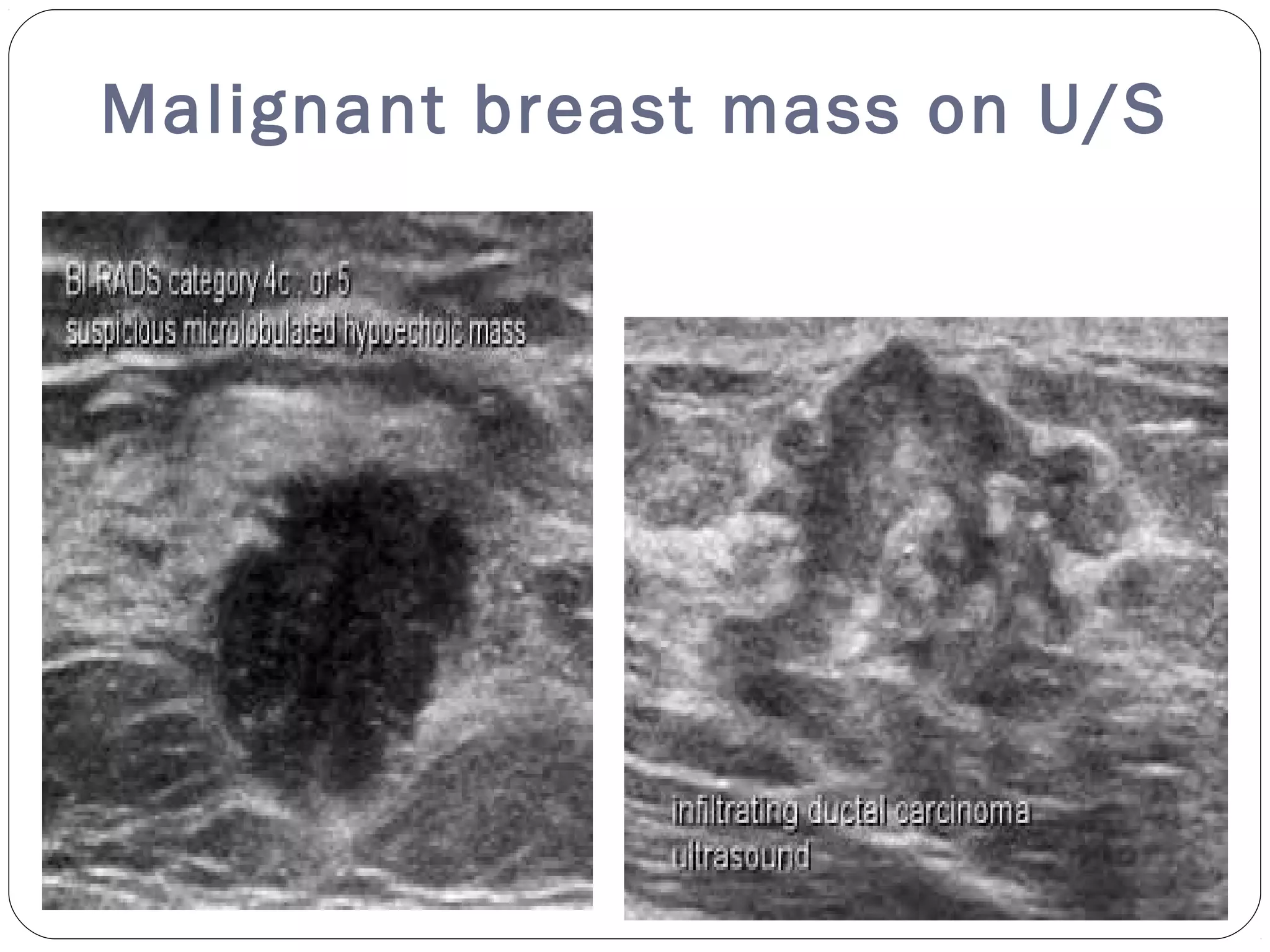
Dr REKHA KHARE
MD. RADIOLOGY
U/S characterisation of breast
mass
Lump in Breast
A lump in the breast is a cause of great concern.
High frequency, high-resolution USG helps in its evaluation.
This is exemplified in women with dense breast tissue where
USG is useful in detecting small breast cancers that are not
seen on mammography
The American College of Radiology has also brought out a
BIRADS-US classification system for categorizing focal breast
lesion
Breast cancer
Breast cancer is among the most common causes of cancer
deaths today, coming fifth after lung, stomach, liver and
colon cancers.
 Refinement of high-frequency technology, particularly with
7.5–13 MHz probes, has brought out a totally new facet in
USG breast imaging
Ultrasound for breast mass
Harmonic imaging and real-time compounding has been shown to
improve image resolution and lesion characterization.
 More recently, USG elastography seems to be quite promising. Initial
results indicate that it can improve the specificity and positive
predictive value of USG in the characterization of breast masses
The reason why any lesion is visible on mammography or USG is the
relative difference in the density and acoustic impedance of the lesion,
respectively, as compared to the surrounding breast tissue.
This is exemplified in women with dense breast tissue, where USG is
useful in detecting small breast cancers that are not detected on
mammography
Indication of breast
ultrasound
Whatever is the indication
Goal of breast u/s is clear
Normal breast parenchymal
patterns
In the young non-lactating breast, the parenchyma is primarily
composed of fibro glandular tissue,
with little or no subcutaneous fat.
With increasing age and parity, more and more fat gets deposited
in both the subcutaneous and retromammary layers
Normal breast parenchyma
U/S in lactating breast
Abnormal breast parenchymal
pattern
Simple cyst
Complex cyst
Chronic abscess
Galactocoel
Fibrocystic disease
Duct ectasia
Fibroadenoma
Cystosarcoma phyllodes
Lipoma
Simple Cyst
Breast cysts are the commonest cause of breast lumps in women
between 35 and 50 years of age.
 A cyst occurs when fluid accumulates due to obstruction of the
extra lobular terminal ducts,
either due to fibrosis or because of intra ductal
epithelial proliferation.
 A cyst is seen on USG as a well-defined, round or oval,
single or multiple anechoic structure
with a thin wall .
Cyst on U/S
Cyst on U/S and
mammogram
Complex cyst
When internal echoes or debris
are seen, the cyst is called a
complex cyst.
 These internal echoes may be
caused by floating cholesterol
crystals, pus, blood or milk of
calcium crystals
Chronic abscess of the
breast
Patients may present with fever, pain, tenderness to touch and
increased white cell count.
Abscesses are most commonly located in the central or subareolar
area.
 An abscess may show an ill-defined or a well-defined
outline. It may be anechoic or may reveal low-level
internal echoes and posterior enhancement
Abscess on U/S
Galactocoel
lacteal cyst or milk cyst
I t is a retention cyst containing milk or a milky substance that is
usually located in the mammary gland
It is caused by a protein plug that blocks off the outlet
It is seen in lactating women on cessation of lactation
Patient typically presents with a painless breast lump occuring
over weeks to month
Galactocoel
internal contents moving with change in position of
breast
Fibrocystic disease
This condition is referred to by many different names:
fibrocystic disease, fibrocystic change, cystic disease, chronic
cystic mastitis or mammary dysplasia.
 About half of these breast masses are usually classified as
indeterminate and will eventually require a biopsy.
Fibrocystic disease on U/S
extremely variable depends on the stage and extent of
morphological changes.
In the early stages, the USG appearance may be normal, even
though lumps may be palpable on clinical examination
There may be focal areas of thickening of the parenchyma, with or
without patchy increase in echogenicity .
Discrete single cysts or clusters of small cysts may be seen in some
 Focal fibrocystic changes may appear as solid masses or thin-
walled cysts.
Fibrocystic disease on U/S
Duct ectasia
This lesion has a variable appearance.
Typically, duct ectasia may appear as a single or
multiple tubular structure filled with fluid
 Old cellular debris may appear as echogenic content. If the debris
fills the lumen, it can be sometimes mistaken for a solid mass,
unless the tubular shape is identified
Ductal Ectasia on U/S
Fibroadenoma
Fibroadenoma is an estrogen-induced tumor that forms in
adolescence. It is the third most common breast lesion after
fibrocystic disease and carcinoma
 It usually presents as a firm, smooth, oval-shaped, freely movable
mass ( breast mouse) on palpation.
 It is rarely tender or painful. The size is usually under 5 cm,
though larger fibroadenomas are known. Fibroadenomas are
multiple in 10–20% and bilateral in 4% of cases. Calcifications
may occur.
Fibroadenoma on U/S
A well-defined lesion. A capsule can usually be identified.
The echotexture is usually homogenous and hypoechoic as
compared to the breast parenchyma, and there may be low-level
internal echoes
 Typically, the transverse diameter is greater than the antero-
posterior diameter
Fibroadenoma on U/S and
Mammography
Cystosarcoma phyllodes
This is a large lesion that presents in older women
Some authors consider it to be a giant fibroadenoma
The mass may involve the whole of the breast. It usually
reveals well-defined margins and an inhomogeneous
echo texture, sometimes with variable cystic areas.
C.Phylloides on U/S
Lipoma in breast parenchyma
Lipoma is a slow-growing, well-defined tumor.
It may be a chance finding or the patient may present with
complaints of increase in the size of the involved breast, though no
discretely palpable mass can be made out.
 The tumor is soft and can be deformed by compression
with the transducer.
Lipoma on U/S and
Mammography
 A thin capsulated
echo genic mass
with a stippled or
lamellar
appearance
Breast ultrasound
criteria for benign lesions
Smooth and well circumscribed
Hyper echoic, iso echoic or mildly hypo echoic
Thin echogenic capsule
Ellipsoid shape, with the maximum diameter being in the
transverse plane
Three or fewer gentle lobulations
Absence of any malignant findings
Breast ultrasound
criteria for malignant lesion
Malignant lesions are commonly hypo echoic nodular lesions with ill-
defined borders, which is ‘taller than broader’ and has spiculated
margins, posterior acoustic shadowing and micro calcifications
 Three-dimensional scanners with the capability of reproducing high-
resolution images in the coronal plane provide additional important
information.
 It was initially believed that color Doppler scanning would add to the
specificity of USG examination, but this has not proven to be very
efficacious
Malignant breast mass on U/S
Benign versus malignant
Gynecomastia
enlarged male breast tissue due to hormonal
imbalance
It could be uni or
bilateral
Oestrogen and
Testosterone are
not adequately
balance
Discussion
Although it may be impossible to distinguish
all benign from all malignant solid breast nodules
using USG criteria, a reasonable goal for
breast USG is to identify a subgroup of solid nodules
that has such a low risk of being malignant that
the option of short-interval follow-up can be
offered as a viable alternative to biopsy.
Combined studies
Combined studies, which included USG and mammography, have
demonstrated nearly 100% negative predictive value for palpable
breast lesions, when both are used together.
In a study based on characterization of breast masses according to
BIRADS-US criteria, people have
found no statistical differences between fine-needle aspiration cytology
and USG with regard to sensitivity and Negative Predictive value (P >
0.05).
It is also found USG characterization of breast lesions using BIRADS-
US criteria to be highly accurate.
Have a nice time
Dr Rekha Khare
Thank you

Ultrasound breast mass

  • 1.
    Dr REKHA KHARE MD.RADIOLOGY U/S characterisation of breast mass
  • 2.
    Lump in Breast Alump in the breast is a cause of great concern. High frequency, high-resolution USG helps in its evaluation. This is exemplified in women with dense breast tissue where USG is useful in detecting small breast cancers that are not seen on mammography The American College of Radiology has also brought out a BIRADS-US classification system for categorizing focal breast lesion
  • 3.
    Breast cancer Breast canceris among the most common causes of cancer deaths today, coming fifth after lung, stomach, liver and colon cancers.  Refinement of high-frequency technology, particularly with 7.5–13 MHz probes, has brought out a totally new facet in USG breast imaging
  • 4.
    Ultrasound for breastmass Harmonic imaging and real-time compounding has been shown to improve image resolution and lesion characterization.  More recently, USG elastography seems to be quite promising. Initial results indicate that it can improve the specificity and positive predictive value of USG in the characterization of breast masses The reason why any lesion is visible on mammography or USG is the relative difference in the density and acoustic impedance of the lesion, respectively, as compared to the surrounding breast tissue. This is exemplified in women with dense breast tissue, where USG is useful in detecting small breast cancers that are not detected on mammography
  • 5.
  • 6.
    Whatever is theindication Goal of breast u/s is clear
  • 7.
    Normal breast parenchymal patterns Inthe young non-lactating breast, the parenchyma is primarily composed of fibro glandular tissue, with little or no subcutaneous fat. With increasing age and parity, more and more fat gets deposited in both the subcutaneous and retromammary layers
  • 8.
  • 9.
  • 10.
    Abnormal breast parenchymal pattern Simplecyst Complex cyst Chronic abscess Galactocoel Fibrocystic disease Duct ectasia Fibroadenoma Cystosarcoma phyllodes Lipoma
  • 11.
    Simple Cyst Breast cystsare the commonest cause of breast lumps in women between 35 and 50 years of age.  A cyst occurs when fluid accumulates due to obstruction of the extra lobular terminal ducts, either due to fibrosis or because of intra ductal epithelial proliferation.  A cyst is seen on USG as a well-defined, round or oval, single or multiple anechoic structure with a thin wall .
  • 12.
  • 13.
    Cyst on U/Sand mammogram
  • 14.
    Complex cyst When internalechoes or debris are seen, the cyst is called a complex cyst.  These internal echoes may be caused by floating cholesterol crystals, pus, blood or milk of calcium crystals
  • 15.
    Chronic abscess ofthe breast Patients may present with fever, pain, tenderness to touch and increased white cell count. Abscesses are most commonly located in the central or subareolar area.  An abscess may show an ill-defined or a well-defined outline. It may be anechoic or may reveal low-level internal echoes and posterior enhancement
  • 16.
  • 17.
    Galactocoel lacteal cyst ormilk cyst I t is a retention cyst containing milk or a milky substance that is usually located in the mammary gland It is caused by a protein plug that blocks off the outlet It is seen in lactating women on cessation of lactation Patient typically presents with a painless breast lump occuring over weeks to month
  • 18.
    Galactocoel internal contents movingwith change in position of breast
  • 19.
    Fibrocystic disease This conditionis referred to by many different names: fibrocystic disease, fibrocystic change, cystic disease, chronic cystic mastitis or mammary dysplasia.  About half of these breast masses are usually classified as indeterminate and will eventually require a biopsy.
  • 20.
    Fibrocystic disease onU/S extremely variable depends on the stage and extent of morphological changes. In the early stages, the USG appearance may be normal, even though lumps may be palpable on clinical examination There may be focal areas of thickening of the parenchyma, with or without patchy increase in echogenicity . Discrete single cysts or clusters of small cysts may be seen in some  Focal fibrocystic changes may appear as solid masses or thin- walled cysts.
  • 21.
  • 22.
    Duct ectasia This lesionhas a variable appearance. Typically, duct ectasia may appear as a single or multiple tubular structure filled with fluid  Old cellular debris may appear as echogenic content. If the debris fills the lumen, it can be sometimes mistaken for a solid mass, unless the tubular shape is identified
  • 23.
  • 24.
    Fibroadenoma Fibroadenoma is anestrogen-induced tumor that forms in adolescence. It is the third most common breast lesion after fibrocystic disease and carcinoma  It usually presents as a firm, smooth, oval-shaped, freely movable mass ( breast mouse) on palpation.  It is rarely tender or painful. The size is usually under 5 cm, though larger fibroadenomas are known. Fibroadenomas are multiple in 10–20% and bilateral in 4% of cases. Calcifications may occur.
  • 25.
    Fibroadenoma on U/S Awell-defined lesion. A capsule can usually be identified. The echotexture is usually homogenous and hypoechoic as compared to the breast parenchyma, and there may be low-level internal echoes  Typically, the transverse diameter is greater than the antero- posterior diameter
  • 26.
    Fibroadenoma on U/Sand Mammography
  • 27.
    Cystosarcoma phyllodes This isa large lesion that presents in older women Some authors consider it to be a giant fibroadenoma The mass may involve the whole of the breast. It usually reveals well-defined margins and an inhomogeneous echo texture, sometimes with variable cystic areas.
  • 28.
  • 29.
    Lipoma in breastparenchyma Lipoma is a slow-growing, well-defined tumor. It may be a chance finding or the patient may present with complaints of increase in the size of the involved breast, though no discretely palpable mass can be made out.  The tumor is soft and can be deformed by compression with the transducer.
  • 30.
    Lipoma on U/Sand Mammography  A thin capsulated echo genic mass with a stippled or lamellar appearance
  • 31.
    Breast ultrasound criteria forbenign lesions Smooth and well circumscribed Hyper echoic, iso echoic or mildly hypo echoic Thin echogenic capsule Ellipsoid shape, with the maximum diameter being in the transverse plane Three or fewer gentle lobulations Absence of any malignant findings
  • 32.
    Breast ultrasound criteria formalignant lesion Malignant lesions are commonly hypo echoic nodular lesions with ill- defined borders, which is ‘taller than broader’ and has spiculated margins, posterior acoustic shadowing and micro calcifications  Three-dimensional scanners with the capability of reproducing high- resolution images in the coronal plane provide additional important information.  It was initially believed that color Doppler scanning would add to the specificity of USG examination, but this has not proven to be very efficacious
  • 33.
  • 34.
  • 35.
    Gynecomastia enlarged male breasttissue due to hormonal imbalance It could be uni or bilateral Oestrogen and Testosterone are not adequately balance
  • 36.
    Discussion Although it maybe impossible to distinguish all benign from all malignant solid breast nodules using USG criteria, a reasonable goal for breast USG is to identify a subgroup of solid nodules that has such a low risk of being malignant that the option of short-interval follow-up can be offered as a viable alternative to biopsy.
  • 37.
    Combined studies Combined studies,which included USG and mammography, have demonstrated nearly 100% negative predictive value for palpable breast lesions, when both are used together. In a study based on characterization of breast masses according to BIRADS-US criteria, people have found no statistical differences between fine-needle aspiration cytology and USG with regard to sensitivity and Negative Predictive value (P > 0.05). It is also found USG characterization of breast lesions using BIRADS- US criteria to be highly accurate.
  • 38.
    Have a nicetime Dr Rekha Khare Thank you