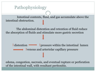
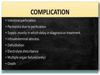
1. P R E P A R E D B Y
M E H W I S H J A M I L
Intestinal obstruction
2. Objectives
At the of this lecture students will be able to:
Define intestinal obstructions.
Explain pathophysiology of intestinal obstruction.
Explain risk factors of intestinal obstruction.
Enlist sign and symptom of intestinal obstruction.
Explain complications of intestinal obstruction.
Describe assessment and diagnostic findings of intestinal
obstructions.
Explain medical and surgical interventions of intestinal
obstruction.
4. Introduction
Two types of processes can impede this flow:
• Mechanical obstruction: An intraluminal obstruction
or a mural obstruction from pressure on the intestinal
wall occurs.
Examples are:
intussusception, polypoid tumors and neoplasms,
stenosis, strictures, adhesions, hernias, and abscesses.
5. Conti…
• Functional obstruction:
The intestinal musculature cannot propel the
contents along the bowel.
Examples are :
amyloidosis, muscular dystrophy, endocrine disorders
such as diabetes mellitus, or neurologic disorders such as
Parkinson’s disease.
The blockage also can be temporary and the result of the
manipulation of the bowel during surgery.
6.
7. Pathophysiology
Intestinal contents, fluid, and gas accumulate above the
intestinal obstruction.
The abdominal distention and retention of fluid reduce
the absorption of fluids and stimulate more gastric secretion
distention pressure within the intestinal lumen
venous and arteriolar capillary pressure
edema, congestion, necrosis, and eventual rupture or perforation
of the intestinal wall, with resultant peritonitis.
8. Causes
Mechanical obstructions are when something physically
blocks your intestine. In the small intestine, this can be due
to:
• adhesions, which are made of fibrous tissue that can
develop after any abdominal or pelvic surgery or after
severe inflammation
• volvulus, or twisting of the intestines
• intussusception, a “telescoping,” or pushing, of one
segment of intestine into the next section
• malformations of the intestine, often in newborns, but can
also occur in children and teens
• tumors within your small intestine
9. Conti…
• tumors within your small intestine
• gallstones, although they rarely cause obstructions
• swallowed objects, especially in children
• hernias, which involve a portion of your intestine pushing
outside the muscle or tissue in your body or into another
part of your body
• inflammatory bowel disease, such as Crohn’s disease
10. Conti…
Causes for an ileus include:
• abdominal or pelvic surgery
• infections, such as gastroenteritis or appendicitis
• some medications, including opioid pain medications
• electrolyte imbalances
Intestinal pseudo-obstruction can be caused by:
• Parkinson’s disease, multiple sclerosis, and other nerve and
muscle disorders
• Hirschsprung’s disease, a disorder in which there’s a lack of
nerves in sections of the large intestine
• disorders that cause nerve injury, such as diabetes mellitus
• hypothyroidism, or an underactive thyroid gland
11. Sign and Symptom
• severe bloating
• abdominal pain The initial symptom is usually crampy pain that
is wavelike and colicky.
• decreased appetite
• nausea
• Vomiting If the obstruction is complete, the peristaltic waves
initially become extremely vigorous and eventually assume a
reverse direction, with the intestinal contents propelled toward
the mouth instead of toward the rectum.
• inability to pass gas or stool
• constipation
• diarrhea
• severe abdominal cramps
• abdominal swelling
12. Conti…
First, the patient vomits the stomach contents, then the bile
stained contents of the duodenum and the jejunum, and
finally, with each paroxysm of pain, the darker, fecal-like
contents of the ileum.
The signs of dehydration become evident:
intense thirst
drowsiness
generalized malaise
aching
a parched tongue
mucous membranes
13. Complications
Treatment is essential to reduce complications such as:
• dehydration
• electrolyte imbalances
• perforation, or a hole that forms in your intestines, which leads to
infections
• kidney failure
If the obstruction is preventing blood from getting to a segment of
intestine, this can lead to:
• infection
• tissue death
• intestinal perforation
• sepsis, a life threatening blood infection
• multiple organ failure
• death
14. Assessment and Diagnosis
Abdominal x-ray and CT findings include abnormal
quantities of gas, fluid, or both in the intestines.
Laboratory studies (i.e., electrolyte studies and a complete
blood cell count) reveal a picture of dehydration, loss of
plasma volume, and possible infection
• colonoscopy, a flexible tube with a light that your doctor
uses to look at your large intestine
• enema with contrast
15. Medical Management
Decompression of the bowel through a nasogastric tube
Surgery
• antibiotics to reduce infection
• anti-nausea medicines to keep you from vomiting
• pain relievers
16.
17. Nursing Interventions
Nursing management of the nonsurgical patient with a
small bowel obstruction includes maintaining the function
of the nasogastric tube, assessing and measuring the
nasogastric output, assessing for fluid and electrolyte
imbalance, monitoring nutritional status, and assessing
improvement.
The nurse reports discrepancies in intake and output,
worsening of pain or abdominal distention, and increased
nasogastric output.
If the patient’s condition does not improve, the nurse
prepares him or her for surgery. Nursing care of the patient
after surgical repair of a small bowel obstruction is similar
to that for other abdominal surgeries.
18. Large Bowel Obstruction
large bowel obstruction results in an
accumulation of intestinal contents, fluid, and gas
proximal to the obstruction. It can lead to severe
distention and perforation unless some gas and fluid
can flow back through the ileal valve.
19. Conti…
Large bowel obstruction, even if complete, may be
undramatic if the blood supply to the colon is not
disturbed.
if the blood supply is cut off, intestinal strangulation and
necrosis occur; this condition is life-threatening.
In the large intestine, dehydration occurs more slowly than
in the small intestine because the colon can absorb its fluid
contents and can distend to a size considerably beyond its
normal full capacity.
20. causes / Sign and Symptom
Adenocarcinoid tumors
diverticulitis. Sigmoid volvulus and cecal volvulus
Sign and Symptom:
In patients with obstruction in the sigmoid colon or the rectum,
constipation may be the only symptom for months.
shape of the stool is altered
Blood loss in the stool may result in iron deficiency anemia.
weakness
weight loss
anorexia
Abdominal distention
Crampy lower abdominal pain
fecal vomiting develops
21.
22. Diagnostic Findings
Abdominal x-ray and abdominal CT or MRI findings
reveal a distended colon and pinpoint the site of the
obstruction.
Barium studies are contraindicated.
23. Medical Treatment
Restoration of intravascular volume
correction of electrolyte abnormalities
nasogastric aspiration and decompression
Colonoscopy
Cecostomy
A rectal tube may be used to decompress an area that is
lower in the bowel.
surgical resection
temporary or permanent colostomy
ileoanal anastomosis
24. Nursing Intervention
The nurse’s role is to monitor the patient for symptoms that
indicate that the intestinal obstruction is worsening and to
provide emotional support and comfort.
The nurse administers IV fluids and electrolytes as
prescribed.
If the patient’s condition does not respond to nonsurgical
treatment, the nurse prepares the patient for surgery.
This preparation includes preoperative teaching as the
patient’s condition indicates.
After surgery, general abdominal wound care and routine
postoperative nursing care are provided.