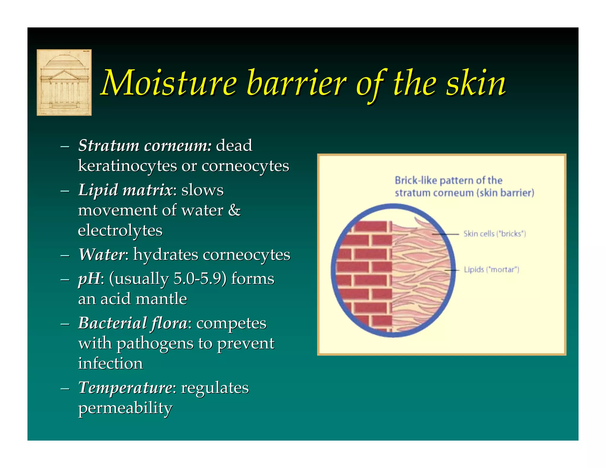
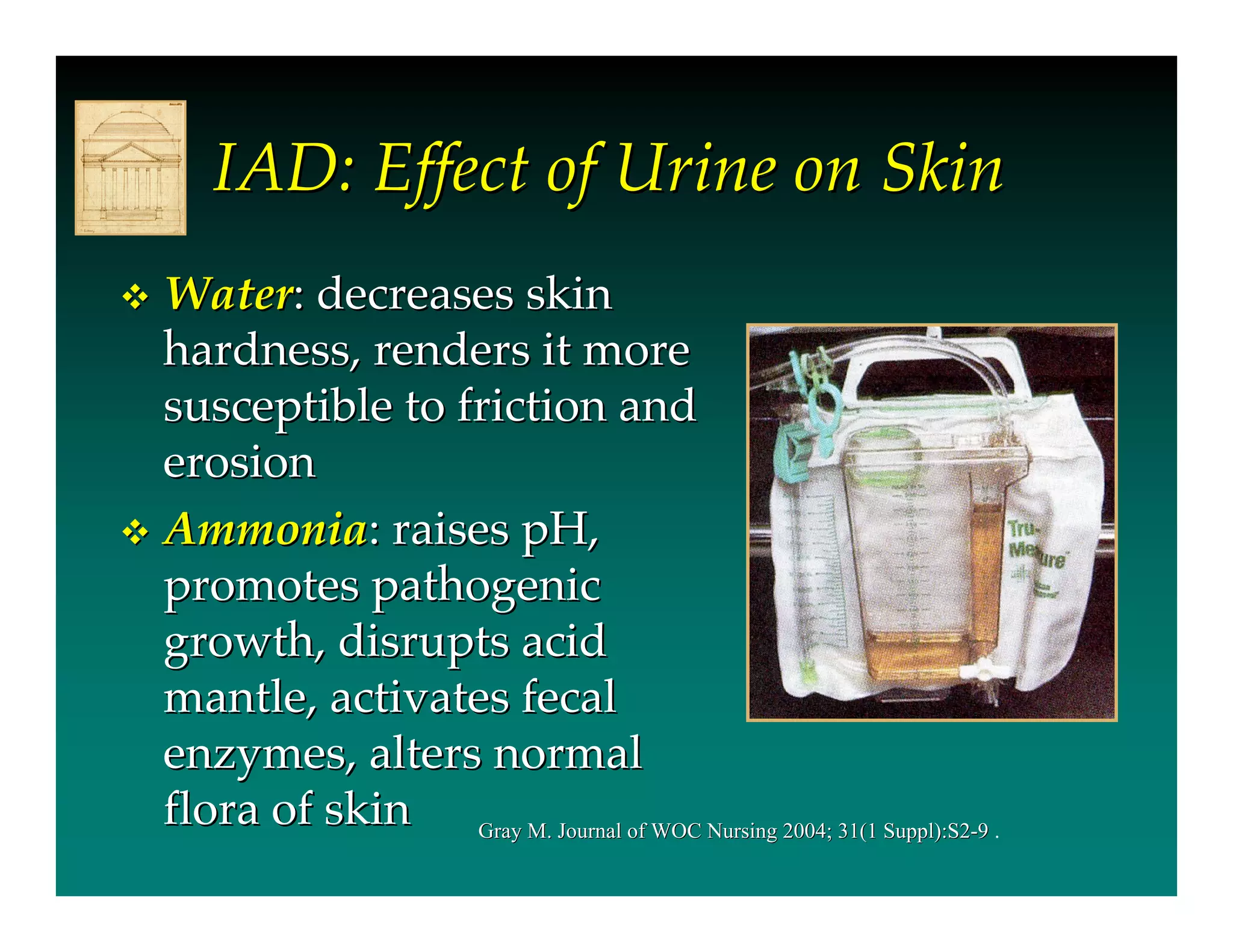
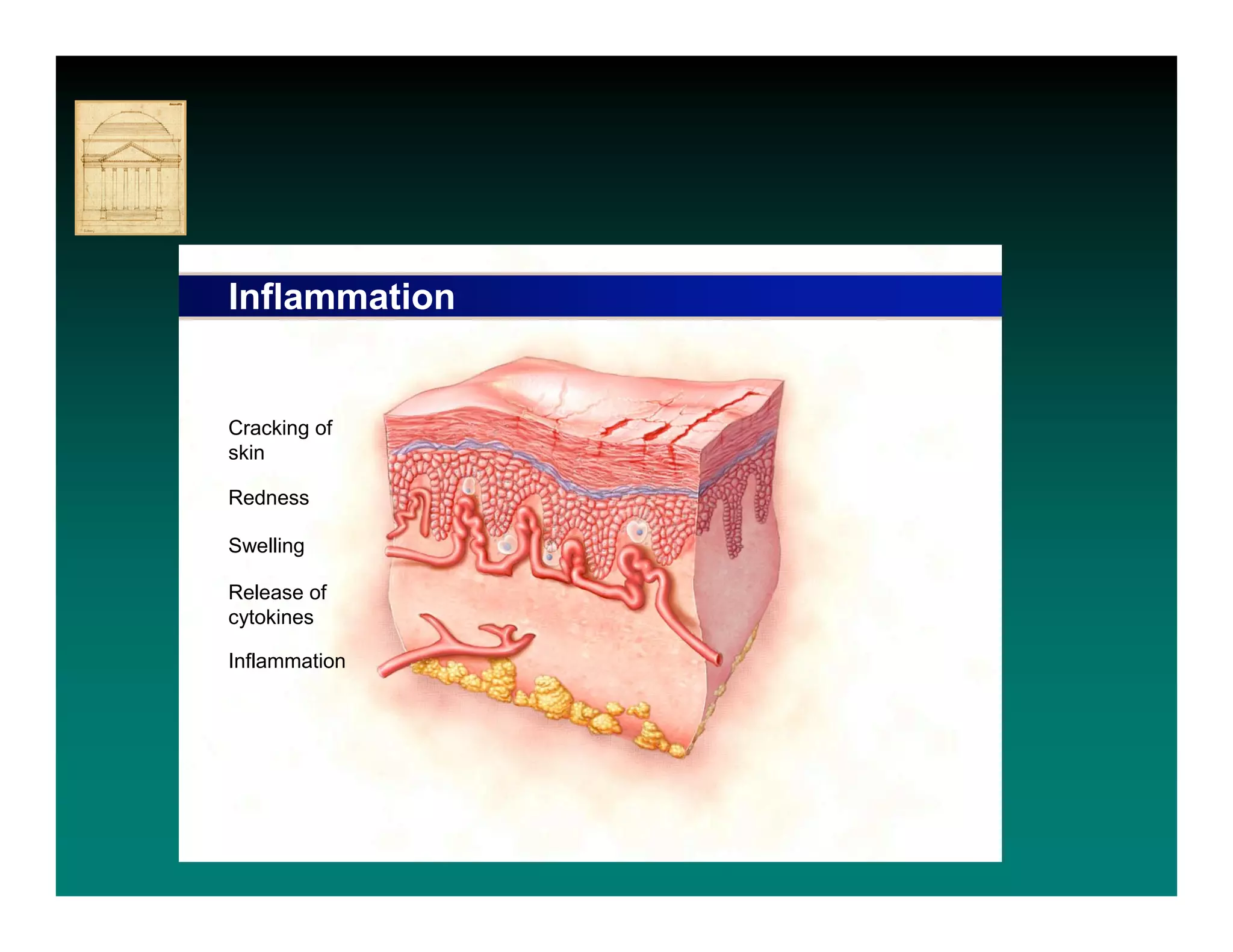
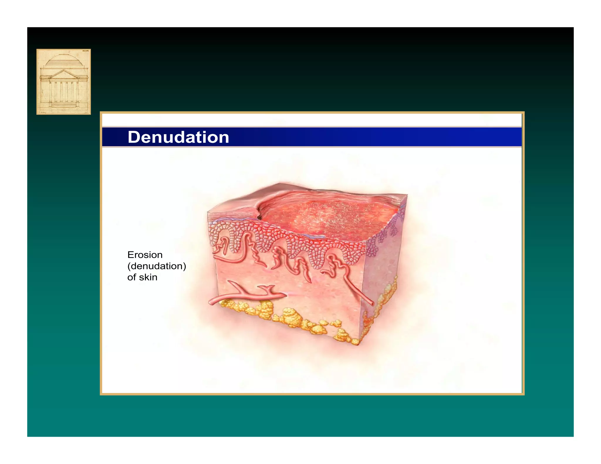
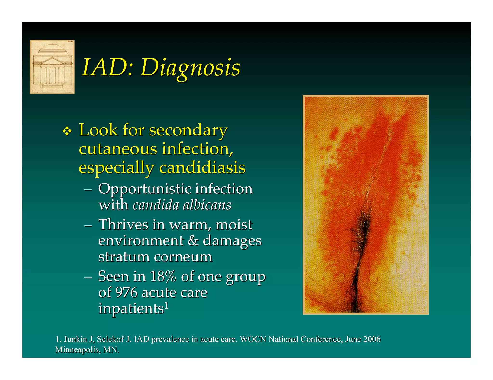
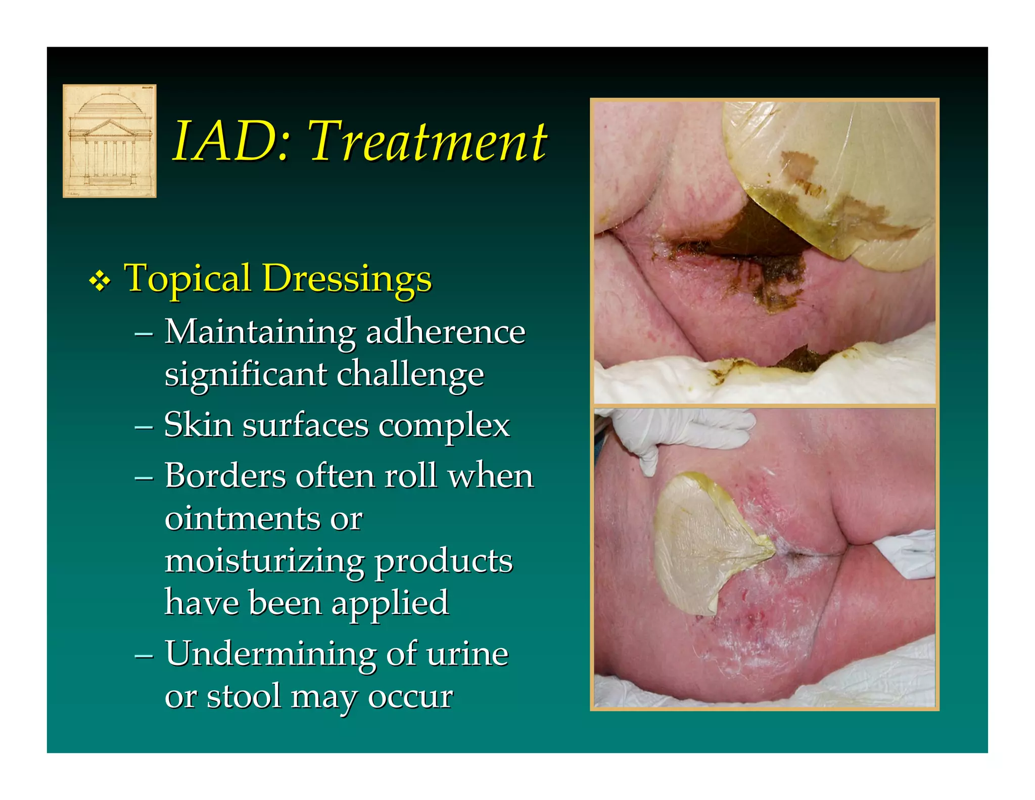
- IAD is skin damage caused by exposure to urine and/or stool, and is exacerbated by the use of absorbent containment products. It affects 5.6-50% of long-term care residents and 10.9-54% of incontinent hospital patients.
- Urine and stool impact the skin's moisture barrier, increasing pH and promoting bacterial/fungal growth. Absorbent products further overhydrate the skin.
- IAD is diagnosed by inspecting the skin for redness,
This document defines incontinence-associated dermatitis (IAD) as inflammation and skin breakdown caused by contact with urine or feces. It describes the skin's structure and functions, and explains that IAD occurs when constant contact with urine and feces disrupts the skin's barrier and natural pH. Predisposing factors include age, medications, mobility issues, and improper hygiene. Diagnosis involves skin assessment to differentiate IAD from pressure ulcers. Treatment focuses on cleansing after incontinence, applying effective skin barriers, and monitoring progress with emollients as needed.
Pressure ulcers, also known as bedsores, develop when skin and underlying tissue is damaged due to prolonged pressure, friction, or moisture. They typically form over bony areas of the body. Factors that increase risk include immobility, incontinence, poor nutrition, aging, and chronic diseases. Pressure ulcers are staged from I to IV based on the depth of tissue damage. Prevention focuses on relieving pressure through repositioning, special beds and cushions, and keeping skin clean and dry. Treatment may include dressings, debridement, and medications to promote healing.
The document discusses various topics related to skin and wound care including:
- The layers of skin and types of wounds
- Staging criteria for pressure ulcers which describes the level of tissue damage
- Risk factors for pressure ulcers like limited mobility and incontinence
- Guidelines for preventing pressure ulcers including repositioning, maintaining nutrition, and managing moisture
- Common skin conditions like fungal infections, abrasions, and vascular wounds
Proper wound care is necessary to prevent infection, assure there are no other associated injuries, and to promote healing of the skin. An additional goal, if possible, is to have a good cosmetic result after the wound has completely healed. This wound care article is designed to present information on wounds involving mainly the skin; it is not meant to cover all wounds (for example, gunshot, degloving wounds, tendon lacerations, and others).
An ideal wound dressing provides protection, maintains moisture, reduces pain, and absorbs exudate. It should not induce pain or itching, be easy to change, allow gaseous exchange, and be inexpensive and readily available. Common types include gauze, tulle, hydrocolloid, hydrogel, alginate, and foam dressings. Vacuum assisted closure (VAC) uses negative pressure to contract the wound and remove exudate, promoting healing through micro and macrostrain. The appropriate dressing depends on the wound characteristics, with dry wounds suited to hydrocolloid or hydrogel and exudating wounds to hydrocolloid or foam.
Pressure ulcers, also known as decubitus ulcers or bedsores, are localized injuries to the skin and underlying tissue that are usually caused by pressure over bony prominences. They are commonly seen in immobilized or bedridden patients and are associated with increased costs of care and risk of litigation. The Braden Scale is commonly used to assess pressure ulcer risk based on factors like mobility, sensation, moisture, activity, nutrition, and friction/shear. Treatment focuses on pressure redistribution through support surfaces, wound care including debridement and moist dressings, and managing pain and infection. Staging systems classify ulcers by depth of tissue damage from non-blanchable erythema to full thickness tissue loss.
This document provides information on nursing care for patients with wounds. It begins by defining wounds and describing the wound healing process. It then classifies wounds by intent, whether open or closed, degree of contamination, and depth. Various types of wounds like incisions, contusions, and lacerations are described. Stages of pressure ulcers are defined. The document outlines factors that influence wound healing and potential complications. It discusses assessing wounds by location, appearance, drainage, and for pressure ulcers, the tissue type. Nursing diagnoses, care planning, interventions, and dressing considerations are presented for different wound stages. Cleaning and securing dressings is demonstrated. References are provided.
This document provides an overview of wound care management. It discusses the anatomy and physiology of the skin, the phases of wound healing, factors affecting wound healing, different types of wound assessment tools, and the process of wound assessment. It also covers identifying wound aetiology, types of wounds, wound classification, complications of wounds, and selecting appropriate wound products and cleansing solutions. The overall aim is to understand wound care and management.
This document provides an overview of wound care including wound healing processes, types of wounds, evaluation and documentation of wounds, and wound management. It discusses the stages of wound healing including hemostasis, inflammation, proliferation, and remodeling. Different types of wounds such as incisions, lacerations, abrasions, and puncture wounds are described along with factors to evaluate like location, size, exudate, signs of infection, and surrounding tissue condition. Methods of documentation including photography and standardized descriptions are covered. The management section addresses wound care plans, cleaning, debridement, exudate management using various dressing materials, and treatment of infections.
Wound care clinicians: do you need to practice your pressure injury staging skills? View a photo and then click forward to see the correct stage according to the 2016 National Pressure Ulcer Advisory Panel (NPUAP) staging system.
This document provides an overview of pressure sores including their definition, epidemiology, pathogenesis, risk factors, staging classifications, prevention, and management. Pressure sores, also known as bedsores or decubitus ulcers, are localized skin or tissue injuries caused by unrelieved pressure. They are common in hospital and nursing home patients and costly to healthcare systems. The document outlines the traditional theories behind their pathogenesis as well as intrinsic and extrinsic risk factors. Staging classifications from the National Pressure Ulcer Advisory Panel are presented along with the Braden and PUSH risk assessment tools. Prevention strategies aim to reduce pressure, shearing forces, and moisture on the skin. Management is based on the stage and may involve wound cleaning, dress
This document defines pressure injuries and discusses their causes, risk factors, prevention, and staging. It notes that pressure injuries result from constant pressure that impairs blood flow to soft tissues over bony areas. The main risk factors are impaired mobility, incontinence, and malnutrition. Prevention focuses on pressure relief through devices, positioning, and skin care. Pressure injuries are staged based on tissue damage depth, from non-blanchable redness to full thickness tissue loss. Incontinence-associated dermatitis involves skin damage from urine and stool exposure.
This document provides information on wound healing and care. It begins with the anatomy and functions of healthy skin. The three layers of skin - epidermis, dermis and subcutaneous tissue - are described. The four phases of wound healing are explained: inflammatory, proliferative, maturation and remodeling. Types of wounds and factors influencing healing are defined. Proper wound observation, cleaning, dressing and drainage are outlined as important for promoting healing.
Pressure ulcers, also known as decubitus ulcers or bed sores, are localized areas of soft tissue damage that result from prolonged pressure. They are staged from 1 to 4 based on depth of tissue damage. Risk factors include older age, immobility, moisture, and poor nutrition. Treatment focuses on relieving pressure, cleaning and debridement of wounds, dressing changes, and surgery if needed to prevent complications like infection.
Lesions and abrasions of the skin can range from superficial to deep. Abrasion injuries occur when skin rubs against a rough surface, removing the outer epidermal layers. They are classified by degree from first to third. Treatment includes cleaning, applying antibiotics to prevent infection, and optionally dressing the wound. Lesions refer broadly to any tissue abnormality or damage, and can be classified by features, location, and other characteristics. Common skin lesions include macules, papules, vesicles, pustules and ulcers. Available treatments can temporarily remove lesions but do not provide a permanent cure.
This document provides information on wound management and wound care. It discusses the different types of wounds including acute and chronic wounds. The goals of wound care are to identify any wounds or complications, prescribe preventative measures to promote skin integrity, and treat any wounds. There are four phases of wound healing: hemostasis, inflammation, proliferation and remodeling. The three types of wound healing and closure are primary, secondary and tertiary. Various wound dressings are also described like hydrocolloid, hydrogel and alginate. The wound care market is large and growing, dominated by major players. Pricing for common wound dressings is also listed.
The document discusses pressure ulcers, including their causes, assessment, stages, treatment, and prevention. Some key points:
- Pressure ulcers are caused by factors like decreased sensation, mobility, nutrition, incontinence, and shear/friction forces.
- Assessment involves visual skin inspection and using a risk assessment scale like the Braden Scale to document factors like moisture, pressure, shear, and friction.
- Pressure ulcers are staged from I to IV based on tissue depth involvement, from non-blanchable redness to full thickness tissue loss with bone/muscle exposure.
- Treatment focuses on relieving pressure, keeping skin clean and dry, using dressings, and managing in
This document provides information on pressure ulcer prevention and management. It defines pressure ulcers and lists objectives of prevention such as assessing risk and providing skin care guidelines. It describes signs and symptoms, risk factors, causes related to pressure, shear and friction. Stages of pressure ulcers from Grade 1 to 4 are defined. Prevention strategies are outlined such as repositioning, nutrition, managing moisture and proper support surfaces. Wound assessment, documentation and staff education are also discussed.
What is cosmetic surgery?
Statistics
History
Some types of cosmetic surgeries
Plastic surgeons said
Celebs Before and After Plastic Surgery
Critical Interpretation and evaluation of culture
Identification of the influences on decisions.
Who stands to gain or lose?
Underlying Commercial Interests
Calling Ideologies to account
Spiritual and Moral Dimension
Caricatures
1) The document discusses current trends in wound management, outlining the phases of wound healing and factors that affect it.
2) Key aspects of wound management covered include wound bed preparation through debridement and dressing, as well as various closure methods and the use of negative pressure wound therapy.
3) Emerging treatments like hyperbaric oxygen therapy and various wound covers including skin substitutes are also summarized.
This document provides guidelines for nurses on performing wound dressings using aseptic technique. It outlines the objectives, standards, purposes and types of dressings used. It then describes the step-by-step procedure for aseptic wound dressing, which involves preparing the necessary items, maintaining sterile technique, applying the appropriate dressing, and documenting wound findings. The overall aim is to promote wound healing through proper dressing while preventing infection.
This document provides an overview of wound assessment techniques. It discusses assessing the patient's history, examining the wound itself using techniques like TIME (tissue, infection, moisture, epithelial edges) and PQRST (provocation, quality, radiation, severity, time) for pain. The document outlines investigating further with tests and making a diagnosis. It also discusses implementing a treatment plan using wound bed preparation principles to manage tissue, infection, moisture and epithelial edges. The goal of wound care is a healed wound through this assessment and treatment cycle approach.
There are several types of wounds including incisions, contusions, abrasions, punctures, and lacerations. Wound drainage can include serous, purulent, or sanguineous exudate. The RYB color code categorizes wounds as red, yellow, or black based on their appearance and healing phase. Red wounds are usually healing and require protection. Yellow wounds contain slough or pus and should be cleansed. Black wounds have necrotic tissue that requires debridement. Wound cleaning involves using isotonic saline to clean or irrigate wounds as needed based on the amount of exudate or foreign material present.
The document discusses wound assessment and management. It outlines the wound care team members and principles of wound management which include identifying underlying causes, controlling impairing factors, and providing local wound care and maintenance therapy. Wound assessment focuses on tissue type, infection/inflammation, moisture imbalance, and wound edge. Various wound dressings and therapies are described for treating different wound characteristics and addressing factors like moisture, infection, and wound edge issues. The wound care services provided are also summarized.
This document discusses bowel and bladder incontinence in elderly patients and its effects on skin health. It defines incontinence and provides epidemiology data. Issues associated with incontinence management include skin maceration, breakdown from moisture, and infections from prolonged catheter or diaper use. Specific skin conditions that can result include incontinence associated dermatitis, moisture associated skin damage from maceration, shear and friction erosions, and unstageable pressure ulcers in immobile patients. The document recommends frequent skin checks, pad changes, and skin protection methods like creams and moisture barriers to prevent and manage incontinence-related skin issues.
Adult diaper rash: Symptoms, Causes and TreatmentUnique Wellness
Rashes are generally caused due to the skin staying wet because of the constant contact with the chemicals of urine and stool. Some of the symptoms, causes and treatment for diaper dermatitis/rashes are given in this presentation. Visit http://wellnessbriefs.com/ for more information.
The document provides an introduction to internal auditing at Morris Heights Health Center. It discusses that the purpose of internal auditing is to independently evaluate an organization's activities as a service to help management meet goals, assess and monitor risks, and ensure compliance. The internal audit process involves planning, performing fieldwork and testing, documenting results in a report. It describes how internal audit can help by objectively assessing operations and sharing best practices to improve controls, processes and risk management. Finally, it notes that building trust with auditees is important by keeping commitments and through collaboration.
this topic is on bed sores. discusses the definition, etiology , pathophysiology of bed sore development as well as prevention and managemene of pressure sores
Pressure ulcers, also known as bedsores or decubitus ulcers, are localized areas of tissue necrosis that occur when soft tissue is compressed between a bony prominence and an external surface for a prolonged period. They are commonly staged from Stage 1 to Stage 4 based on depth of tissue damage. Key risk factors include immobility, moisture, malnutrition, and aging. Prevention focuses on risk assessment, pressure relief, skin care, and nutrition. Treatment involves debridement, dressings, management of bacterial infection, and surgery for advanced cases. Complications can include infection, osteomyelitis, and rarely, cancer.
The document discusses fecal incontinence (FI), including its causes, evaluation, and treatment options. It defines FI and lists factors involved in continence. Evaluation involves clinical history, examinations, and investigations like endoscopy and imaging. Treatment includes non-surgical options like diet, medication, rehabilitation and plugs, as well as surgical procedures like sphincteroplasty, graciloplasty, and sacral nerve stimulation. The aim is to discuss dimensions of FI and clarify diagnosis and management.
Best Practice Statement Principles of wound management in paediatric patientsGNEAUPP.
This document provides guidance on wound management principles for pediatric patients. It discusses key differences in pediatric and neonatal skin that require special considerations in wound care. The causes of wounds in pediatric patients include trauma, medical devices, and certain skin conditions. A thorough assessment of the wound and any factors that could delay healing is important. Wound documentation and management should be tailored to the individual patient. Regular reassessment is needed to monitor progress and watch for signs of infection. Managing pain and anxiety is also a priority, given their impact on the healing process.
This document summarizes a presentation on fecal incontinence and its treatments. It begins with an overview of fecal incontinence, including its prevalence, impact on quality of life, and typical treatment pathway. It then discusses conservative treatments like diet and medication. Surgical options are explored in more detail, including sphincter repair and augmentation techniques like bulking agents and radiofrequency. Outcomes data is presented for these approaches. The document also summarizes sacral nerve stimulation, including its mechanism of action and results from multiple studies showing its effectiveness in reducing incontinence episodes and improving quality of life. Risks associated with sacral nerve stimulation like infection and device issues are also reviewed.
Bowel incontinence is the inability to control bowel movements and involuntarily pass stool. It can be caused by problems with the rectum, sphincter muscles, or nerves controlling the bowel. Common etiologies include constipation, diarrhea, childbirth complications, rectal surgery, diabetes, and spinal cord injuries. The pathophysiology involves anything interfering with anal sphincter pressure, rectal storage, or sensation. Clinical features include diarrhea, abdominal pain, lower back pain, bloating, and emotional effects. Management includes pharmacologic treatments, surgical procedures like sphincteroplasty or artificial anal sphincters, and non-surgical options like biofeedback exercises, dietary changes, and planned bowel movements.
The Science of Neuromodulation and Neuromodulation Therapies Eyad Kishawi
As a biomedical engineer, Eyad Kishawi helps develop instruments and devices that treat a range of medical issues and conditions. Mr. Kishawi is particularly interested in neuroscience and neuromodulation and practical medical research in these fields.
Neuromodulazione tibiale: indicazioni, risultati e limitiGLUP2010
PTNS, or percutaneous tibial nerve stimulation, is a neuromodulation technique that involves electrically stimulating the posterior tibial nerve. It has shown efficacy in treating overactive bladder (OAB) symptoms, with 71% of OAB patients improving after 10-12 PTNS sessions. Long term follow up studies found that most patients who responded initially continued to experience symptom improvement even with intermittent PTNS treatment over subsequent years. Predictors of success include being female and having fewer involuntary detrusor contractions. PTNS provides an alternative treatment option for OAB when antimuscarinic medications are ineffective.
The document outlines the definition, prevalence, etiology, diagnosis, and treatment of overactive bladder (OAB). It defines OAB based on symptoms as urgency, usually accompanied by frequency and nocturia, in the absence of infection or other pathology. OAB affects approximately 17% of adults, with higher rates in women and increasing with age. Potential causes include neurological factors, changes to detrusor muscle, and increased sensitivity of bladder nerves. Diagnosis is based on symptoms through history and exams. Treatments include behavioral changes, medications, botulinum toxin injections, and neuromodulation procedures.
Epistaxis, or nosebleed, is bleeding from inside the nose. It is a common condition, with 5-10% of people experiencing an episode each year. Anterior nosebleeds are more common in children and young adults, while posterior nosebleeds are more common in older adults with hypertension or arteriosclerosis. The majority of epistaxis cases have no identifiable cause. When a cause is identified, it is often local trauma, inflammation, or a bleeding disorder. Treatment involves first controlling the bleeding, then treating any underlying causes through cauterization, nasal packing, or ligation of blood vessels.
This case report describes a 55-year-old man who presented with a swelling below his eye for 5 months. Surgical exploration and biopsy revealed pus and bone erosion. Histopathology showed chronic osteomyelitis due to fungal infection, identified as Aspergillus species. The patient's wound continued draining despite antibiotics. Further imaging and debridement surgery confirmed destruction of the zygomatic arch and maxilla from chronic osteomyelitis. The patient was treated with antifungal medication for 6 weeks.
This document is a presentation by DDX, an Australian digital solutions company. It introduces DDX, highlighting their 7 years of experience in digital services, 500+ projects completed, and expertise in areas like user experience design, rich internet applications, web development, and hosting. The presentation provides an overview of DDX's capabilities and services, case studies, client testimonials, associations, and examples of past work. It is intended to promote DDX's services and expertise to potential clients.
This document discusses the symptoms and treatment of dandruff. It lists common symptoms like white flakes on shoulders, itchy scalp, and dry facial skin. It explains that dandruff is caused by skin cells shedding from the scalp faster than normal, which allows oil and cells to clump together into white flakes. It recommends treating dandruff by regularly shampooing the scalp with anti-dandruff products and using over-the-counter cortisone for skin inflammation. Natural remedies like washing with white vinegar are also listed.
Visit our site http://www.rashonfaceblog.net for more information on Rash On Face.The use of homely Rash On Face treatment becomes necessary if you have rashes on your face and other parts of body due to various reasons including lack of skin maintenance, excessive sweating, dryness on skin or sensitivity of the skin.
1040122 oab diagnosis, management and current trend of therapyAlex Chen
This document discusses overactive bladder (OAB). It defines OAB as a symptom syndrome characterized by urgency, with or without urge incontinence, usually accompanied by frequency and nocturia. The prevalence of OAB is estimated to be around 16-17% globally. Common causes include problems with the pelvic floor muscles, nervous system issues, and other factors. OAB can negatively impact quality of life by limiting physical, sexual, occupational, social, domestic, and psychological activities. Treatment involves behavioral modifications, pharmacotherapy such as anticholinergic medications, and in some cases neuromodulation procedures or surgery.
This document discusses overactive bladder (OAB), including its definition, symptoms, prevalence, impact, and treatment options. It defines OAB as a syndrome characterized by urgency, with or without urge incontinence, usually accompanied by frequency and nocturia. It notes that OAB is very common, affecting 11-22% of adults over 40, and is more prevalent in older individuals and females. While over half of females with OAB seek treatment, far fewer males do. Non-pharmacological treatments discussed include behavior modification, diet and fluid management, and pelvic floor exercises.
The document is a presentation about optimizing oral antibiotic treatment for acne vulgaris. It discusses DORYX (doxycycline hyclate delayed-release tablets), which is indicated as adjunctive therapy for severe acne. The presentation covers topics such as the epidemiology and classification of acne vulgaris, its impact on quality of life, current treatment options including oral antibiotics, and the efficacy and safety profile of doxycycline specifically.
The normal microbial flora of the human body includes diverse populations of microorganisms inhabiting different body sites. The composition of the flora varies by body region and is determined by environmental factors like pH, moisture and nutrients. The flora plays important roles in host health, aiding digestion and competing against pathogens. While usually harmless, elements of the flora can cause disease in immunocompromised hosts. The skin flora in particular is dominated by staphylococci and micrococci that vary in concentration between dry and moist skin sites.
Fournier’s Gangrene in a 9 Yrs. Old Patient; A Rare Presentation in Paediatri...semualkaira
Necrotizing fasciitis of the perineum and external genitalia is a
life-threatening infective gangrene, primarily seen in adults but
relatively rare in children. We present a nine-year-old male child
with spinal bifida and double incontence who was admitted at our
hospital due to gangrenous right hemi-scrotal ulcer extending to
the right thigh. It was proceeded with painful swollen hemi-scrotum 2wks prior to admission. We treated him aggressively with
broad spectrum antibiotics and early surgical debridement. Being
paraplegic with double incontinence hence spending most of the
time dressed with diapers we therefore think of poor hygiene and
the diaper rash as the etiological factors. Early surgical debridement with appropriate antibiotics and aggressive supportive care
usually gave good results.
The document discusses the skin microbiome and its role in atopic dermatitis (eczema). It notes that the skin hosts trillions of bacteria that create an ecosystem. Factors like location on the body, moisture levels, and individual characteristics can influence the types of microbes present. In eczema, skin barrier defects allow overgrowth of bacteria like Staphylococcus aureus that secrete toxins exacerbating inflammation. Studies show shifts in skin microbiota during eczema flares and treatment. A healthy microbiome including species like Staphylococcus epidermidis promotes skin immunity, while dysbiosis may increase eczema risk.
AN OUTLINE ON HERPESVIRAL DISEASES IN MARINE TURTLESICAR-CIFE
Abstract: Marine turtles are calm, long-living reptiles with complicated lifestyle. They have their attendance all over the globe. The survival of turtles became so pathetic by various means of human development exercises. Now, on the other hand, emerging diseases came to the platform. Disease indications are alarming high in the coastal belts where human agitation is heavier. Herpesviridae family members are the top most pathogenic agents that are disturbing the survival of these long-living animals. They are a total of five herpesviruses that are associated with diseases in turtles; in them, Fibropapillomatosis is the catastrophic disease, which is characterized by tumors over smooth surfaces of the body. Disease had turned to be an upcoming invader for the maritime turtle’s community. The establishment and potential of this disease is under the hands of environmental factors. The longevity of marine turtles, coupled with their close association with inshore habitats and seagrass meadows and coral reefs in these habitats, has led to the proposal that they may act as sentinel indicators of marine ecosystem health.
This document discusses a study investigating the effect of temperature and salinity on infection intensity of Bonamia ostreae, a parasite that infects European flat oysters (Ostrea edulis). The study found that infection intensity increased by 43% in oysters kept at 20°C compared to 12°C, and decreased by 69% in oysters kept at 28‰ salinity compared to 34‰ salinity. These results have implications for disease management, as culturing oysters at lower temperatures and salinities could help reduce parasite impact. The study also found the primary PCR method unreliable for detection and recommends using nested PCR.
Mucocutaneous Involvement in Behcets Diseasenavasreni
Behçet’s disease is a chronic inflammatory disease characterized by its clinical polymorphism associating mucocutaneous involvement to systemic manifestations. The mucocutaneous lesions are considered the hallmark of the disease, being the most common symptoms presenting at the onset of disease. Our objective was to determine the characteristics of this skin involvement during Behçet’s disease. We conducted a descriptive study over a period of 30 years, having collected all patients with Behçet’s disease.
Mucocutaneous Involvement in Behcets Diseasepateldrona
This document summarizes a study on mucocutaneous involvement in Behcet's disease. The study analyzed 98 patients over 30 years. All patients showed mucocutaneous lesions. Oral aphthosis was present in all patients, and genital ulcers were seen in 81% of patients. Other common lesions included pseudofolliculitis in 61% of patients and erythema nodosum in 7% of patients. All patients were treated with colchicine, while some severe cases also received corticosteroids or immunosuppressants. The main findings were that mucocutaneous lesions are very common in Behcet's disease, especially oral aphthosis and genital ul
Mucocutaneous Involvement in Behcets Diseasekomalicarol
Behçet's disease is a chronic inflammatory disease characterized by
its clinical polymorphism associating mucocutaneous involvement
to systemic manifestations. The mucocutaneous lesions are
considered the hallmark of the disease, being the most common
symptoms presenting at the onset of disease. Our objective was
to determine the characteristics of this skin involvement during
Behçet's disease. We conducted a descriptive study over a period
of 30 years, having collected all patients with Behçet's disease.
These were 98 patients. A male predominance was observed in
our studied population with a Sex Ratio of 2.5. The mean age at
diagnosis was 34 years. Mucocutaneous involvement was observed
in all patients. Oral aphthosis was constant and genital ulcers, were
observed in 81 cases. The other mucocutaneous manifestations
were: pseudofolliculitis (61 cases), erythema nodosum (7 cases),
skin ulcers (4 cases), acneiform lesions (2 cases), perianal ulcers (1
case), skin ulceration (1 case) and erythema multiforme. (1 case).
All of our patients were treated with colchicine. Corticosteroids
and non-steroidal anti-inflammatory drugs were each indicated in
one case for resistant forms
Mucocutaneous Involvement in Behcets Diseasesemualkaira
Behçet's disease is a chronic inflammatory disease characterized by
its clinical polymorphism associating mucocutaneous involvement
to systemic manifestations. The mucocutaneous lesions are
considered the hallmark of the disease, being the most common
symptoms presenting at the onset of disease. Our objective was
to determine the characteristics of this skin involvement during
Behçet's disease. We conducted a descriptive study over a period
of 30 years, having collected all patients with Behçet's disease.
These were 98 patients. A male predominance was observed in
our studied population with a Sex Ratio of 2.5. The mean age at
diagnosis was 34 years. Mucocutaneous involvement was observed
in all patients. Oral aphthosis was constant and genital ulcers, were
observed in 81 cases. The other mucocutaneous manifestations
were: pseudofolliculitis (61 cases), erythema nodosum (7 cases),
skin ulcers (4 cases), acneiform lesions (2 cases), perianal ulcers (1
case), skin ulceration (1 case) and erythema multiforme. (1 case).
All of our patients were treated with colchicine. Corticosteroids
and non-steroidal anti-inflammatory drugs were each indicated in
one case for resistant forms.
Relationships between onchocerca volvulus microfilaraemia and the clinical ma...Alexander Decker
This study examined the relationship between Onchocerca volvulus microfilariae and clinical manifestations of onchocerciasis in two communities in southeastern Nigeria. Skin snips and clinical exams were performed on residents and microfilarial prevalence and intensity were analyzed in relation to skin changes, nodules, and visual problems. Microfilarial prevalence and intensity were higher in those with chronic skin damage, leopard skin, subcutaneous nodules, and visual issues. The differences were statistically significant, indicating O. volvulus microfilariae were closely associated with clinical manifestations, especially in older age groups, in this meso-endemic area.
Cutaneous Larva Migrans: A Case Report in a Traveler Childpateldrona
Cutaneous Larva Migrans (CLM) is the most common skin disease of tropical origincaused by hookworms larvae, occurring in international travelers. Typical manifestations consist of erythematous, serpiginous slightly elevated linear cutaneous lesions.
Cutaneous Larva Migrans: A Case Report in a Traveler Childnavasreni
Cutaneous Larva Migrans (CLM) is the most common skin disease of tropical origincaused by hookworms larvae, occurring in international travelers. Typical manifestations consist of erythematous, serpiginous slightly elevated linear cutaneous lesions. We describe the case of an 8-year-old boy, with CLM infection acquired during travel to Burkina-Faso, and successfully treated...
Cutaneous Larva Migrans: A Case Report in a Traveler ChildSarkarRenon
This case report describes an 8-year-old boy who presented with cutaneous larva migrans (CLM), a skin infection caused by hookworm larvae, after traveling to Burkina Faso. The boy had a serpiginous rash on his foot. He was diagnosed with CLM based on his travel history and clinical presentation. He was treated successfully with oral ivermectin. CLM is a common infection in travelers to tropical regions who come into contact with contaminated soil. It presents as migrating skin lesions caused by hookworm larvae that are unable to fully develop in human skin. Early diagnosis and treatment can prevent complications.
Cutaneous Larva Migrans: A Case Report in a Traveler Childclinicsoncology
Cutaneous Larva Migrans (CLM) is the most common skin disease of tropical origincaused by hookworms larvae, occurring in international travelers. Typical manifestations consist of erythematous, serpiginous slightly elevated linear cutaneous lesions
Cutaneous Larva Migrans: A Case Report in a Traveler Childkomalicarol
Cutaneous Larva Migrans (CLM) is the most common skin disease of tropical origincaused by
hookworms larvae, occurring in international travelers. Typical manifestations consist of erythematous, serpiginous slightly elevated linear cutaneous lesions. We describe the case of an
8-year-old boy, with CLM infection acquired during travel to Burkina-Faso, and successfully
treated with Ivermectin. Epidemiology, clinicaldiagnosis and therapeutic are debated
Cutaneous Larva Migrans: A Case Report in a Traveler ChildAnonIshanvi
Cutaneous Larva Migrans (CLM) is the most common skin disease of tropical origincaused by hookworms larvae, occurring in international travelers. Typical manifestations consist of erythematous, serpiginous slightly elevated linear cutaneous lesions.
Cutaneous Larva Migrans: A Case Report in a Traveler Childgeorgemarini
Cutaneous Larva Migrans (CLM) is the most common skin disease of tropical origincaused by hookworms larvae, occurring in international travelers. Typical manifestations consist of erythematous, serpiginous slightly elevated linear cutaneous lesions
Melioidosis- An overview, covers the Aetiology, Epidemiology, World as well as Indian Scenario of Meliodosis, Its public health impact, control strategy and Indian Research prospects of the disease.
All credit goes to Dr. Gazanfar Abass, MVSc Scholar at Division of Veterinary Public Health, Indian Veterinary Research Institute, izatnagar UP, India
Similar to Incontinence Associated Dermatitis by Prof Dr Mikel Gray (20)
Our backs are like superheroes, holding us up and helping us move around. But sometimes, even superheroes can get hurt. That’s where slip discs come in.
Histololgy of Female Reproductive System.pptxAyeshaZaid1
Dive into an in-depth exploration of the histological structure of female reproductive system with this comprehensive lecture. Presented by Dr. Ayesha Irfan, Assistant Professor of Anatomy, this presentation covers the Gross anatomy and functional histology of the female reproductive organs. Ideal for students, educators, and anyone interested in medical science, this lecture provides clear explanations, detailed diagrams, and valuable insights into female reproductive system. Enhance your knowledge and understanding of this essential aspect of human biology.
TEST BANK For Community Health Nursing A Canadian Perspective, 5th Edition by...Donc Test
TEST BANK For Community Health Nursing A Canadian Perspective, 5th Edition by Stamler, Verified Chapters 1 - 33, Complete Newest Version Community Health Nursing A Canadian Perspective, 5th Edition by Stamler, Verified Chapters 1 - 33, Complete Newest Version Community Health Nursing A Canadian Perspective, 5th Edition by Stamler Community Health Nursing A Canadian Perspective, 5th Edition TEST BANK by Stamler Test Bank For Community Health Nursing A Canadian Perspective, 5th Edition Pdf Chapters Download Test Bank For Community Health Nursing A Canadian Perspective, 5th Edition Pdf Download Stuvia Test Bank For Community Health Nursing A Canadian Perspective, 5th Edition Study Guide Test Bank For Community Health Nursing A Canadian Perspective, 5th Edition Ebook Download Stuvia Test Bank For Community Health Nursing A Canadian Perspective, 5th Edition Questions and Answers Quizlet Test Bank For Community Health Nursing A Canadian Perspective, 5th Edition Studocu Test Bank For Community Health Nursing A Canadian Perspective, 5th Edition Quizlet Test Bank For Community Health Nursing A Canadian Perspective, 5th Edition Stuvia Community Health Nursing A Canadian Perspective, 5th Edition Pdf Chapters Download Community Health Nursing A Canadian Perspective, 5th Edition Pdf Download Course Hero Community Health Nursing A Canadian Perspective, 5th Edition Answers Quizlet Community Health Nursing A Canadian Perspective, 5th Edition Ebook Download Course hero Community Health Nursing A Canadian Perspective, 5th Edition Questions and Answers Community Health Nursing A Canadian Perspective, 5th Edition Studocu Community Health Nursing A Canadian Perspective, 5th Edition Quizlet Community Health Nursing A Canadian Perspective, 5th Edition Stuvia Community Health Nursing A Canadian Perspective, 5th Edition Test Bank Pdf Chapters Download Community Health Nursing A Canadian Perspective, 5th Edition Test Bank Pdf Download Stuvia Community Health Nursing A Canadian Perspective, 5th Edition Test Bank Study Guide Questions and Answers Community Health Nursing A Canadian Perspective, 5th Edition Test Bank Ebook Download Stuvia Community Health Nursing A Canadian Perspective, 5th Edition Test Bank Questions Quizlet Community Health Nursing A Canadian Perspective, 5th Edition Test Bank Studocu Community Health Nursing A Canadian Perspective, 5th Edition Test Bank Quizlet Community Health Nursing A Canadian Perspective, 5th Edition Test Bank Stuvia
These lecture slides, by Dr Sidra Arshad, offer a quick overview of the physiological basis of a normal electrocardiogram.
Learning objectives:
1. Define an electrocardiogram (ECG) and electrocardiography
2. Describe how dipoles generated by the heart produce the waveforms of the ECG
3. Describe the components of a normal electrocardiogram of a typical bipolar lead (limb II)
4. Differentiate between intervals and segments
5. Enlist some common indications for obtaining an ECG
6. Describe the flow of current around the heart during the cardiac cycle
7. Discuss the placement and polarity of the leads of electrocardiograph
8. Describe the normal electrocardiograms recorded from the limb leads and explain the physiological basis of the different records that are obtained
9. Define mean electrical vector (axis) of the heart and give the normal range
10. Define the mean QRS vector
11. Describe the axes of leads (hexagonal reference system)
12. Comprehend the vectorial analysis of the normal ECG
13. Determine the mean electrical axis of the ventricular QRS and appreciate the mean axis deviation
14. Explain the concepts of current of injury, J point, and their significance
Study Resources:
1. Chapter 11, Guyton and Hall Textbook of Medical Physiology, 14th edition
2. Chapter 9, Human Physiology - From Cells to Systems, Lauralee Sherwood, 9th edition
3. Chapter 29, Ganong’s Review of Medical Physiology, 26th edition
4. Electrocardiogram, StatPearls - https://www.ncbi.nlm.nih.gov/books/NBK549803/
5. ECG in Medical Practice by ABM Abdullah, 4th edition
6. Chapter 3, Cardiology Explained, https://www.ncbi.nlm.nih.gov/books/NBK2214/
7. ECG Basics, http://www.nataliescasebook.com/tag/e-c-g-basics
These lecture slides, by Dr Sidra Arshad, offer a simplified look into the mechanisms involved in the regulation of respiration:
Learning objectives:
1. Describe the organisation of respiratory center
2. Describe the nervous control of inspiration and respiratory rhythm
3. Describe the functions of the dorsal and respiratory groups of neurons
4. Describe the influences of the Pneumotaxic and Apneustic centers
5. Explain the role of Hering-Breur inflation reflex in regulation of inspiration
6. Explain the role of central chemoreceptors in regulation of respiration
7. Explain the role of peripheral chemoreceptors in regulation of respiration
8. Explain the regulation of respiration during exercise
9. Integrate the respiratory regulatory mechanisms
10. Describe the Cheyne-Stokes breathing
Study Resources:
1. Chapter 42, Guyton and Hall Textbook of Medical Physiology, 14th edition
2. Chapter 36, Ganong’s Review of Medical Physiology, 26th edition
3. Chapter 13, Human Physiology by Lauralee Sherwood, 9th edition
Clinic ^%[+27633867063*Abortion Pills For Sale In Tembisa Central19various
Clinic ^%[+27633867063*Abortion Pills For Sale In Tembisa Central Clinic ^%[+27633867063*Abortion Pills For Sale In Tembisa CentralClinic ^%[+27633867063*Abortion Pills For Sale In Tembisa CentralClinic ^%[+27633867063*Abortion Pills For Sale In Tembisa CentralClinic ^%[+27633867063*Abortion Pills For Sale In Tembisa Central
Rasamanikya is a excellent preparation in the field of Rasashastra, it is used in various Kushtha Roga, Shwasa, Vicharchika, Bhagandara, Vatarakta, and Phiranga Roga. In this article Preparation& Comparative analytical profile for both Formulationon i.e Rasamanikya prepared by Kushmanda swarasa & Churnodhaka Shodita Haratala. The study aims to provide insights into the comparative efficacy and analytical aspects of these formulations for enhanced therapeutic outcomes.
8 Surprising Reasons To Meditate 40 Minutes A Day That Can Change Your Life.pptxHolistified Wellness
We’re talking about Vedic Meditation, a form of meditation that has been around for at least 5,000 years. Back then, the people who lived in the Indus Valley, now known as India and Pakistan, practised meditation as a fundamental part of daily life. This knowledge that has given us yoga and Ayurveda, was known as Veda, hence the name Vedic. And though there are some written records, the practice has been passed down verbally from generation to generation.
8 Surprising Reasons To Meditate 40 Minutes A Day That Can Change Your Life.pptx
Incontinence Associated Dermatitis by Prof Dr Mikel Gray
1. Incontinence
Associated Dermatitis
Mikel Gray, PhD, FNP, PNP, CUNP, CCCN, FAAN
Professor & Nurse Practitioner
University of Virginia Department of Urology
2. Anatomy & Physiology
Largest organ (6 pounds or 3,000 sq inches); its
thickness varies from 0.5mm – 6 mm
Functions:
– Barrier: against toxins in external environment and for
the prevention of excessive fluid & electrolyte loss
from internal environment
– Thermoregulation
– Sensory organ/ communication
– Immune functions
– Vitamin D metabolism
Burns T et al. Textbook of Dermatology, 2004. Mass: Blackwell Science
3. Moisture barrier of the skin
– Stratum corneum: dead
keratinocytes or corneocytes
– Lipid matrix: slows
movement of water &
electrolytes
– Water: hydrates corneocytes
– pH: (usually 5.0-5.9) forms
an acid mantle
– Bacterial flora: competes
with pathogens to prevent
infection
– Temperature: regulates
permeability
4. How do Clinicians & Researchers
Measure the skin’s Moisture Barrier?
No clinical test for measuring
moisture barrier
Researchers measure
Transepidermal water loss (TEWL);
which is the rate of passive diffusion
of H20 from internal environment to
external environment (differs from
perspiration)
The perineal skin and scrotum have
the highest TEWL os any surfaces of
the body, skin over back is the
lowest
Loffler H, Hautarzt. 50(11):769-78, 1999
Hautarzt.
5. Perineal Skin at the
Extremes of Life
Barrier function in the neonate
– Less robust than adults, particularly premature infants
Higher TEWL
Higher rates of percutaneous absorption
Greater risk for erosion, stripping, pressure injury
– Cornification of skin begins about GW 20
– Vernix contains FFA, cholesterol & ceramides, thus acting as
proxy while skin develops
– Full-term skin contains 10-20 layers of stratum corneum, skin
in premature baby has 2-3
Lund C et al. JOGNN 1999; 28(3): 241.
6. Perineal Skin at the
Extremes of Life
Aging Skin: gradual decline
in barrier function
– ↑ TEWL
– Overall thickness declines
– ↓ Collagen & elastin
– Local changes in capillary
beds reflect systemic
changes in
microcirculation
Ghadially R. American J Contact Dermatitis 1998; 9(3): 162.
7. Searching for an appropriate name:
Perineal Dermatitis?
Perineum: region between the thighs, in the female
between the vulva and the anus, in males, between the
scrotum and the anus1
Dermatitis: inflammation of the skin1, itself a broad term
may be divided into2
– Atopic (eczema)
– Allergic
– Irritant
– Multiple other terms used, dermatoses used to describe
“well defined endogenous skin dysfunction”2
1. Online Medical Dictionary, http://cancerweb.ncl.ac.uk/cgi-bin/omd?action=Home&query
http://cancerweb.ncl.ac.uk/cgi-bin/omd?action=
2. Burns T et al. Textbook of Dermatology, 2004. Mass: Blackwell Science.
8. Searching for an appropriate name:
Diaper or Nappy Dermatitis?
Strengths
– Clearly associated with incontinence and use of
one type of containment device, infant diaper
(often called nappy in UK) or adult
containment brief
Limitations
– Unfairly blames one type of containment
device as cause of the problem itself
– Possible pejorative interpretation when applied
to adults
9. Searching for an appropriate name:
Incontinence Associated Dermatitis
Name selected from alternatives at
consensus conference held in Chicago,
IL summer of 2005, results of conference
published in JWOCN, 20071
Describes etiology and outcome of
condition
* Supported by unrestricted educational grand from SAGE, Inc.
1. Gray M, Bliss DZ, Doughty DB< Ermer-Seltun K, Kennedy-Evans KL, Palmer MH. JWOCN
34(1): 57-69.
10. Moisture Associated Skin Damage
(MASD)
IAD is part of larger etiological framework
called MASD
– Intertrigo: inflammation in skin folds related to
perspiration, friction and bacterial/ fungal
bioburden
– Periwound maceration: skin breakdown from
wound exudate, related to volume, constituents
or exudate & bacterial bioburden
– IAD: urine, stool, containment device, secondary
cutaneous infection – typically fungal
11. Epidemiology of IAD
Long-term care literature reports
– Prevalence of 5.6%-50%
– Incidence of 3.4%-25%
Acute-care
– Incontinence prevalence: 20%
– IAD prevalence was 10.9% of the general
hospital population
– IAD prevalence was 54% in incontinent
patients in 3 acute-care hospitals
Lyder, et al., 1992; Bale, et al., 2004; Bliss, et al., 2005; Junkin, More-Lisi, Selekof, 2005
Selekof,
12. 2005 IAD Prevalence Study
976
Total number of
patients surveyed
• 27% had IAD
20.3% (198)
35% had • 33% had a pressure
prevalence of
Foley catheter ulcer
incontinence
(deemed continent) • 18% had a probable
urine or stool
fungal Infection
21% had more than 1 type of injury
Junkin J, Selekof J. IAD prevalence in acute care. WOCN National Conference, June 2006 Minneapolis, MN.
13. IAD: Effect of Urine on Skin
Water: decreases skin
hardness, renders it more
susceptible to friction and
erosion
Ammonia: raises pH,
promotes pathogenic
growth, disrupts acid
mantle, activates fecal
enzymes, alters normal
flora of skin Gray M. Journal of WOC Nursing 2004; 31(1 Suppl):S2-9 .
14. Impact of Stool on Skin
Intestinal colonization acts as a reservoir for
potential pathogenic substances1
– VRE
– MRSA
– Clostridium difficile
– Antibiotic resistant Staphylococcus aureus
– Multiple other antimicrobial resistant gram-
negative bacilli
Steifel & Doskey, 2004; Current Infectious Disease Report 2004; 6:420.
Doskey,
15. Impact of Stool on Skin
Disruption of the usual microflora provides
opportunity for pathogenic colonization1
– Normal colon: 1012 CFU per Gm with obligate
anaerobe counts exceeding parasitic organisms
~1000:1; important defense against pathogens
– Antimicrobials that are excreted into the intestinal
tract disrupt this balance
– Result in skin contamination in 83% and
environmental surface contamination in 67%,
diarrhea and fecal incontinence magnify risk2
1. Steifel & Doskey, 2004; Current Infectious Disease Report 2004; 6:420.
Doskey,
2. Donskey et al. NEJM 2000; 343: 1925.
16. Impact of Stool on Skin
Disruption of gastric acid content in stomach
– Healthy individual: >99% of coliform bacteria
ingested killed within 30 minutes because of
gastric acid secretion1
– Use of medications that inhibit stomach acid
production associated with C. difficile, S. aureus,
VRE and antibiotic resistant gam negative
infections2
1.Donskey, Clinical infectious Disease 2004; 39: 219.
2. Cunningham et al., J. Hospital Infection 2003; 36: 149.
17. Pathophysiology
Use of absorptive containment devices
– Exacerbate overhydration by promoting perspiration
& retaining urine and stool; with padding alone:
TEWL increases 3-4 fold within days
CO2 emission increases > 4 fold
pH increases from 4.4 to 7.1 (without incontinence)
– Emerging data supports direct role in PU risk…
1. Grove GL et al. Clinical Problems in Dermatology 1998; 26:183
2. Zimmerer RE et al. Pediatric Dermatology 1986; 3: 95.
3. Zhai H et al. Skin Research & Technology 2002; 8:13.
18. IAD & Pressure Ulceration
Precise nature of association not understood
Fecal incontinence strongly associated with PU
risk, UI is not1-4
Analysis rarely based on PU stage, few articles
that use stage associate FI/ UI with stage I & II3
Both FI & UI associated with increased time and
cost to wound healing5
1. Maklebust J & Magnan MA Advances in Wound Care 1994; 7(6): 25.
2. Gunninberg L. Journal of Wound Care 2004; 13(7): 286.
3. Fader M et al. Journal of Clinical Nursing 2003; 12(3):374.
4. Berlowitz DR et al. Journal of the American Geriatrics Society 2001; 49(7):866-71.
5. Narayan S et al. Jounal of WOCN 2005; 32(3): 163.
19. IAD & Pressure Ulceration
Does FI or UI indirectly contribute to pressure
ulcer risk?
– Skin wetted with synthetic urine or water shows a
significant decrease in hardness, temperature, and
blood flow during pressure load when compared to
dry sites1
– Absorbent products may enhance the risk for
pressure ulceration by creating areas of increased
interface pressure, even when used in conjunction
with a pressure reducing or relieving device2
1. Mayrovitz HN, Sims N Adv Skin Wound Care 2001;14(6):302.
2. Fader M et al. Journal of Advanced Nursing 2004; 48(6): 569.
27. IAD: Diagnosis
Inspect the skin for
erythema, redness,
cracking, swelling,
vesicles
Determine location
of skin damage –
does it lie in skin
fold or over bony
prominence,
underneath
containment
device?
28. IAD: Diagnosis
Look in Skin Folds
– Opposing skin surfaces trap
moisture
– Warm moist environment
encourages bacterial and
fungal colonization,
overgrowth and infection
– Friction created as skin folds
rub against one another
29. IAD: Diagnosis
Look for erosion of
skin
Partial thickness
erosion common
Full thickness wound
implies pressure or
shear and pressure
ulceration
30. IAD: Diagnosis
Look for secondary
cutaneous infection,
especially candidiasis
– Opportunistic infection
with candida albicans
– Thrives in warm, moist
environment & damages
stratum corneum
– Seen in 18% of one group
of 976 acute care
inpatients1
1. Junkin J, Selekof J. IAD prevalence in acute care. WOCN National Conference, June 2006
Minneapolis, MN.
33. IAD: Prevention
Principles of Prevention: 1) cleanse, 2)
moisturize, 3) protect
– Gentle cleansing: NO scrubbing
– Select a cleanser with acceptable pH
& no irritants
– Moisturize dried areas to maximize
lipid barrier
– Apply moisture barrier as indicated
34. Hospital Disposable
Washcloth Vs. Washcloth
Basin Sage
35. Preventive Skin Care:
Cleanse
Soap & Water
– What is the clinical evidence for soap &
water as a perineal skin cleanser
alkaline pH raises pH more than cleansing with
pH ‘balanced’ cleansers; alkaline pH associated
with skin irritation and severity of IAD1
cleansing requires significantly more time than
with cleansers1,2
2 small RCT have not demonstrated greater risk
for dermatitis in frail elder patients1,2
1. Byers et al. JWOCN, 1995, 187.
2. Lewis-Byers et al. OWM, 2002, 44.
36. Preventive Skin Care:
Cleanse
Incontinence skin cleansers
– ‘pH Balanced’ designed to maintain the
acid mantle of perineal skin
– Many described as “no rinse” (no water
required)
– Require significantly less time than
traditional cleansing with soap and water
– Many contain emollients (skin softeners) or
moisturizers to preserve lipid barrier, thus
combining 2 steps into a single action
37. Preventive Skin Care:
Perineal Skin Cleansers
Product Key Components Notes
Aloe-Vesta 2-n-1 Cleanser, moisturizer* (aloe 3-n-1 adds
and 3- n-1 vera), emollient emollient, lemon
scented
Sensi-care Cleanser, emollient, No scents, no
moisturizer preservatives
Cavilon 1-step Cleanser, moisturizer*, Labeled as “Skin
emollient, moisture barrier care lotion”
Cavilon Cleanser Cleanser, moisturizer, Humectant acts as
humectant moisture barrier
38. Preventive Skin Care:
Perineal Skin Cleansers
Product Key Components Notes
DermaRite 3 in 1 Cleanser, moisturizer Advocates use as
shampoo as well
Peri-Fresh Cleanser, moisturizer* “Fresh fruit” fragrance
Perigene Cleanser, moisturizer No alcohol, fragrances,
preservatives, dyes
Provon Perineal P Wash: cleanser, vit. E, Wash has “herbal”
Wash & moisturizer*, fragrance, AB has
Antibacterial antibacterial in one “deodorizer”
preparation
41. Preventive Skin Care
Typical Protocol
– Routine daily cleansing for
everyone
– Cleanse & moisturize with
each major incontinent
episode
– Apply moisture barrier for
significant UI, fecal or double
incontinence
– Comfort Shield: cleanser,
moisturizer, 3% dimethicone
skin protectant
42. Risk Factors
for Pressure Ulcer Development
“…The odds of having a pressure ulcer were
22 times greater for hospitalized adult patients
with fecal incontinence compared to hospitalized
patients without fecal incontinence…and 37.5 times
greater in patients who had both impaired mobility
and fecal incontinence”
JoAnn Maklebust, MSN, RN, CS, NP and Morris A. Magnan, MSN, RN,
“Risk Factors Associated with Having a Pressure Ulcer: A Secondary Data Analysis”, Advances in Wound Care, November 1994
43. Facts About Pressure Ulcers
80% of pressure ulcers in hospital are Stage I or Stage II.1
Almost half of all pressure ulcers form on the sacrum (36.9%) and
ischium (8.0%).2
A healthcare facility will spend between $400K and $700K
annually on pressure ulcer treatment.3
JACHO lists prevention of health care associated pressure ulcers
as a patient safety goal.4
1. Whittington KT, Briones R, “National Prevalence and Incidence Study: 6-Year Sequential Acute Care Data,” Adv Skin Wound Care.
2004 Nov/Dec;17(9):490-4. 2. Amlung SR, Miller WL, Bosley LM, Adv. Skin Wound Care. 2001 Nov/Dec; 14(6): 297-301. 3.
Robinson, C; Gioekner, M; Bush, S; Copas, J; et al. Determining the efficacy of a pressure ulcer prevention program by collecting
prevalence and incidence data: a unit-based effort. Ostomy Wound Manage. 2003. May: 49(5):44-6. 48-51. 4.
http://www.jointcommission.org/PatientSafety/NationalPatientSafetyGoals/07_ltc_npsgs.htm
44. Clever et al. - Pressure Ulcer Study
“Evaluating the Efficacy of a Uniquely Delivered Skin Protectant
and Its Effect on the Formation of Sacral/Buttock Pressure Ulcers”*
Average Monthly Incidence of Sacral/Buttock Pressure Ulcers
Old standard of care vs.
4.7%
using Comfort Shield®
as preventative in new Reduction in Incidence
standard of care Of sacral/buttock pressure
0.5% ulcers
Old Standard of Care New Standard of Care
7/00 – 3/01 5/01 – 7/01
2/02 – 4/02
*Comfort Shield® was used on all incontinent patients and was the only variable changed from the control period.
Clever K, Smith G, Bowser C, Monroe K
Long-Term Care Unit, Fulton County Medical Center, McConnellsburg, PA, Ostomy/Wound Management. Dec 2002;48(12):60-7.
45. “The Development of Cost-Effective
Quality Care for the Patient with
Incontinence”
Group A = Cleansing spray, washcloths, skin barrier
(multi- step process and the current practice).
Group B = Shield Barrier Cloths.
Group C = Disposable washcloth without dimethicone.
Results:
• Group A = $6.13 per patient per day; 10% skin breakdown.
• Group B = $5.40 per patient per day; 8% skin breakdown.
• Group C = Discontinued in week 4 due to 29% skin breakdown.
• 2003 72 consults due to IAD and 2004 10 consults due to IAD.
http://www.sageproducts.com/education/shSymposiaPres.asp
Dieter L, Drolshagen C, Blum K, Cost-effective, quality care for the patient with incontinence. Research Poster Abstract presented at
WOCN , Minneapolis, MN June 2006
46. “Developing a Comprehensive Fecal
Incontinence Management Program…
(for IAD)”
Program, guidelines and algorithm for clinical
decision making to include: Protection, Treatment
and Containment devices.
33% of hospitalized patients have fecal incontinence.
Fecal Incontinence increases PU risk 22 times and
30% if immobile.
Shield Barrier cloths for prevention of IAD;
Xenaderm for Treatment of IAD and guidelines for
external and internal fecal containment devices.
http://www.sageproducts.com/education/shSymposiaPres.asp
Gray DP, Developing a comprehensive fecal incontinence management program.
Practice Innovation Poster Abstract presented at WOCN, Minneapolis, MN June 2006.
47.
48. Treat Underlying Incontinence
Consider Diversion of
Stool When Indicated
Anal Pouch
– Synthetic, adhesive
skin barrier attached to
pouch
Bowel Management
System
– Zassi BMS or Flexiseal
Nasal Trumpet
– Off label use
49. Treat Underlying Incontinence
Temporary Diversion for UI:
Indwelling Catheter
– Indications
UI complicated by urinary retention,
obstruction & only when CIC not feasible
Stage 3-4 PU for transient diversion only
– Selection criteria
Siliconeor Lubricath
Smaller French size
53. IAD: Treatment
Goals
– Establish or continue cleansing/
moisturization/ skin barrier program
– Restore epidermal integrity
– Minimize exposure to irritants (Manage
UI or Fecal incontinence)
– Treat secondary cutaneous infections
– Create environment for wound healing
54. IAD: Treatment
Inert Skin Barriers
– Deflect drainage and
provides moisture
barrier
Most common
contain
– Petrolatum
– Dimethicone
– Zinc oxide
55. IAD: Treatment
Inert moisture barriers
– No evidence base could
be identified supporting
efficacy for existing IAD
– Ample anecdotal
evidence supports role in
mild to moderate cases in
outpatient/ home setting
– Disadvantages include
removal (zinc oxide in
particular)
56. IAD: Treatment
Topical Dressings
– Hydrocolloids
– Thin film dressings
Act as barrier to urine &
stool
Promote moist environment
for wound healing
Can be combined with
topical treatments
57. IAD: Treatment
Topical Dressings
– Maintaining adherence
significant challenge
– Skin surfaces complex
– Borders often roll when
ointments or
moisturizing products
have been applied
– Undermining of urine
or stool may occur
58. IAD: Treatment
BCT agents
BCT Ointment (Xenaderm)
– Balsam Peru, Castor Oil, Trypsin in
ointment base
– Applied to dermatitis twice daily or with
major cleansing
BCT gel (Optase)
NOTE: FDA has ruled out further
reimbursement pending documentation
of efficacy
59. IAD Treatment:
Secondary Complications
Candidiasis
– Topical antifungals are effective for the
treatment of cutaneous infections
– Effective agents include the polyene
antibiotics, azoles and the allylamines1
– Resistance to antifungals is emerging,
careful monitoring of research literature is
essential
1. Evans & Gray, JWOCN, 30(1), 2003
60. IAD and IHI as it relates to Sage
Facilities need to follow the Six Elements
of Pressure Ulcer Prevention (from IHI)
– Asssess the skin upon admission
– Reassess the skin daily
– Inspect the skin daily
– Manage moisture
– Optimize nutrition and hydration
– Minimize pressure
61. Summary: Manage Moisture: Keep
the Patient Dry and Moisturize Skin
Provide supplies at the bedside of each at-risk patient who is
incontinent. This provides the staff with the supplies that they need to
immediately clean, dry, and protect the patient’s skin after each
episode of incontinence.
Provide under-pads that pull the moisture away from the skin, and
limit the use of disposable briefs or containment garments if at all
possible.
Provide pre-moistened, disposable barrier wipes to help cleanse,
moisturize, deodorize, and protect patients from perineal dermatitis
due to incontinence.
http://www.ihi.org/IHI/Programs/Campaign/