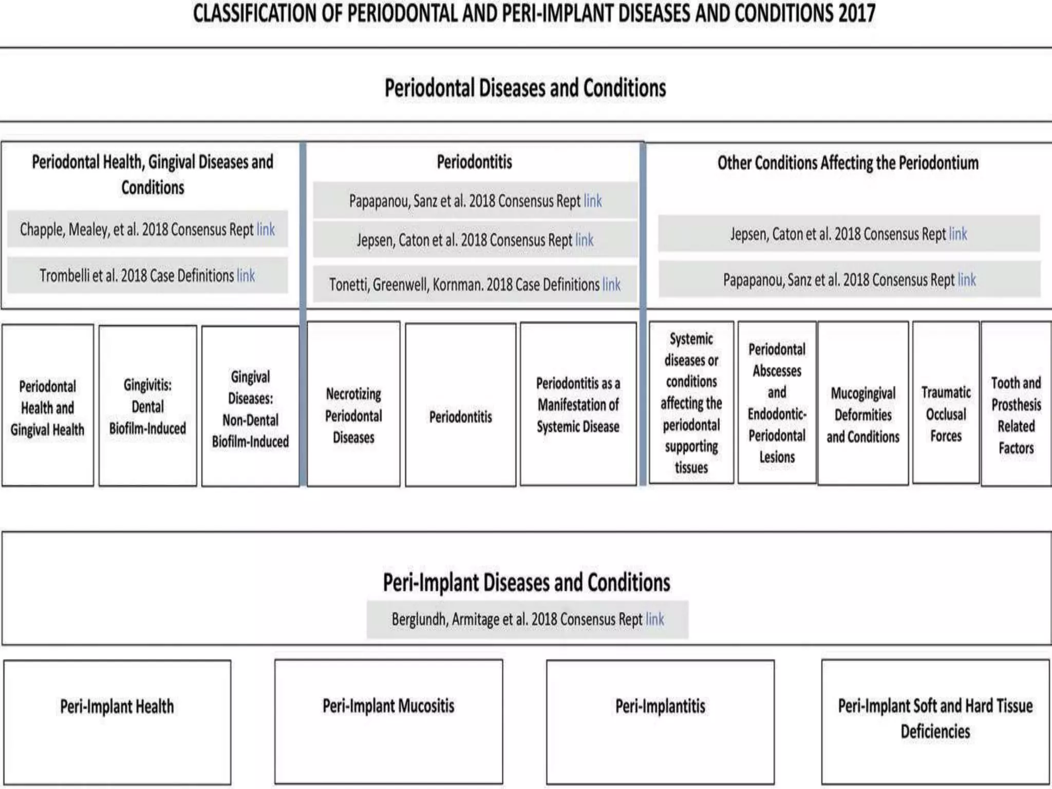
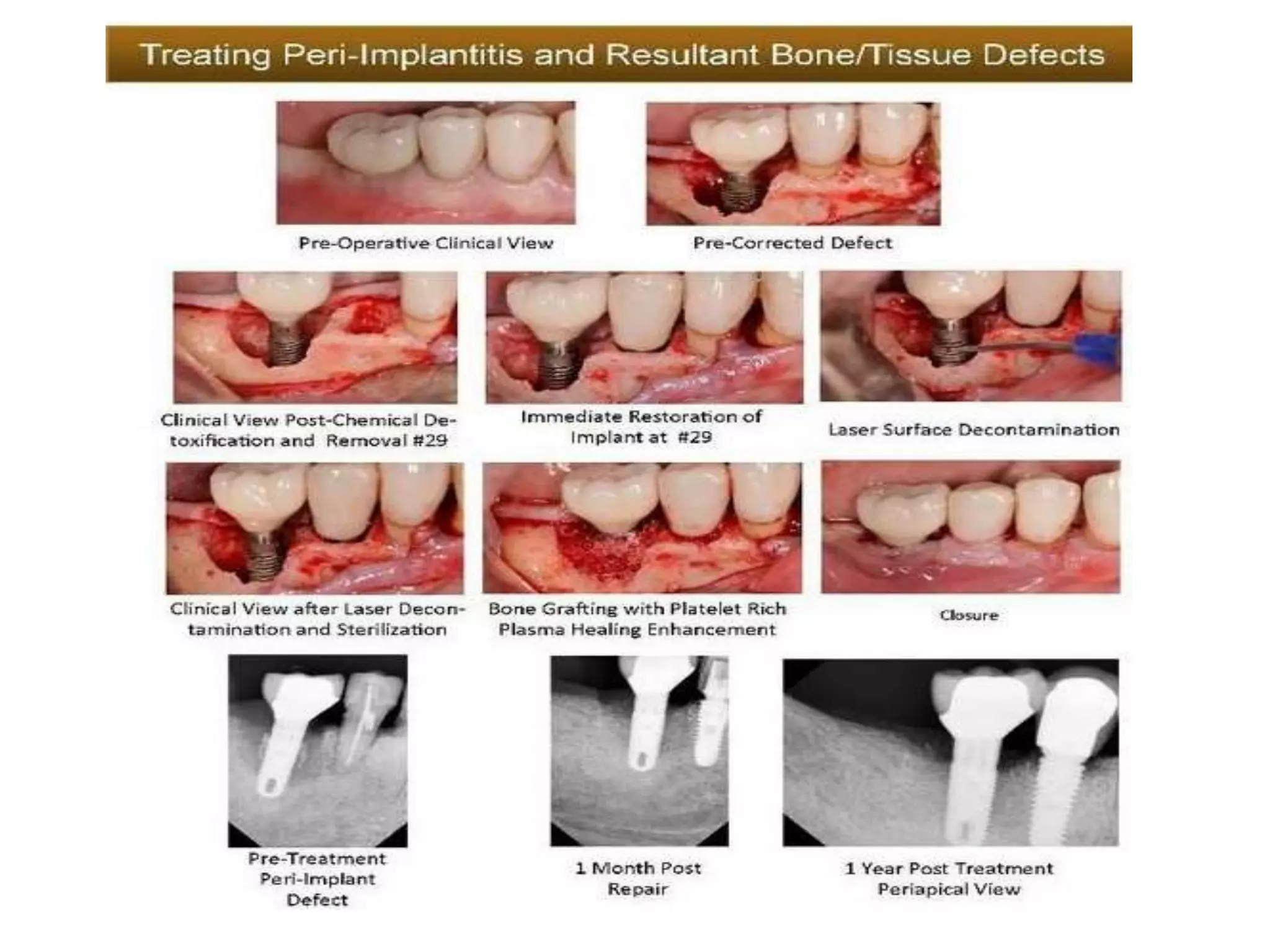
Implant Maintenance
Prof. Dr. Enas Elgendy
Professor of Oral Medicine,
Periodontology& Oral Diagnosis, Faculty
of Dentistry
Kafrelsheikh University
Maintenance of functional
implant
Periodic evaluation of implants, surrounding tissue and
oral hygiene, vital to the long-term success of the dental
implant
For the first year after treatment, recall maintenance
visits should be scheduled at 3-month intervals and then
adjusted to suit the patient's needs.
Once the implants have been placed in the edentulous
region routine maintenance, recall evaluations and
radiographs are necessary to insure the long life of
these restorations, and this need the team of dental
implant specialists.
These procedures are usually performed at selected
intervals to assist the patient in maintaining oral
implant health.
With time, the emphasis for long-term success of
implant has changed from a focus on the surgical phase
of treatment to obtaining osseointegration and, now,
recently, towards the long-term maintenance health of
the peri-implant hard and soft tissues.
The long-term success of implants is dependent upon
both the patient's maintenance of effective home care
and on the dental team's administration of professional
prophylaxis procedures in the dental office.
I-Patient history
II-Clinical examination
III-Investigation and X-ray
Examination of Implant
Step by Step
• Medical and Dental History Look for changes in
systemic risk factors (i.e. diabetes, smoking,
medications)
• New restorations, missing teeth which may
change occlusal relationships.
• Extraoral clinical examination
• Intraoral clinical examination oral soft tissue
evaluation tooth mobility, fremitus, occlusion
caries restorative factors (fracture or defective)
other factors (open contacts)
Step by step
Dental implant examination:
• Presence of plaque and calculus
• Probing depths
• Bleeding on probing
• Implant stability
• Occlusal evaluation
• Other signs and symptoms of disease.
• Radiographic examination (the level of the peri-
implant crestal bone)
1- Evaluation of Biofilm Control
• Poor biofilm control is associated with peri-
implant disease.
• Visually
• The importance of good oral hygiene should be
stressed even before implants are placed, and
peri-implant oral hygiene for biofilm control
should begin as early as possible after the
implant is exposed to the oral cavity.
Methods for Patient Oral Hygiene
• A cotton tip, cotton gauze, or soft toothbrush can
be used to gently remove biofilm from healing
abutments or provisional restorations during the
early postoperative phase of healing.
• Before implant osseointegration, the use of
powered toothbrushes should be avoided.
The rubber tip stimulator can be used to stimulate blood flow.
• AIRFLOW® is the unique approach for regular implant
maintenance providing efficient and safe biofilm
removal.
• AIRFLOW® with PLUS powder is minimally invasive with
no risk of scratching implant surfaces, abutments or
prosthesis.
• AIRFLOW® using PLUS powder is comfortable and safe
around soft tissues.
2- Evaluation of Peri-Implant Health
and Disease
• Peri-implant mucosal health is characterized by
pink, firm, and well-adapted gingival tissue.
• Peri-implant disease is associated with clinical
erythema, edema, and loss of tissue tightness
around the implant.
• In the setting of keratinized, attached mucosa, a
gingival seal or gingival cuff is established around
the implant.
• However, the presence of keratinized, attached
gingiva, which can facilitate oral hygiene, is not a
requisite for periimplant health if biofilm is well
controlled.
Peri-implant Probing
Bleeding on probing at implant sites can indicate inflammation
in the peri-implant mucosa.
Scaling
• Removal of dental biofilm and calculus from implant
components exposed to the oral environment.
• All metal instruments, including metal curettes and
scalers, and ultrasonic scalers increase the surface
roughness of polished titanium.
• The use of plastic, Teflon-coated, and carbon and
gold- coated curettes and nonmetal ultrasonic tips.
Titanium implant scalers
Polishing
• The prosthesis and abutments may be selectively
polished with a rubber cup and nonabrasive
polishing paste such as aluminum oxide, tin
oxide, acidulated phosphate sodium fluoride
(APF) -free prophy paste, and low abrasive
dentifrice after hard deposits have been removed
3- Stability Measures
• The assessment of implant stability or mobility is
an important measure for determining whether
osseointegration is being maintained.
• An implant can exhibit significant bone loss and
remain stable.
• Conversely, if implant mobility is detected, it is
likely that the implant is not surrounded by bone;
• Mobility is highly specific for the detection of
implant failure or lack of osseointegration.
3- Stability Measures
• Two noninvasive techniques for evaluating implant
stability are impact resistance (e.g., Periotest) and
resonance frequency analysis (RFA).
• Originally designed to evaluate tooth mobility
quantitatively, the Periotest is a noninvasive
electronic device that provides an objectaive
measurement of the reaction of the periodontium
to a defined impact load applied to the tooth
crown.
3- Stability Measures
• Another noninvasive method used to
measure the stability of implants is
resonance frequency analysis , which
uses a transducer that is attached to
the implant or abutment.
• A steady-state signal is applied to the
implant through the transducer, and
the response is measured.
• An increase in RFA value indicates
increased implant stability, whereas a
decrease indicates loss of stability.
Mobility remains the cardinal sign of implant failure, and
detecting mobility is therefore an important parameter.
4- Implant Percussion
• Tapping an implant's healing abutment or
restoration with an instrument produces a
sound that can help determine its
osseointegration.
• A solid resonating sound and the absence of
pain usually indicate osseointegration.
• A dull sound can indicate that the implant is
fibrous encapsulated
5- Evaluation of Implant Restorations
• Implant superstructures, and restorations
should be fabricated to accommodate and
facilitate oral hygiene (embrasure spaces
made to allow passage of a proxy brush)
• After delivery, cement retained implant
restorations should be thoroughly evaluated
for residual excess cement, which must be
removed.
5- Evaluation of Implant Restorations
• During follow-up visits, implant
restorations should be carefully
examined for heavy contacts,
fractures, loose screws.
• Occlusion should be adjusted
accordingly to prevent implant
overload and fractures of implant
parts.
• In patients with oral parafunctions
and heavy occlusal forces, occlusal
guards are recommended to
protect implants and restorations.
6- Radiographic Examination
• Intraoral periapical radiographs should be taken at implant
placement, at abutment connection, and at final restoration.
• The radiographic examination remains one of the primary tools
for detection of failed or failing implants in routine clinical
evaluations.
• To monitor marginal or peri-implant bone changes.
• Periapical radiographs have excellent resolution and, when taken
perpendicular to an implant, can provide valuable details
Peri-Implant Mucositis and Peri-Implantitis
• Peri-mucositis is the inflammatory reaction of the
soft tissues surrounding an implant, with no signs
of loss of supporting bone. This reversible
condition is clinically characterized by the
presence of bleeding on probing and/or
suppuration, which are usually associated with
probing depths ≥4 mm.
• Peri-implantitis has been described as a
destructive inflammatory process around an
osseointegrated implant that leads to peri-
implant pocket formation and progressive loss of
supporting bone.
Peri-Implant Mucositis
• Peri-implant mucositis can be effectively
treated with nonsurgical mechanical therapy.
• Treatment requires complete removal of
supramucosal and submucosal biofilm,
calculus, and deposits using curettes,
ultrasonic scalers, and polishing cups with
prophy paste.
• Antimicrobials (e.g., chlorhexidine irrigation,
mouthrinse) can be used with mechanical
debridement to enhance treatment outcome.
Peri-Implantitis
• The treatment of peri-implantitis includes
nonsurgical and surgical interventions
• Nonsurgical interventions consist of antimicrobial
rinse and irrigation, local antibiotics, ultrasonic
debridement, mechanical debridement.
• Surgical treatment includes full-thickness flap
elevation for access, followed by degranulation,
surface debridement by mechanical instruments,
surface decontamination with laser or
antimicrobials, and bone augmentation.
• for peri-implantitis is most effective or to allow
specific recommendations for the use of locally or
systematically administered antibiotics.
Implant maintenance
Implant maintenance
Implant maintenance
Implant maintenance

Implant maintenance

  • 1.
    Implant Maintenance Prof. Dr.Enas Elgendy Professor of Oral Medicine, Periodontology& Oral Diagnosis, Faculty of Dentistry Kafrelsheikh University
  • 2.
    Maintenance of functional implant Periodicevaluation of implants, surrounding tissue and oral hygiene, vital to the long-term success of the dental implant For the first year after treatment, recall maintenance visits should be scheduled at 3-month intervals and then adjusted to suit the patient's needs.
  • 3.
    Once the implantshave been placed in the edentulous region routine maintenance, recall evaluations and radiographs are necessary to insure the long life of these restorations, and this need the team of dental implant specialists. These procedures are usually performed at selected intervals to assist the patient in maintaining oral implant health. With time, the emphasis for long-term success of implant has changed from a focus on the surgical phase of treatment to obtaining osseointegration and, now, recently, towards the long-term maintenance health of the peri-implant hard and soft tissues. The long-term success of implants is dependent upon both the patient's maintenance of effective home care and on the dental team's administration of professional prophylaxis procedures in the dental office.
  • 4.
  • 5.
    Step by Step •Medical and Dental History Look for changes in systemic risk factors (i.e. diabetes, smoking, medications) • New restorations, missing teeth which may change occlusal relationships. • Extraoral clinical examination • Intraoral clinical examination oral soft tissue evaluation tooth mobility, fremitus, occlusion caries restorative factors (fracture or defective) other factors (open contacts)
  • 6.
    Step by step Dentalimplant examination: • Presence of plaque and calculus • Probing depths • Bleeding on probing • Implant stability • Occlusal evaluation • Other signs and symptoms of disease. • Radiographic examination (the level of the peri- implant crestal bone)
  • 7.
    1- Evaluation ofBiofilm Control • Poor biofilm control is associated with peri- implant disease. • Visually • The importance of good oral hygiene should be stressed even before implants are placed, and peri-implant oral hygiene for biofilm control should begin as early as possible after the implant is exposed to the oral cavity.
  • 8.
    Methods for PatientOral Hygiene • A cotton tip, cotton gauze, or soft toothbrush can be used to gently remove biofilm from healing abutments or provisional restorations during the early postoperative phase of healing. • Before implant osseointegration, the use of powered toothbrushes should be avoided.
  • 9.
    The rubber tipstimulator can be used to stimulate blood flow.
  • 10.
    • AIRFLOW® isthe unique approach for regular implant maintenance providing efficient and safe biofilm removal. • AIRFLOW® with PLUS powder is minimally invasive with no risk of scratching implant surfaces, abutments or prosthesis. • AIRFLOW® using PLUS powder is comfortable and safe around soft tissues.
  • 11.
    2- Evaluation ofPeri-Implant Health and Disease • Peri-implant mucosal health is characterized by pink, firm, and well-adapted gingival tissue. • Peri-implant disease is associated with clinical erythema, edema, and loss of tissue tightness around the implant. • In the setting of keratinized, attached mucosa, a gingival seal or gingival cuff is established around the implant. • However, the presence of keratinized, attached gingiva, which can facilitate oral hygiene, is not a requisite for periimplant health if biofilm is well controlled.
  • 12.
    Peri-implant Probing Bleeding onprobing at implant sites can indicate inflammation in the peri-implant mucosa.
  • 13.
    Scaling • Removal ofdental biofilm and calculus from implant components exposed to the oral environment. • All metal instruments, including metal curettes and scalers, and ultrasonic scalers increase the surface roughness of polished titanium. • The use of plastic, Teflon-coated, and carbon and gold- coated curettes and nonmetal ultrasonic tips.
  • 14.
  • 15.
    Polishing • The prosthesisand abutments may be selectively polished with a rubber cup and nonabrasive polishing paste such as aluminum oxide, tin oxide, acidulated phosphate sodium fluoride (APF) -free prophy paste, and low abrasive dentifrice after hard deposits have been removed
  • 16.
    3- Stability Measures •The assessment of implant stability or mobility is an important measure for determining whether osseointegration is being maintained. • An implant can exhibit significant bone loss and remain stable. • Conversely, if implant mobility is detected, it is likely that the implant is not surrounded by bone; • Mobility is highly specific for the detection of implant failure or lack of osseointegration.
  • 17.
    3- Stability Measures •Two noninvasive techniques for evaluating implant stability are impact resistance (e.g., Periotest) and resonance frequency analysis (RFA). • Originally designed to evaluate tooth mobility quantitatively, the Periotest is a noninvasive electronic device that provides an objectaive measurement of the reaction of the periodontium to a defined impact load applied to the tooth crown.
  • 18.
    3- Stability Measures •Another noninvasive method used to measure the stability of implants is resonance frequency analysis , which uses a transducer that is attached to the implant or abutment. • A steady-state signal is applied to the implant through the transducer, and the response is measured. • An increase in RFA value indicates increased implant stability, whereas a decrease indicates loss of stability. Mobility remains the cardinal sign of implant failure, and detecting mobility is therefore an important parameter.
  • 19.
    4- Implant Percussion •Tapping an implant's healing abutment or restoration with an instrument produces a sound that can help determine its osseointegration. • A solid resonating sound and the absence of pain usually indicate osseointegration. • A dull sound can indicate that the implant is fibrous encapsulated
  • 20.
    5- Evaluation ofImplant Restorations • Implant superstructures, and restorations should be fabricated to accommodate and facilitate oral hygiene (embrasure spaces made to allow passage of a proxy brush) • After delivery, cement retained implant restorations should be thoroughly evaluated for residual excess cement, which must be removed.
  • 21.
    5- Evaluation ofImplant Restorations • During follow-up visits, implant restorations should be carefully examined for heavy contacts, fractures, loose screws. • Occlusion should be adjusted accordingly to prevent implant overload and fractures of implant parts. • In patients with oral parafunctions and heavy occlusal forces, occlusal guards are recommended to protect implants and restorations.
  • 22.
    6- Radiographic Examination •Intraoral periapical radiographs should be taken at implant placement, at abutment connection, and at final restoration. • The radiographic examination remains one of the primary tools for detection of failed or failing implants in routine clinical evaluations. • To monitor marginal or peri-implant bone changes. • Periapical radiographs have excellent resolution and, when taken perpendicular to an implant, can provide valuable details
  • 26.
    Peri-Implant Mucositis andPeri-Implantitis • Peri-mucositis is the inflammatory reaction of the soft tissues surrounding an implant, with no signs of loss of supporting bone. This reversible condition is clinically characterized by the presence of bleeding on probing and/or suppuration, which are usually associated with probing depths ≥4 mm. • Peri-implantitis has been described as a destructive inflammatory process around an osseointegrated implant that leads to peri- implant pocket formation and progressive loss of supporting bone.
  • 27.
    Peri-Implant Mucositis • Peri-implantmucositis can be effectively treated with nonsurgical mechanical therapy. • Treatment requires complete removal of supramucosal and submucosal biofilm, calculus, and deposits using curettes, ultrasonic scalers, and polishing cups with prophy paste. • Antimicrobials (e.g., chlorhexidine irrigation, mouthrinse) can be used with mechanical debridement to enhance treatment outcome.
  • 28.
    Peri-Implantitis • The treatmentof peri-implantitis includes nonsurgical and surgical interventions • Nonsurgical interventions consist of antimicrobial rinse and irrigation, local antibiotics, ultrasonic debridement, mechanical debridement. • Surgical treatment includes full-thickness flap elevation for access, followed by degranulation, surface debridement by mechanical instruments, surface decontamination with laser or antimicrobials, and bone augmentation. • for peri-implantitis is most effective or to allow specific recommendations for the use of locally or systematically administered antibiotics.