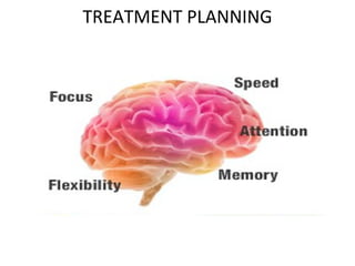
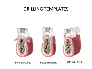
TREATMENT PLANNING
IMPLANTS ADD LIGHT TO LIFE!!
ALGORITHM OF IMPLANT THERAPY
Surgery
Bone healing
period
Second stage
surgery
Early loading period
Recall and Maintainance
Prosthetic treatment
Full functional loading
period
Diagnosis and treatment planning
CASE SELECTION & TREATMENT PLANNING
• Entire chronological sequence of clinical steps
necessary to bring about comprehensive and
successful patient care.
TREATMENT PLANNING AND CASE
SELECTION
• A planned schedule of procedures and
appointments designed to restore patient's
oral health step by step.
The plan contains the advantages,
disadvantages, costs, alternatives, and
sequelae of treatment.
TREATMENT PLANNING AND CASE
SELECTION
• Treatment does not guarantee results
• Not all patients are implant candidates FPD,
RPD &CD still have indications based on
anatomic and other conditions
CRITERIA OF SUCCESS
• Thorough study of stomatoganathic system.
• Supervised learning.
• Customized treatment planning.
STOMATOGANATHIC SYSTEM
• Stomatoganathic system consists of mouth,
jaw and closely associated
structures(anatomy).
• Structures involved in speech.
• Reception, mastication, and deglutition of
food.
TREATMENT PLANNING
TREATMENT PLANNING
TREATMENT PLANNING
TREATMENT PLANNING
TREATMENT PLANNING
TREATMENT PLANNING
TREATMENT PLANNING
TREATMENT PLANNING
TREATMENT PLANNING
IMPLANT SELECTION
TREATMENT PLANNING
TREATMENT PLANNING
BROKEN SCREW
FIRST VISIT
• Reason for the patients visit
• Primary selection of the patient
• Information and motivation
• Medical questionnaire
• Patient examination
EXAMINATION OF PATIENT
• Extra oral examination
• Intra oral
• Study models
• Complimentary examination
1.Medical imaging eg : IOPA,OPG,Scan
2.Bio-chemical tests
EXTRA ORAL EXAMINATION
• Psychologically , functionally & anatomically
• Patients general behaviour
• Palpation for glands and lymph nodes
• Lip line & cheek support
• TMJ disorders
• Facial symmetry
INTRA ORAL EXAMINATION
Visual examination
• Patient’s edentulous status
• Width and length of the operative site
• Malocclusion
• Caries
• Periodontal condition
• Amount of attached gingiva
• Mouth opening
• Gagging reflex
• Teeth attrition
• Oral infections and growths
• Muscle attachment
INTRA ORAL EXAMINATION
Manual palpation
• Palpate edentulous area
• Thickness of soft tissue
• Convexities and concavities
PHYSICAL EXAMINATION
• Evaluation of Mouth opening
• Health of the intraoral soft tissues
• Soft tissue attachment of the floor of the
mouth
• Attachment of mentalis musculature
• Width of the band of keratinized gingival (KG)
on the alveolar crest
• Distance from the crest to the junction of the
attached and unattached mucosa
• Soft tissue examination for vestibuloplasty.
PHYSICAL EXAMINATION
• Locations of the submandibular ducts
• Locations of the mental foramina are palpated and
transferred to a diagnostic cast.
• The slopes of the labial and lingual cortices.
• The height of the mandible
• The location of the genial tubercles.
• In a relaxed vertical position of the jaws, the
relationship of the anterior mandible to the maxilla is
observed.
• Confirmed by radiographic evaluation.
• Discuss need for an adjunctive soft tissue procedure
such as a simultaneous vestibuloplasty.
PHYSICAL EXAMINATION
COMPLIMENTARY EXAMINATION
• Medical history
• Medical imaging
a. IOPA
b. OPG
c. DENTA SCAN
• Bio-chemical tests
• Consent form
BIO-CHEMICAL TESTS
• Serum glucose
Fasting - 80 to 100 mg/dl
Post prandial – 110 to 140 mg/dl
• S. creatinine – 0.8-1.2 mg/dl
• S.urea – 20 to 40 mg/dl
• S. uric acid – 4 to 7 mg/dl
• S. chlolesterol – 150 to 250 mg/dl
• S. bilirubin – 0.2 to 1 mg/dl
• SGOT - Serum Glutamic Oxalacetic .
Transaminase – 5 to 35 IU
BIO-CHEMICAL TESTS
• SGPT –Serum Glutamic Pyruvic
Transaminase
• Gamma glutamic transpeptidase - <25 mU
• Alkaline phosphatase – 13 to 39 IU
• Serum calcium – 10mg/dl
• Serum phosphates – 1.5 to 6.8 mg/dl
a. Reciprocal relationship with calcium one>-other<????????
b. Ratio of phosphates to calcium is 4 : 10
• CBC
• Blood tests for hemorrhagic conditions
STUDY CASTS
• To study patients occlusion especially
combination syndrome
• Bone mapping
• To plan prosthetic driven surgery
• Cast open to discussion with colleagues and
patient
• To design surgical templates
• To keep patients record
• To educate & motivate patient
BONE MAPPING
BONE MAPPING
BONE MAPPING
BONE MAPPING
BONE MAPPING
BONE MAPPING
BONE MAPPING
STUDY CASTS
STUDY CASTS
Surgical template
Surgical template
TREATMENT PLANNING
DRILLING TEMPLATES
DRILLING TEMPLATES
• Mucosa – supported (polymer resin)templates
• Tooth – supported templates
• Bone – supported templates
• Implant supported templates
DRILLING TEMPLATES
Bone supported Tissue supported Tooth supported
TREATMENT PLANNING FOR EDENTULOUS JAW
• Options :
1. Conventional denture
2. Tissue-borne - implant-supported
prosthesis
3. Implant-supported prosthesis
TREATMENT PLANNING FOR EDENTULOUS JAW
• The tissue-borne removable prosthesis -
2 – 4 implants
• Implant-supported prosthesis -
4 – 10 implants (meticulous planning).
SIMPLE SOLUTIONS
RADIOLOGIC EVALUATION
• Focused on determining the vertical height and
slopes of the cortices in relation to the opposite arch.
• OPG & lateral Ceph. is typically used.
• Usually magnified.
• Ball bearings of known diameter can be placed in a
stent in the positions that are prescribed to receive
implants, and then magnification error can be
determined and the correct vertical dimension
calculated.
• If the mandible is greater then 15 mm, the
panoramic radiograph is the only film needed.
• If width is doubtful, then Dental scans are preferred.
Radiographic template
LATERAL CEPH
LATERAL CEPH
DETERMINING CANINE POSITION
Alla of the nose depicts the mesial position of the canine.
Corner of the mouth depicts the distal aspect of the canine.
CB D
EA
TREATMENT OPTIONS FOR EDENTULOUS
MANDIBULAR RIDGE
OPTION 1
C
B D
E
A
OPTION 2
CB D
EA
OPTION 3
CB D
EA
OPTION 4
CB D
EA
OPTION 5
CB D
EA
OPTION 6
CB D
EA
3 mm
7 mm 8 mm 8 mm 7 mm
3 mm
TREATMENT OPTIONS FOR EDENTULOUS
MAXILLARY RIDGE
TREATMENT OPTIONS FOR EDENTULOUS
MAXILLARY RIDGE
TREATMENT OPTIONS FOR EDENTULOUS
MAXILLARY RIDGE
TREATMENT PLANNING
TREATMENT PLANNING
TREATMENT PLANNING
O-ring attachment Maxilla
Telescopic attachment Maxilla
Bar attachment Maxilla
TREATMENT PLANNING
TREATMENT PLANNING
TREATMENT PLANNING
Denture duplication
DRILLING TEMPLATE
DRILLING TEMPLATE
DRILLING TEMPLATE
Surgical site
INCISION DESIGN
• Crestal
• Vesibular Incision
ATROPHIC MANDIBLE 8-12MM
• If thin band of KG is lingually positioned. Soft tissue grafting
fails.
• Implants placed lingual to the crest of the ridge, lingual to the
attachment of the muscles.
• If labially chronic irritation causes soreness.
• Mucosal recession.
• No vestibuloplasty.(witch like appearance)
ATROPHIC MANDIBLE
• Incision crestal or vestibular .
• Incision bisecting the KG allows it to remain on the
labial surface
• Vestibuloplasty if needed.
MANDIBLE >12MM
PREMATURE BREAKDOWN OF INCISION
• excessive pressure from the removable prosthesis,
• supracrestal profile of the implant with cover screw in place
• surgical trauma to the tissues,
• poor tissue quality and
• poor healing.
• Poor incision design.
• Poor incision & suturing.
• If the alveolar crest is thin with the band of KG over the thin
portion of the crest, bisecting the KG may create a difficult
dissection of the flaps because the gingiva will be thin over
the thin crest.
• In case of traumatic dissection- Layered suturing.
• Avoid re-suturing.
INCISION BREAKDOWN
INCISION BREAKDOWN
PARTS OF A DENTAL IMPLANT
IMPLANT BODY
OFTEN REFERRED TO AS AN IMPLANT.
PARTS OF A DENTAL IMPLANT
COVER SCREW
PREVENTS BONE INGRESS INTO IMPLANT HEAD.
PARTS OF A DENTAL IMPLANT
HEALING ABUTMENT
PLACED TEMPORARILY ON THE IMPLANT BODY
TO MAINTAIN POTENCY OF THE MUCOSAL PENETRATION.
PARTS OF A DENTAL IMPLANT
TEMPORARY COMPONENTS
PRE-MANUFACTURED COMPONENTS
USED TO MAKE TEMPORARY
CROWNS AND BRIDGES FOR
FITTING ON DENTAL IMPLANTS AND
ABUTMENTS.
PARTS OF A DENTAL IMPLANT
IMPRESSION COPING
USED TO TRANSFER THE LOCATION
OF THE IMPLANT BODY
OR ABUTMENT TO A DENTAL CAST.
PARTS OF A DENTAL IMPLANT
LABORATORY ANALOGUE
A BASE METAL REPLICA OF
IMPLANT BODY.
PARTS OF A DENTAL IMPLANT
INSERTION OF HEALING COLLAR
SEVEN DAYS LATER…
SEVEN DAYS LATER…
PARTS OF A DENTAL IMPLANT
ABUTMENT
LINKS THE IMPLANT BODY TO THE MOUTH.
Bone quantity & quality
Type-A
Type-A
Type-B
Type-C
Type-D
SURGICAL KITS
INTRODUCTION
MIS
IMPLANT SURGICAL KIT
SURGICAL PROCEDURE
INCISION ROUND MARKER DRILL PILOT DRILL
SURGICAL STOPS
PILOT DRILL
SURGICAL PROCEDURE
PARALLELING PIN WIDENING DRILL
SURGICAL DRILLS
WIDENING DRILL
WIDENING DRILL
SURGICAL DRILLS
WIDENING DRILL WIDENING DRILL WIDENING DRILL
SURGICAL PROCEDURE
REAMING TAPPING
SURGICAL PROCEDURE
IMPLANT PLACEMENT
SURGICAL PROCEDURE
PLACEMENT OF COVER SCREW
&
CLOSURE
Rule of 1, 2 , 3 , 7
Sat sri akal

Case selection & treatment planning

Editor's Notes

  • #6 Length of crown Missing papilla Broad crowns Food impaction Speech Hygiene Adjacent teeth are preserved Dental implants help to preserve the health of natural teeth as they share the occlusion load. Preserves occlusion Hygienic One additional and very important benefit can be reduction or elimination of bone shrinkage or atrophy commonly related to tooth loss Maintenance of VDO Regained proprioception Increased stability, retention and support of soft tissue Maintenance of muscles of mastication and facial expression Improved phonetics and psychological health SMILE WITH CONFIDENCE Prosthesis over implants lasts longer Replacement of teeth look, feel and function more like natural teeth . Improve taste and appetite (less coverage of the palate ) Improve cosmetic appearance. Ability to chew without pain and gum irritation . Foods that were unable to eat are back in the diet. No longer need to use the distasteful adhesives.
  • #28 Limitations in opening may affect the treatment plan in extreme conditions. The presence of undiagnosed pathologic and dental infection, as well as mucosal infections, must be treated to completion before implant placement.
  • #33 The prothrombin time (PT) and its derived measures of prothrombin ratio (PR) and international normalized ratio (INR) are measures of the extrinsic pathway of coagulation. They are used to determine the clotting tendency of blood, in the measure of warfarin dosage, liver damage, and vitamin K status. PT measures factors I, II, V, VII, and X. It is used in conjunction with the activated partial thromboplastin time (aPTT) which measures the intrinsic pathway. he reference range for prothrombin time is usually around 11–16 seconds; the normal range for the INR is 0.8–1.2. Clinicians desiring therapeutic anticoagulation may aim for a higher INR - in many cases 2.5 - using anticoagulants such as warfarin.[1 The prothrombin time can be prolonged as a result of deficiencies in vitamin K, which can be caused by warfarin, malabsorption, or lack of intestinal colonization by bacteria (such as in newborns). In addition, poor factor VII synthesis (due to liver disease) or increased consumption (in disseminated intravascular coagulation) may prolong the PT.
  • #54 The surgical placement of implants for a full-arch crown and bridge prosthesis with individual porcelain teeth requires meticulous planning and placement of implants to locate them within the confines of the crown, avoiding the embrasure spaces.
  • #55 Retentive elements
  • #56 ). The lateral cephalogram demonstrates the slopes of the cortices of the mandible and the skele­tal ridge relationships of the mandible to the maxilla, and it provides a simple and inexpensive radiograph­ic assessment of anterior height. If desired, foil can be placed over the anterior teeth in a set of dentures, which if worn during exposure of the lateral cephalogram, can demonstrate clearly the relationship of the teeth to the ridge and the ridge relationships. This provides insight as to angulation of the implants to place then in ideal locations for the implant supported or tissue supported prostheses.
  • #68 The fixed implant rehabilitation of edentulous maxillas with favorable anatomy and favorable arch position, favorable anatomy, and unfavorable arch position, and unfavorable anatomy and favorable arch position.
  • #85 If the attachment of the mentalis muscle is 3 mm or more labial to the location of the attached gingiva on the alveolar crest, a crestal incision can be If the location of the mentalis muscle is adjacent to the alveolar crest, which would result in mobile unattached gingiva directly against the implant abutment when restored, a vestibular incision is used. A type of lipswitch vestibuloplasty is performed to reposition the muscle attachments infe­riorly, resulting in nonmobile tissue on the labial -sur­face of the implant abutment complex. Difficult dissection. lipswitch vestibuloplasty contraindicated because displacement of the mentalis musculature in an atrophic mandible will result in a drooping, &amp;quot;witch­like&amp;quot; chin deformity. 10-15mm Mentalis muscle attachment is must
  • #86 For the atrophic mandible with 12 to 8 mm vertical bone height, the locations of the incisions and implants and the location of the incision for second-stage surgery are critical for successful restoration. The incision for placement should be placed to avoid loss of KG. Thus the incision placed at the anterior border of the mandibular alveolar crest, typically labial to the KG, allows for an adequate dissection.
  • #87 Attempts to enlarge the band of attached ,KG do not have a high rate of success because the lip muscles tend to displace the graft from the host bed.
  • #88 CRESTAL INCISION AND DISSECTION. The crestal incision should bisect the band of KG. Bisecting the KG is important to avoid a potential soft tissue problem if the incision should open during the healing period. The incision should extend along the alveolar crest posterior to the mental foramen. When the mental forame is on top of the crest, secondary to severe bone resorption, the incision should be stopped anterior to the foramen. After the Periosteum has been flected, the mental foramen is visualized and the crestial incision can then be extended posteriorly along the lingual crest, avoiding trauma to the nerve. Occasionally, vertical release incisions can be used posteriorly. This author avoids a midline verti­cal release incision because of increased patient dis­comfort during the first 2 weeks of healing. After the incision is made through the perios‑teum to the bone, a periosteal elevator is used toreflect subperiosteal flaps both labially and lingually. A clean subperiosteal dissection is important to perform because bleeding is minimal and large lingual blood vessels can be avoided. If muscle attachments are found inserting into the crest, a scalpel is used to sever them cleanly rather than tearing them, which subsequently increases bleeding and trauma to the soft tissue. The reflection of the labial tissues can be tedious because of the firm attachment of the dense fibrous alveolar crestal tissue or if the ridge is narrow. Great care should be taken to raise an intact flap without multiple tears