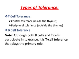
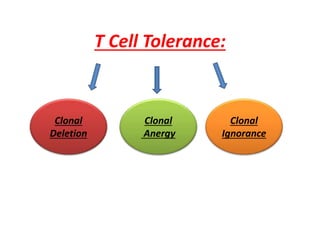
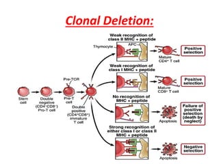
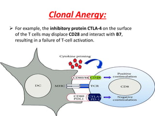
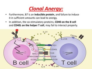
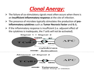
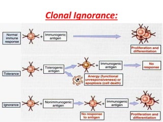
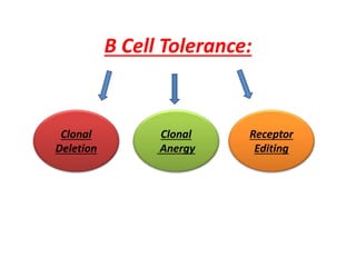
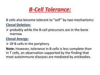
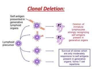
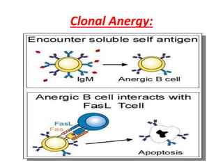
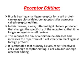
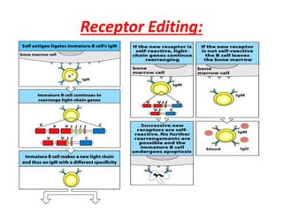
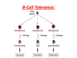
“Tolerance”
Presented by:
Ahmad Aman (Pharm-D)
Riphah Institute of Pharmaceutical
Sciences
Tolerance:
“Tolerance is specific immunologic unresponsiveness
(i.e., an immune response to a certain antigen [or
epitope] does not occur, although the immune system is
otherwise functioning normally).”
 In general, antigens that are present during embryonic
life are considered “self ” and do not stimulate an
immunologic response (i.e., we are tolerant to those
antigens).
 The lack of an immune response in the fetus is caused
by the deletion of self-reactive T-cell precursors in the
thymus.
Types of Tolerance:
T Cell Tolerance
Central tolerance (inside the thymus)
Peripheral tolerance (outside the thymus)
B Cell Tolerance
Note: Although both B cells and T cells
participate in tolerance, it is T-cell tolerance
that plays the primary role.
Types of Tolerance:
Tolerance to “self” acquired within the thymus is
called central tolerance.
Whereas, the tolerance acquired outside the
thymus is called peripheral tolerance. ( mostly in
spleen, lymph nodes)
Peripheral tolerance is necessary because some
antigens are not expressed in the thymus and
therefore some self reactive T cells are not killed
in the thymus.
T Cell Tolerance:
Clonal
Deletion
Clonal
Anergy
Clonal
Ignorance
Clonal Deletion:
T lymphocytes acquires the ability to distinguish
“self” from “non-self” in the fetal thymus by the
process called clonal deletion.
It is the process that involves the killing of T cells
(“negative selection”) that react against antigens
present in the fetus at that time.
The self reactive cells die by a process of
programmed cell death called apoptosis.
For negative selection and clonal deletion to be
efficient, the thymic epithelial cells must display a
vast repertoire of “self ” proteins.
Clonal Deletion:
Clonal Deletion:
 A transcriptional regulator called the autoimmune
regulator (AIRE) enhances the synthesis of this array of
self proteins.
 The AIRE transcription factor also functions in the
peripheral lymphoid organs such as the spleen and
lymph nodes, where it contributes to peripheral
tolerance.
 Mutations in the gene encoding the AIRE protein result
in the development of an autoimmune disease called
autoimmune polyendocrinopathy.
Polyendocrinopathy:
 APS type 1, also known as APECED (autoimmune
polyendocrinopathy-candidiasis-ectodermal dystrophy).
 MEDAC (multiple endocrine deficiency autoimmune
candidiasis syndrome).
 Juvenile autoimmune polyendocrinopathy.
 Whitaker's syndrome.
Polyendocrinopathy:
a) Vitiligo b) Alopecia Areata:
Clonal Anergy:
“Clonal anergy is the term used to describe self-
reactive T cells that are not activated because proper
co-stimulation does not occurs.”
Mechanism of clonal anergy:
 Inappropriate presentation of antigen, leading to a
failure of interleukin-2 (IL-2) production.
 Inappropriate presentation is due to a failure of
“co-stimulatory signals”
 For instance, sufficient amounts of IL-1 might not
be made, or cell surface proteins, such as CD28 on
the T cell and B7 on the B cell, might not interact
properly, leading to a failure of signal transduction
by RAS proteins.
Normal Activation of T cell:
Clonal Anergy:
 For example, the inhibitory protein CTLA-4 on the surface
of the T cells may displace CD28 and interact with B7,
resulting in a failure of T-cell activation.
Clonal Anergy:
• Furthermore, B7 is an inducible protein, and failure to induce
it in sufficient amounts can lead to anergy.
• In addition, the co-stimulatory proteins, CD40 on the B cell
and CD40L on the helper T cell, may fail to interact properly.
Clonal Anergy:
 The failure of co-stimulatory signals most often occurs when there is
an insufficient inflammatory response at the site of infection.
 The presence of microbes typically stimulates the production of pro-
inflammatory cytokines such as Tumor Necrosis Factor and IL-1.
 If the inflammatory response is insufficient (i.e. adjuvant effect of
the cytokines is inadequate, the T cells will not be activated).
Clonal Ignorance:
“Clonal ignorance refers to self-reactive T cells that
ignore self antigens.”
 These self-reactive T cells are either kept ignorant by
physical separation from the target antigens (e.g.,
the blood–brain barrier).
 Or ignorance to self antigens may occurs if the
antigens are present in small amounts.
Clonal Ignorance:
B Cell Tolerance:
Clonal
Deletion
Clonal
Anergy
Receptor
Editing
B-Cell Tolerance:
B cells also become tolerant to “self” by two mechanisms:
Clonal Deletion:
 probably while the B-cell precursors are in the bone
marrow.
Clonal Anergy:
 Of B cells in the periphery.
Note: However, tolerance in B cells is less complete than
in T cells, an observation supported by the finding that
most autoimmune diseases are mediated by antibodies.
Clonal Deletion:
Clonal Anergy:
Receptor Editing:
 B cells bearing an antigen receptor for a self protein
can escape clonal deletion (apoptosis) by a process
called receptor editing.
 In this process, a new, different light chain is produced
that changes the specificity of the receptor so that it no
longer recognizes a self protein.
 This reduces the risk of autoimmune diseases and
increases the repertoire of B cells that can react against
foreign proteins.
 It is estimated that as many as 50% of self-reactive B
cells undergo receptor editing. T cells do not undergo
receptor editing.
Receptor Editing:
B-Cell Tolerance:
Induction Of Tolerance:
Whether an antigen will induce tolerance rather than an
immunologic response is largely determined by the following:
The immunologic maturity of the host:
 Neonatal animals are immunologically immature and do
not respond well to foreign antigens.
The structure and dose of the antigen:
 A very simple molecule induces tolerance more readily
than a complex one.
 Very high or very low doses of antigen may result in
tolerance instead of an immune response.
 Purified polysaccharides or amino acid copolymers injected
in very large doses result in “immune paralysis” —a lack of
response.
Induction Of Tolerance:
Other aspects of the induction or maintenance of
tolerance are as follows:
 T cells become tolerant more readily and remain
tolerant longer than B cells.
 Administration of a cross-reacting antigen tends to
terminate tolerance.
 Administration of immunosuppressive drugs
enhances tolerance (e.g., in patients who have
received organ transplants).
 Tolerance is maintained best if the antigen to which
the immune system is tolerant continues to be
present.
Immunological tolerance

Immunological tolerance

  • 1.
    “Tolerance” Presented by: Ahmad Aman(Pharm-D) Riphah Institute of Pharmaceutical Sciences
  • 2.
    Tolerance: “Tolerance is specificimmunologic unresponsiveness (i.e., an immune response to a certain antigen [or epitope] does not occur, although the immune system is otherwise functioning normally).”  In general, antigens that are present during embryonic life are considered “self ” and do not stimulate an immunologic response (i.e., we are tolerant to those antigens).  The lack of an immune response in the fetus is caused by the deletion of self-reactive T-cell precursors in the thymus.
  • 3.
    Types of Tolerance: TCell Tolerance Central tolerance (inside the thymus) Peripheral tolerance (outside the thymus) B Cell Tolerance Note: Although both B cells and T cells participate in tolerance, it is T-cell tolerance that plays the primary role.
  • 4.
    Types of Tolerance: Toleranceto “self” acquired within the thymus is called central tolerance. Whereas, the tolerance acquired outside the thymus is called peripheral tolerance. ( mostly in spleen, lymph nodes) Peripheral tolerance is necessary because some antigens are not expressed in the thymus and therefore some self reactive T cells are not killed in the thymus.
  • 5.
  • 6.
    Clonal Deletion: T lymphocytesacquires the ability to distinguish “self” from “non-self” in the fetal thymus by the process called clonal deletion. It is the process that involves the killing of T cells (“negative selection”) that react against antigens present in the fetus at that time. The self reactive cells die by a process of programmed cell death called apoptosis. For negative selection and clonal deletion to be efficient, the thymic epithelial cells must display a vast repertoire of “self ” proteins.
  • 7.
  • 8.
    Clonal Deletion:  Atranscriptional regulator called the autoimmune regulator (AIRE) enhances the synthesis of this array of self proteins.  The AIRE transcription factor also functions in the peripheral lymphoid organs such as the spleen and lymph nodes, where it contributes to peripheral tolerance.  Mutations in the gene encoding the AIRE protein result in the development of an autoimmune disease called autoimmune polyendocrinopathy.
  • 9.
    Polyendocrinopathy:  APS type1, also known as APECED (autoimmune polyendocrinopathy-candidiasis-ectodermal dystrophy).  MEDAC (multiple endocrine deficiency autoimmune candidiasis syndrome).  Juvenile autoimmune polyendocrinopathy.  Whitaker's syndrome.
  • 10.
  • 11.
    a) Vitiligo b)Alopecia Areata:
  • 12.
    Clonal Anergy: “Clonal anergyis the term used to describe self- reactive T cells that are not activated because proper co-stimulation does not occurs.” Mechanism of clonal anergy:  Inappropriate presentation of antigen, leading to a failure of interleukin-2 (IL-2) production.  Inappropriate presentation is due to a failure of “co-stimulatory signals”  For instance, sufficient amounts of IL-1 might not be made, or cell surface proteins, such as CD28 on the T cell and B7 on the B cell, might not interact properly, leading to a failure of signal transduction by RAS proteins.
  • 13.
  • 14.
  • 15.
    Clonal Anergy:  Forexample, the inhibitory protein CTLA-4 on the surface of the T cells may displace CD28 and interact with B7, resulting in a failure of T-cell activation.
  • 16.
    Clonal Anergy: • Furthermore,B7 is an inducible protein, and failure to induce it in sufficient amounts can lead to anergy. • In addition, the co-stimulatory proteins, CD40 on the B cell and CD40L on the helper T cell, may fail to interact properly.
  • 17.
    Clonal Anergy:  Thefailure of co-stimulatory signals most often occurs when there is an insufficient inflammatory response at the site of infection.  The presence of microbes typically stimulates the production of pro- inflammatory cytokines such as Tumor Necrosis Factor and IL-1.  If the inflammatory response is insufficient (i.e. adjuvant effect of the cytokines is inadequate, the T cells will not be activated).
  • 18.
    Clonal Ignorance: “Clonal ignorancerefers to self-reactive T cells that ignore self antigens.”  These self-reactive T cells are either kept ignorant by physical separation from the target antigens (e.g., the blood–brain barrier).  Or ignorance to self antigens may occurs if the antigens are present in small amounts.
  • 19.
  • 20.
  • 21.
    B-Cell Tolerance: B cellsalso become tolerant to “self” by two mechanisms: Clonal Deletion:  probably while the B-cell precursors are in the bone marrow. Clonal Anergy:  Of B cells in the periphery. Note: However, tolerance in B cells is less complete than in T cells, an observation supported by the finding that most autoimmune diseases are mediated by antibodies.
  • 22.
  • 23.
  • 24.
    Receptor Editing:  Bcells bearing an antigen receptor for a self protein can escape clonal deletion (apoptosis) by a process called receptor editing.  In this process, a new, different light chain is produced that changes the specificity of the receptor so that it no longer recognizes a self protein.  This reduces the risk of autoimmune diseases and increases the repertoire of B cells that can react against foreign proteins.  It is estimated that as many as 50% of self-reactive B cells undergo receptor editing. T cells do not undergo receptor editing.
  • 25.
  • 26.
  • 27.
    Induction Of Tolerance: Whetheran antigen will induce tolerance rather than an immunologic response is largely determined by the following: The immunologic maturity of the host:  Neonatal animals are immunologically immature and do not respond well to foreign antigens. The structure and dose of the antigen:  A very simple molecule induces tolerance more readily than a complex one.  Very high or very low doses of antigen may result in tolerance instead of an immune response.  Purified polysaccharides or amino acid copolymers injected in very large doses result in “immune paralysis” —a lack of response.
  • 28.
    Induction Of Tolerance: Otheraspects of the induction or maintenance of tolerance are as follows:  T cells become tolerant more readily and remain tolerant longer than B cells.  Administration of a cross-reacting antigen tends to terminate tolerance.  Administration of immunosuppressive drugs enhances tolerance (e.g., in patients who have received organ transplants).  Tolerance is maintained best if the antigen to which the immune system is tolerant continues to be present.