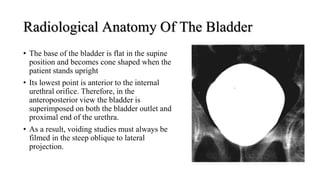
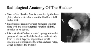
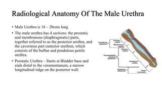
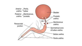
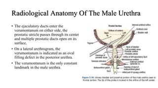
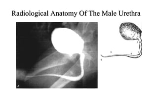
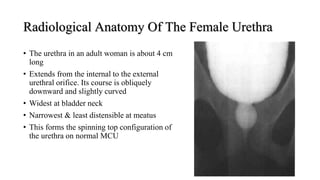
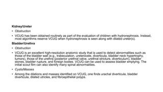
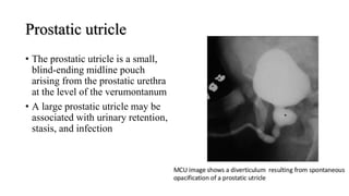
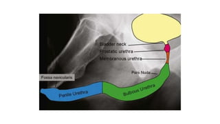
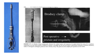
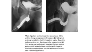
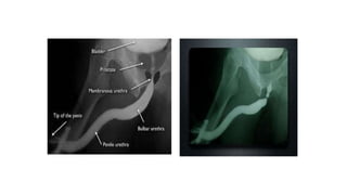
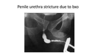
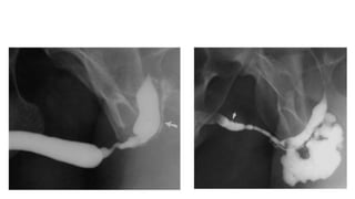
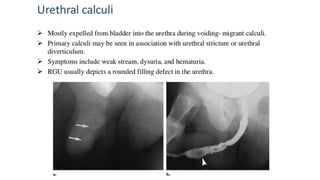
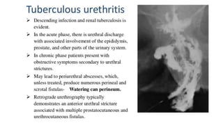
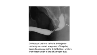
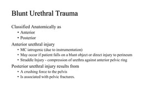
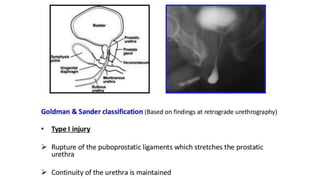
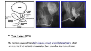
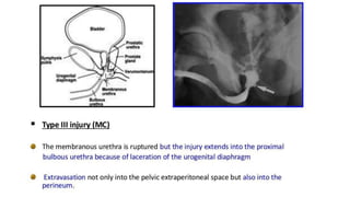
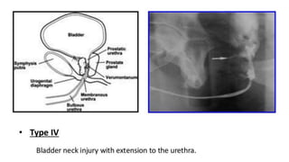
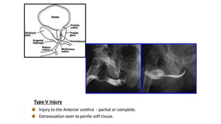
MCU & AUG
Radiological Anatomy Of The Bladder
โ€ข In a newborn infant the bladder lies above the
symphysis pubis , but as the child grows, the
bladder descends, until at about 5 years of age
the bladder floor lies at or somewhat below the
level of the symphysis pubis.
โ€ข Shopfner and Hutch divide the bladder into two
units: the vault and the bladder floor.
โ€ข When the patient is in either the supine or the
upright position, the top of the bladder appears
round.
โ€ข Sometimes an indentation is seen at both sides of
the distended bladder and gives the appearance
of a โ€œwaistโ€
Radiological Anatomy Of The Bladder
โ€ข The base of the bladder is flat in the supine
position and becomes cone shaped when the
patient stands upright
โ€ข Its lowest point is anterior to the internal
urethral orifice. Therefore, in the
anteroposterior view the bladder is
superimposed on both the bladder outlet and
proximal end of the urethra.
โ€ข As a result, voiding studies must always be
filmed in the steep oblique to lateral
projection.
Radiological Anatomy Of The Bladder
โ€ข Most of the bladder floor is occupied by the base
plate, which is circular when the bladder is full
and at rest.
โ€ข It consists of an anterior and posterior trigonal
plate with the vesicourethral junction slightly
anterior to its center.
โ€ข It is best identified on a lateral cystogram as the
posteroinferior wall of the bladder and extends
from its most dependent point to a small
indentation representing the inter-ureteric ridge,
which is part of the trigone
Radiological Anatomy Of The Male Urethra
โ€ข Male Urethra is 18 โ€“ 20cms long
โ€ข The male urethra has 4 sections: the prostatic
and membranous (diaphragmatic) parts,
together referred to as the posterior urethra, and
the cavernous part (anterior urethra), which
consists of the bulbar and pendulous penile
urethra.
โ€ข Prostatic Urethra โ€“ Starts at Bladder base and
ends distal to the verumontanum, a narrow
longitudinal ridge on the posterior wall.
Radiological Anatomy Of The Male Urethra
โ€ข The ejaculatory ducts enter the
verumontanum on either side, the
prostatic utricle passes through its center,
and multiple prostatic ducts open on its
surface.
โ€ข On a lateral urethrogram, the
verumontanum is indicated as an oval
filling defect in the posterior urethra.
โ€ข The verumontanum is the only constant
landmark in the male urethra.
Radiological Anatomy Of The Male Urethra
Radiological Anatomy Of The Female Urethra
โ€ข The urethra in an adult woman is about 4 cm
long
โ€ข Extends from the internal to the external
urethral orifice. Its course is obliquely
downward and slightly curved
โ€ข Widest at bladder neck
โ€ข Narrowest & least distensible at meatus
โ€ข This forms the spinning top configuration of
the urethra on normal MCU
CYSTOURETHEROGRAM
Voiding Cystourethrogram
โ€ข VCU is the most widely used examination for radiological evaluation of the
bladder and urethra in both children and adults
Indications
Children
โ€ข Recurrent UTI
โ€ข Voiding difficulties
โ€ข VUR
โ€ข Meningomyelocele, sacral
agenesis, Rectal anomalies
โ€ข Baseline study prior to lower
urinary tract surgery
โ€ข Post-op evaluation of ureteric
abnormalities
โ€ข Pelvic trauma
โ€ข In renal failure to exclude reflux
โ€ข Posterior urethral valve or polyp
โ€ข Congenital anomalies of bladder
& urethra.
Indications
โ€ข Trauma to urethra
โ€ข Urethral stricture
โ€ข Urethral diverticulum
โ€ข Recurrent UTI
โ€ข Reflux nephropathy prior to renal transplant
โ€ข Follow up patients of spinal cord injury
โ€ข Stress incontinence
Kidney/Ureter
โ€ข Obstruction
โ€ข VCUG has been obtained routinely as part of the evaluation of children with hydronephrosis. Instead,
most algorithms reserve VCUG when hydronephrosis is seen along with dilated ureter(s).
Bladder/Urethra
โ€ข Obstruction
โ€ข VCUG is an excellent high-resolution anatomic study that is used to detect abnormalities such as
those of the bladder wall (e.g., trabeculation, ureterocele, diverticula, bladder neck hypertrophy,
tumors), those of the urethra (posterior urethral valve, urethral stricture, diverticulum), bladder
stones, bladder rupture, and foreign bodies. VCUG can be used to assess bladder emptying. The
initial scout film can also identify many spinal abnormalities.
โ€ข Cysts/Masses
โ€ข Among the dilations and masses identified on VCUG, one finds urachal diverticula, bladder
diverticula, dilated utricles, and fibroepithelial polyps.
Technique
โ€ข No preparation is needed; no cleansing enema, fasting, or anesthesia is
required
โ€ข To rule out UTI
โ€ข Bladder capacity
โ€ข Less than one year,
โ€ข Weight (kg) x 7 = capacity (ml)
โ€ข Less than two years,
โ€ข (2 x age in years + 2) x 30 = capacity (ml)
โ€ข More than two years,
โ€ข (Age in years/2 + 6) x 30 = capacity (ml)
Technique
โ€ข With sterile technique, a catheter is introduced into the bladder (Infants 5-7 Fr
feeding tube and Foleys for adults)
โ€ข A No. 5 feeding tube with side holes is used for babies, and in older children, No. 8
or 10 polyethylene or soft rubber catheters with end holes are quite suitable
โ€ข Once the catheter has safely reached the bladder and residual urine has been
removed, filling with contrast material can begin.
Technique
โ€ข Methods of Instillation
โ€ข Hand-injected
โ€ข Gravity method, which is usually less uncomfortable for the child.
โ€ข An infusion set is connected to the catheter, and the contrast- filled bottle is suspended above
the table to a height of 35 to 40 cm
โ€ข Capacity is reached when the child becomes uncomfortable and restless or begins
voiding around the catheter or when bladder and infusion pressure becomes equal
and flow ceases or reverses. Forceful flexion of the toes sometimes signifies that
bladder capacity has been reached
โ€ข In Adults, The patient can manually compress the meatus to improve visualization of
the urethra.
โ€ข Bladder trabeculation, vesicoureteral reflux and narrowing of the bladder neck or
urethra, if present, will be successfully demonstrated.
Alternative Techniques
โ€ข Suprapubic bladder puncture
โ€ข Sometimes in PUV & pelvic trauma - not possible to catheterize.
โ€ข Urethrocystography
โ€ข Contrast medium introduced into the bladder during RGU.
โ€ข Excretion MCU ( MCU followed by IVU )
โ€ข Advantage - avoid catheterization and related risk of infection.
โ€ข Disadvantage - VUR can not be visualized properly takes longer time
Posterior urethral Valve
โ€ข Congenital thick folds of mucous membrane located in the posterior
urethra (prostatic + membranous) distal to the verumontanum.
โ€ข Most common cause of severe obstructive uropathy in children.
โ€ข Almost exclusively in males.
โ€ข Leading cause of end stage renal disease in boys.
โ€ข Now rare for them to present with severe UTI and septicaemia -
diagnosis is generally made in early infancy and antenatal period.
โ€ข Procedure of choice for defining the valves.
Prostatic utricle
โ€ข The prostatic utricle is a small,
blind-ending midline pouch
arising from the prostatic urethra
at the level of the verumontanum
โ€ข A large prostatic utricle may be
associated with urinary retention,
stasis, and infection
Complications
โ€ข Contrast reaction
โ€ข Contrast induced cystitis
โ€ข UTI
โ€ข Catheter trauma
โ€ข Bladder perforation - overfilling
โ€ข Retention of a foley catheter
โ€ข Catheterisation of vagina / ectopic ureter
โ€ข Radiation exposure
RETROGRADE
URETHROGRAPHY
Retrograde Urethrography
โ€ข A retrograde urethrogram is a study meant to evaluate the anterior and
posterior urethra.
โ€ข Retrograde urethrography may be particularly beneficial in
demonstrating the total length of a urethral stricture, which cannot be
negotiated by cystoscopy.
โ€ข Retrograde urethrography also demonstrates the anatomy of the urethra
distal to a stricture, which may not be assessable by voiding
cystourethrography.
Indications
โ€ข Evaluation of urethral stricture
disease
โ€ข Location of stricture
โ€ข Length of stricture
โ€ข Assessment for foreign bodies
โ€ข Evaluation of penile or urethral
penetrating trauma
โ€ข Evaluation of traumatic gross
hematuria
โ€ข Urethral foreign body
โ€ข Periurethral abscess
โ€ข Fistula/ false passage
โ€ข Urethral mucosal tumor
โ€ข Postoperative evaluation
Retrograde Urethrography
โ€ข Penis glans and urethral meatus should be cleaned with antiseptic
โ€ข The Foley catheter, with care taken to place the balloon in the fossa navicularis,
which is the most distal part of the urethra.
โ€ข The balloon, distended with 1 to 2 mL of saline, should effectively close off the
urethral meatus.
โ€ข We do not use a local anesthetic because it may cause mucosal edema,
increased permeability, and vascular stasis, as well as poor retention of the
balloon, which may slip out of the urethra, or leakage of contrast material.
โ€ข Contrast material can now be injected and will visualize the anterior urethra
facilitated by resistance of the external sphincter.
Retrograde Urethrography
Position
โ€ข Exposures are made with the patient
in a 45-degree oblique position lying
on either side and the dependent
thigh acutely flexed with penis
placed in slight tension.
Interpretation
โ€ข If the radiopaque contrast is injected properly, the entire anterior and
posterior urethra should be filled with contrast and seen to jet into the
bladder neck.
โ€ข The verumontanum is seen as an ovoid filling defect in the posterior
urethra
โ€ข The distal end of the verumontanum marks the proximal boundary of
the membranous urethra and constitutes the urethra that passes through
the urogenital diaphragm.
Role of urethrography in stricture
โ€ข Accurately delineates the anatomy of urethra.
โ€ข Location, number and extent of the strictures are very well displayed
โ€ข Delineation of the bladder neck and urethra is best achieved on the
MCU in the oblique projection.
โ€ข Secondary changes in the bladder.
โ€ข To demonstrate the VUR
โ€ข Visualization of any associated fistulas.
Blunt Urethral Trauma
Classified Anatomically as
โ€ข Anterior
โ€ข Posterior
Anterior urethral injury
โ€ข MC iatrogenic (due to instrumentation)
โ€ข May occur if patient falls on a blunt object or direct injury to perineum
โ€ข Straddle Injury - compression of urethra against anterior pelvic ring
Posterior urethral injury results from
โ€ข A crushing force to the pelvis
โ€ข Is associated with pelvic fractures.
Thank you

MCU and AUG - CYSTOURETHEROGRAM Urology.pptx

  • 1.
  • 2.
    Radiological Anatomy OfThe Bladder โ€ข In a newborn infant the bladder lies above the symphysis pubis , but as the child grows, the bladder descends, until at about 5 years of age the bladder floor lies at or somewhat below the level of the symphysis pubis. โ€ข Shopfner and Hutch divide the bladder into two units: the vault and the bladder floor. โ€ข When the patient is in either the supine or the upright position, the top of the bladder appears round. โ€ข Sometimes an indentation is seen at both sides of the distended bladder and gives the appearance of a โ€œwaistโ€
  • 3.
    Radiological Anatomy OfThe Bladder โ€ข The base of the bladder is flat in the supine position and becomes cone shaped when the patient stands upright โ€ข Its lowest point is anterior to the internal urethral orifice. Therefore, in the anteroposterior view the bladder is superimposed on both the bladder outlet and proximal end of the urethra. โ€ข As a result, voiding studies must always be filmed in the steep oblique to lateral projection.
  • 4.
    Radiological Anatomy OfThe Bladder โ€ข Most of the bladder floor is occupied by the base plate, which is circular when the bladder is full and at rest. โ€ข It consists of an anterior and posterior trigonal plate with the vesicourethral junction slightly anterior to its center. โ€ข It is best identified on a lateral cystogram as the posteroinferior wall of the bladder and extends from its most dependent point to a small indentation representing the inter-ureteric ridge, which is part of the trigone
  • 5.
    Radiological Anatomy OfThe Male Urethra โ€ข Male Urethra is 18 โ€“ 20cms long โ€ข The male urethra has 4 sections: the prostatic and membranous (diaphragmatic) parts, together referred to as the posterior urethra, and the cavernous part (anterior urethra), which consists of the bulbar and pendulous penile urethra. โ€ข Prostatic Urethra โ€“ Starts at Bladder base and ends distal to the verumontanum, a narrow longitudinal ridge on the posterior wall.
  • 7.
    Radiological Anatomy OfThe Male Urethra โ€ข The ejaculatory ducts enter the verumontanum on either side, the prostatic utricle passes through its center, and multiple prostatic ducts open on its surface. โ€ข On a lateral urethrogram, the verumontanum is indicated as an oval filling defect in the posterior urethra. โ€ข The verumontanum is the only constant landmark in the male urethra.
  • 8.
    Radiological Anatomy OfThe Male Urethra
  • 9.
    Radiological Anatomy OfThe Female Urethra โ€ข The urethra in an adult woman is about 4 cm long โ€ข Extends from the internal to the external urethral orifice. Its course is obliquely downward and slightly curved โ€ข Widest at bladder neck โ€ข Narrowest & least distensible at meatus โ€ข This forms the spinning top configuration of the urethra on normal MCU
  • 10.
  • 11.
    Voiding Cystourethrogram โ€ข VCUis the most widely used examination for radiological evaluation of the bladder and urethra in both children and adults
  • 12.
    Indications Children โ€ข Recurrent UTI โ€ขVoiding difficulties โ€ข VUR โ€ข Meningomyelocele, sacral agenesis, Rectal anomalies โ€ข Baseline study prior to lower urinary tract surgery โ€ข Post-op evaluation of ureteric abnormalities โ€ข Pelvic trauma โ€ข In renal failure to exclude reflux โ€ข Posterior urethral valve or polyp โ€ข Congenital anomalies of bladder & urethra.
  • 13.
    Indications โ€ข Trauma tourethra โ€ข Urethral stricture โ€ข Urethral diverticulum โ€ข Recurrent UTI โ€ข Reflux nephropathy prior to renal transplant โ€ข Follow up patients of spinal cord injury โ€ข Stress incontinence
  • 14.
    Kidney/Ureter โ€ข Obstruction โ€ข VCUGhas been obtained routinely as part of the evaluation of children with hydronephrosis. Instead, most algorithms reserve VCUG when hydronephrosis is seen along with dilated ureter(s). Bladder/Urethra โ€ข Obstruction โ€ข VCUG is an excellent high-resolution anatomic study that is used to detect abnormalities such as those of the bladder wall (e.g., trabeculation, ureterocele, diverticula, bladder neck hypertrophy, tumors), those of the urethra (posterior urethral valve, urethral stricture, diverticulum), bladder stones, bladder rupture, and foreign bodies. VCUG can be used to assess bladder emptying. The initial scout film can also identify many spinal abnormalities. โ€ข Cysts/Masses โ€ข Among the dilations and masses identified on VCUG, one finds urachal diverticula, bladder diverticula, dilated utricles, and fibroepithelial polyps.
  • 15.
    Technique โ€ข No preparationis needed; no cleansing enema, fasting, or anesthesia is required โ€ข To rule out UTI โ€ข Bladder capacity โ€ข Less than one year, โ€ข Weight (kg) x 7 = capacity (ml) โ€ข Less than two years, โ€ข (2 x age in years + 2) x 30 = capacity (ml) โ€ข More than two years, โ€ข (Age in years/2 + 6) x 30 = capacity (ml)
  • 16.
    Technique โ€ข With steriletechnique, a catheter is introduced into the bladder (Infants 5-7 Fr feeding tube and Foleys for adults) โ€ข A No. 5 feeding tube with side holes is used for babies, and in older children, No. 8 or 10 polyethylene or soft rubber catheters with end holes are quite suitable โ€ข Once the catheter has safely reached the bladder and residual urine has been removed, filling with contrast material can begin.
  • 17.
    Technique โ€ข Methods ofInstillation โ€ข Hand-injected โ€ข Gravity method, which is usually less uncomfortable for the child. โ€ข An infusion set is connected to the catheter, and the contrast- filled bottle is suspended above the table to a height of 35 to 40 cm โ€ข Capacity is reached when the child becomes uncomfortable and restless or begins voiding around the catheter or when bladder and infusion pressure becomes equal and flow ceases or reverses. Forceful flexion of the toes sometimes signifies that bladder capacity has been reached โ€ข In Adults, The patient can manually compress the meatus to improve visualization of the urethra. โ€ข Bladder trabeculation, vesicoureteral reflux and narrowing of the bladder neck or urethra, if present, will be successfully demonstrated.
  • 18.
    Alternative Techniques โ€ข Suprapubicbladder puncture โ€ข Sometimes in PUV & pelvic trauma - not possible to catheterize. โ€ข Urethrocystography โ€ข Contrast medium introduced into the bladder during RGU. โ€ข Excretion MCU ( MCU followed by IVU ) โ€ข Advantage - avoid catheterization and related risk of infection. โ€ข Disadvantage - VUR can not be visualized properly takes longer time
  • 19.
    Posterior urethral Valve โ€ขCongenital thick folds of mucous membrane located in the posterior urethra (prostatic + membranous) distal to the verumontanum. โ€ข Most common cause of severe obstructive uropathy in children. โ€ข Almost exclusively in males. โ€ข Leading cause of end stage renal disease in boys. โ€ข Now rare for them to present with severe UTI and septicaemia - diagnosis is generally made in early infancy and antenatal period. โ€ข Procedure of choice for defining the valves.
  • 25.
    Prostatic utricle โ€ข Theprostatic utricle is a small, blind-ending midline pouch arising from the prostatic urethra at the level of the verumontanum โ€ข A large prostatic utricle may be associated with urinary retention, stasis, and infection
  • 26.
    Complications โ€ข Contrast reaction โ€ขContrast induced cystitis โ€ข UTI โ€ข Catheter trauma โ€ข Bladder perforation - overfilling โ€ข Retention of a foley catheter โ€ข Catheterisation of vagina / ectopic ureter โ€ข Radiation exposure
  • 27.
  • 28.
    Retrograde Urethrography โ€ข Aretrograde urethrogram is a study meant to evaluate the anterior and posterior urethra. โ€ข Retrograde urethrography may be particularly beneficial in demonstrating the total length of a urethral stricture, which cannot be negotiated by cystoscopy. โ€ข Retrograde urethrography also demonstrates the anatomy of the urethra distal to a stricture, which may not be assessable by voiding cystourethrography.
  • 29.
    Indications โ€ข Evaluation ofurethral stricture disease โ€ข Location of stricture โ€ข Length of stricture โ€ข Assessment for foreign bodies โ€ข Evaluation of penile or urethral penetrating trauma โ€ข Evaluation of traumatic gross hematuria โ€ข Urethral foreign body โ€ข Periurethral abscess โ€ข Fistula/ false passage โ€ข Urethral mucosal tumor โ€ข Postoperative evaluation
  • 30.
    Retrograde Urethrography โ€ข Penisglans and urethral meatus should be cleaned with antiseptic โ€ข The Foley catheter, with care taken to place the balloon in the fossa navicularis, which is the most distal part of the urethra. โ€ข The balloon, distended with 1 to 2 mL of saline, should effectively close off the urethral meatus. โ€ข We do not use a local anesthetic because it may cause mucosal edema, increased permeability, and vascular stasis, as well as poor retention of the balloon, which may slip out of the urethra, or leakage of contrast material. โ€ข Contrast material can now be injected and will visualize the anterior urethra facilitated by resistance of the external sphincter.
  • 31.
    Retrograde Urethrography Position โ€ข Exposuresare made with the patient in a 45-degree oblique position lying on either side and the dependent thigh acutely flexed with penis placed in slight tension.
  • 32.
    Interpretation โ€ข If theradiopaque contrast is injected properly, the entire anterior and posterior urethra should be filled with contrast and seen to jet into the bladder neck. โ€ข The verumontanum is seen as an ovoid filling defect in the posterior urethra โ€ข The distal end of the verumontanum marks the proximal boundary of the membranous urethra and constitutes the urethra that passes through the urogenital diaphragm.
  • 38.
    Role of urethrographyin stricture โ€ข Accurately delineates the anatomy of urethra. โ€ข Location, number and extent of the strictures are very well displayed โ€ข Delineation of the bladder neck and urethra is best achieved on the MCU in the oblique projection. โ€ข Secondary changes in the bladder. โ€ข To demonstrate the VUR โ€ข Visualization of any associated fistulas.
  • 44.
    Blunt Urethral Trauma ClassifiedAnatomically as โ€ข Anterior โ€ข Posterior Anterior urethral injury โ€ข MC iatrogenic (due to instrumentation) โ€ข May occur if patient falls on a blunt object or direct injury to perineum โ€ข Straddle Injury - compression of urethra against anterior pelvic ring Posterior urethral injury results from โ€ข A crushing force to the pelvis โ€ข Is associated with pelvic fractures.
  • 50.