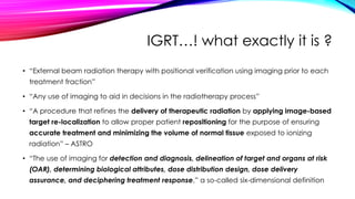
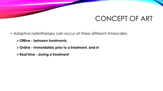
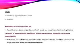
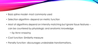
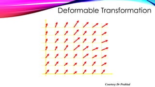
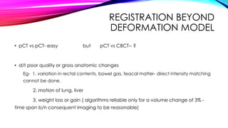
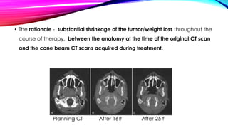
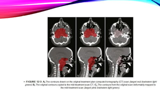
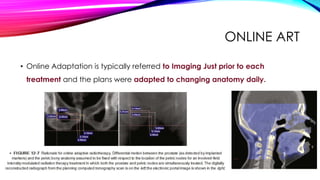
The vmat vs other recent radiotherapy techniquesM'dee Phechudi
VMAT is a new type of intensity-modulated radiation therapy (IMRT) treatment technique that uses the same hardware (i.e. a digital linear accelerator) as used for IMRT or conformal treatment, but delivers the radiotherapy treatment using a rotational or arc geometry rather than several static beams.
This technique uses continuous modulation (i.e. moving the collimator leaves) of the multileaf collimator (MLC) fields, continuous change of the fluence rate (the intensity of the X rays) and gantry rotation speed across a single or multiple 360 degree rotations
1.Aim of Radiotherapy
The goal of radiotherapy is to deliver a prescribed dose of radiation to the Target while sparing surrounding Healthy tissues to the largest extent possible
2.Organ Motion
Intra-fraction motion
during the fraction
Heartbeat
Swallowing
Coughing
Eye movement
Inter-fraction motion
- in between the fractions
Tumour change
Weight gain/loss
Positioning deviation
Breathing
Bowel and rectal filling
Bladder filling
Muscle relaxation/tension
3. Respiratory motion affects:
Respiratory motion affects all tumour sites in the thorax, abdomen and Pelvis. Tumours in the Lung, Liver, Pancreas, Oesophagus, Breast, Kidneys, prostate
Tumour displacement varies depending on the site and organ Location
Lung tumours can move several cm in any direction during irradiation
It is most prevalent and prominent in Lung cancers
4. Problems associated with respiratory motion during RT
Image acquisition limitations
Treatment planning limitations
Radiation delivery limitations
5. Methods to Account for Respiratory Motion
1. Motion encompassing methods
2. Respiratory gating methods
3. Breath hold methods
4. Forced shallow breathing with abdominal compression
5. Real-time tumor tracking methods
Summary:
The management of respiratory motion in radiation oncology is an evolving field
IGRT provides a solution for combating organ motion in radiotherapy
Delivering higher dose to tumor and less dose to normal tissue.
Limited clinical studies, needs to be studied further
IGRT – the future of radiotherapy
1.Stereotactic Radiosurgery (SRS)
SRS is a precise and focused delivery of a single, high dose of irradiation to a small and critically located intracranial volume while sparing normal structure
2.Stereotactic Body Radiation Therapy (SBRT)
SBRT is a treatment procedure similar to SRS, except that it deals extra-cranial radiosurgery
3.Flattening Filter Free (FFF) mode
FFF beam is produced without the use of flattening Filter
In the 1990s, several groups studied about FFF high-energy photon beams. The main interest for that, is to increase the dose rate for radiosurgery or the "physics interest”.
Need of increase in dose rate from traditional 300-600 to 1400-2400MU/min to overcome time-inefficiency and to improve patients comfort specially in SRS/SBRT
Flattening Filter Free (FFF) mode
FFF beam is produced without the use of flattening Filter
In the 1990s, several groups studied about FFF high-energy photon beams. The main interest for that, is to increase the dose rate for radiosurgery or the "physics interest”.
Need of increase in dose rate from traditional 300-600 to 1400-2400MU/min to overcome time-inefficiency and to improve patients comfort specially in SRS/SBRT
Hypofractionation in early breast cancer is no more a research scholars topic. Multiple studies with robust data have proven its utility. It may hold an important role in many countries with constrained resources. This is a short presentation incorporating important completed and ongoing trials. Feel free to use this.
A short overview of Image Guided Radiotherapy process in Lung Cancer presented at TMC Kolkata circa 2016. Basic principles and concepts as well as examples are outlined.
This is a made easy summary of ICRU 89 guidelines for gynecological brachytherapy. Extra practical questions for MD/DNB Radiotherapy exams are also attached.
Adaptive Radiotherapy at GenesisCare UKGenesisCareUK
A discussion about adaptive radiation therapy, a closed-loop radiation treatment process and how it can improve radiation treatment by systematically monitoring treatment variations and incorporating them to re-optimize the treatment plan early on during the course of treatment.
A summary of recent innovations in radiation oncology focussing on the priniciples of different techniques and their application. An overview of clinical results has also been given
Introduction to Medical Imaging, Basics of Medical Imaging, Fundamentals of Digital Image Processing, First chapter of Digital Image Processing Book by Rafael C. Gonzalez.
The vital importance of imaging techniques in radiation oncology now extends beyond diagnostic evaluation and treatment planning. Radiotherapy requires input from imaging for treatment planning and execution, when the treatment target is not located on the surface and, inspection and visual confirmation are not feasible. Traditional radiotherapy practices incorporate use of anatomic surface landmarks as well as radiologic correlation with 2D imaging in the form of port films or fluoroscopic imaging. Targets to be irradiated and normal tissues to be spared are delineated on CT scans in the planning process. Recent technical advances have enabled the integration of various imaging modalities into the everyday practice of radiotherapy directly at the linear accelerator. IGRT seeks to address geometric uncertainties in dose placement for target and normal tissues. It has become a routine part of current RT practice. Safe application of IGRT technology requires additional training and careful integration into the clinical process. IGRT reveals changes in anatomy during treatment which challenges conventional practices. IGRT facilitates the precise application of specialized irradiation techniques with narrow safety margins to radiosensitive organs.
Portal Imaging used to clear setup uncertaintyMajoVJJose
Title: Portal Imaging in Radiotherapy: A Comprehensive Exploration of Techniques, Applications, and Advancements
Introduction
Portal imaging is a critical component of modern radiotherapy, playing a pivotal role in the verification and precision of radiation treatment delivery. This technique involves the acquisition of X-ray images during or immediately after a patient's radiotherapy session, providing valuable information on the alignment of the treatment field with the intended target and surrounding critical structures. In this comprehensive exploration, we delve into the principles, techniques, clinical applications, challenges, and future prospects of portal imaging in the context of radiotherapy.
1. Principles of Portal Imaging
Portal imaging is rooted in the principles of verifying and ensuring the accuracy of radiation therapy delivery. Before each treatment fraction, the patient's position is verified to ensure it aligns precisely with the treatment plan. Portal images are acquired using specialized imaging devices, usually in the form of electronic portal imaging devices (EPIDs) or film-based systems. These images serve as a real-time snapshot of the radiation field, allowing clinicians to assess the actual treatment setup against the planned position.
2. Techniques of Portal Imaging
2.1 Electronic Portal Imaging Devices (EPIDs)
Electronic portal imaging devices, or EPIDs, have become a standard tool in portal imaging due to their real-time imaging capabilities and digital nature. EPIDs consist of a detector panel that captures the transmitted radiation through the patient during treatment. The resulting electronic images are immediately available for review, facilitating prompt decision-making regarding the need for adjustments in patient positioning or treatment parameters.
2.2 Film-Based Portal Imaging
Film-based portal imaging, while less commonly used today, has historical significance and is still employed in certain clinical settings. It involves exposing X-ray film positioned behind the patient during treatment. The film is then developed, and the resulting image is analyzed to verify the alignment of the treatment field. Though the process is not as immediate as with EPIDs, film-based systems may still offer advantages in certain situations.
3. Clinical Applications of Portal Imaging
Portal imaging is integral to the success of radiotherapy across various cancer types and treatment modalities.
3.1 Treatment Verification and Positioning
The primary application of portal imaging is to verify the accuracy of patient positioning and the alignment of the treatment field with the intended target volume. Any discrepancies detected through portal images allow for immediate adjustments to be made, ensuring that the radiation is delivered precisely to the targeted area while minimizing exposure to adjacent healthy tissues.
3.2 Tumor Localization and Changes in Anatomy
Portal imaging aids in localizing tumors, particularly
This seminar is presented as a part of weekly journal club and seminar presented in Apollo Hospital,Kolkata Department of Radiation Oncology.This seminar is moderated by Dr Tanweer Shahid.
TEST BANK for Operations Management, 14th Edition by William J. Stevenson, Ve...kevinkariuki227
TEST BANK for Operations Management, 14th Edition by William J. Stevenson, Verified Chapters 1 - 19, Complete Newest Version.pdf
Recomendações da OMS sobre cuidados maternos e neonatais para uma experiência pós-natal positiva.
Em consonância com os ODS – Objetivos do Desenvolvimento Sustentável e a Estratégia Global para a Saúde das Mulheres, Crianças e Adolescentes, e aplicando uma abordagem baseada nos direitos humanos, os esforços de cuidados pós-natais devem expandir-se para além da cobertura e da simples sobrevivência, de modo a incluir cuidados de qualidade.
Estas diretrizes visam melhorar a qualidade dos cuidados pós-natais essenciais e de rotina prestados às mulheres e aos recém-nascidos, com o objetivo final de melhorar a saúde e o bem-estar materno e neonatal.
Uma “experiência pós-natal positiva” é um resultado importante para todas as mulheres que dão à luz e para os seus recém-nascidos, estabelecendo as bases para a melhoria da saúde e do bem-estar a curto e longo prazo. Uma experiência pós-natal positiva é definida como aquela em que as mulheres, pessoas que gestam, os recém-nascidos, os casais, os pais, os cuidadores e as famílias recebem informação consistente, garantia e apoio de profissionais de saúde motivados; e onde um sistema de saúde flexível e com recursos reconheça as necessidades das mulheres e dos bebês e respeite o seu contexto cultural.
Estas diretrizes consolidadas apresentam algumas recomendações novas e já bem fundamentadas sobre cuidados pós-natais de rotina para mulheres e neonatos que recebem cuidados no pós-parto em unidades de saúde ou na comunidade, independentemente dos recursos disponíveis.
É fornecido um conjunto abrangente de recomendações para cuidados durante o período puerperal, com ênfase nos cuidados essenciais que todas as mulheres e recém-nascidos devem receber, e com a devida atenção à qualidade dos cuidados; isto é, a entrega e a experiência do cuidado recebido. Estas diretrizes atualizam e ampliam as recomendações da OMS de 2014 sobre cuidados pós-natais da mãe e do recém-nascido e complementam as atuais diretrizes da OMS sobre a gestão de complicações pós-natais.
O estabelecimento da amamentação e o manejo das principais intercorrências é contemplada.
Recomendamos muito.
Vamos discutir essas recomendações no nosso curso de pós-graduação em Aleitamento no Instituto Ciclos.
Esta publicação só está disponível em inglês até o momento.
Prof. Marcus Renato de Carvalho
www.agostodourado.com
- Video recording of this lecture in English language: https://youtu.be/lK81BzxMqdo
- Video recording of this lecture in Arabic language: https://youtu.be/Ve4P0COk9OI
- Link to download the book free: https://nephrotube.blogspot.com/p/nephrotube-nephrology-books.html
- Link to NephroTube website: www.NephroTube.com
- Link to NephroTube social media accounts: https://nephrotube.blogspot.com/p/join-nephrotube-on-social-media.html
ARTIFICIAL INTELLIGENCE IN HEALTHCARE.pdfAnujkumaranit
Artificial intelligence (AI) refers to the simulation of human intelligence processes by machines, especially computer systems. It encompasses tasks such as learning, reasoning, problem-solving, perception, and language understanding. AI technologies are revolutionizing various fields, from healthcare to finance, by enabling machines to perform tasks that typically require human intelligence.
Flu Vaccine Alert in Bangalore Karnatakaaddon Scans
As flu season approaches, health officials in Bangalore, Karnataka, are urging residents to get their flu vaccinations. The seasonal flu, while common, can lead to severe health complications, particularly for vulnerable populations such as young children, the elderly, and those with underlying health conditions.
Dr. Vidisha Kumari, a leading epidemiologist in Bangalore, emphasizes the importance of getting vaccinated. "The flu vaccine is our best defense against the influenza virus. It not only protects individuals but also helps prevent the spread of the virus in our communities," he says.
This year, the flu season is expected to coincide with a potential increase in other respiratory illnesses. The Karnataka Health Department has launched an awareness campaign highlighting the significance of flu vaccinations. They have set up multiple vaccination centers across Bangalore, making it convenient for residents to receive their shots.
To encourage widespread vaccination, the government is also collaborating with local schools, workplaces, and community centers to facilitate vaccination drives. Special attention is being given to ensuring that the vaccine is accessible to all, including marginalized communities who may have limited access to healthcare.
Residents are reminded that the flu vaccine is safe and effective. Common side effects are mild and may include soreness at the injection site, mild fever, or muscle aches. These side effects are generally short-lived and far less severe than the flu itself.
Healthcare providers are also stressing the importance of continuing COVID-19 precautions. Wearing masks, practicing good hand hygiene, and maintaining social distancing are still crucial, especially in crowded places.
Protect yourself and your loved ones by getting vaccinated. Together, we can help keep Bangalore healthy and safe this flu season. For more information on vaccination centers and schedules, residents can visit the Karnataka Health Department’s official website or follow their social media pages.
Stay informed, stay safe, and get your flu shot today!
Report Back from SGO 2024: What’s the Latest in Cervical Cancer?bkling
Are you curious about what’s new in cervical cancer research or unsure what the findings mean? Join Dr. Emily Ko, a gynecologic oncologist at Penn Medicine, to learn about the latest updates from the Society of Gynecologic Oncology (SGO) 2024 Annual Meeting on Women’s Cancer. Dr. Ko will discuss what the research presented at the conference means for you and answer your questions about the new developments.
New Drug Discovery and Development .....NEHA GUPTA
The "New Drug Discovery and Development" process involves the identification, design, testing, and manufacturing of novel pharmaceutical compounds with the aim of introducing new and improved treatments for various medical conditions. This comprehensive endeavor encompasses various stages, including target identification, preclinical studies, clinical trials, regulatory approval, and post-market surveillance. It involves multidisciplinary collaboration among scientists, researchers, clinicians, regulatory experts, and pharmaceutical companies to bring innovative therapies to market and address unmet medical needs.
Explore natural remedies for syphilis treatment in Singapore. Discover alternative therapies, herbal remedies, and lifestyle changes that may complement conventional treatments. Learn about holistic approaches to managing syphilis symptoms and supporting overall health.
micro teaching on communication m.sc nursing.pdfAnurag Sharma
Microteaching is a unique model of practice teaching. It is a viable instrument for the. desired change in the teaching behavior or the behavior potential which, in specified types of real. classroom situations, tends to facilitate the achievement of specified types of objectives.
The prostate is an exocrine gland of the male mammalian reproductive system
It is a walnut-sized gland that forms part of the male reproductive system and is located in front of the rectum and just below the urinary bladder
Function is to store and secrete a clear, slightly alkaline fluid that constitutes 10-30% of the volume of the seminal fluid that along with the spermatozoa, constitutes semen
A healthy human prostate measures (4cm-vertical, by 3cm-horizontal, 2cm ant-post ).
It surrounds the urethra just below the urinary bladder. It has anterior, median, posterior and two lateral lobes
It’s work is regulated by androgens which are responsible for male sex characteristics
Generalised disease of the prostate due to hormonal derangement which leads to non malignant enlargement of the gland (increase in the number of epithelial cells and stromal tissue)to cause compression of the urethra leading to symptoms (LUTS
Ethanol (CH3CH2OH), or beverage alcohol, is a two-carbon alcohol
that is rapidly distributed in the body and brain. Ethanol alters many
neurochemical systems and has rewarding and addictive properties. It
is the oldest recreational drug and likely contributes to more morbidity,
mortality, and public health costs than all illicit drugs combined. The
5th edition of the Diagnostic and Statistical Manual of Mental Disorders
(DSM-5) integrates alcohol abuse and alcohol dependence into a single
disorder called alcohol use disorder (AUD), with mild, moderate,
and severe subclassifications (American Psychiatric Association, 2013).
In the DSM-5, all types of substance abuse and dependence have been
combined into a single substance use disorder (SUD) on a continuum
from mild to severe. A diagnosis of AUD requires that at least two of
the 11 DSM-5 behaviors be present within a 12-month period (mild
AUD: 2–3 criteria; moderate AUD: 4–5 criteria; severe AUD: 6–11 criteria).
The four main behavioral effects of AUD are impaired control over
drinking, negative social consequences, risky use, and altered physiological
effects (tolerance, withdrawal). This chapter presents an overview
of the prevalence and harmful consequences of AUD in the U.S.,
the systemic nature of the disease, neurocircuitry and stages of AUD,
comorbidities, fetal alcohol spectrum disorders, genetic risk factors, and
pharmacotherapies for AUD.
These simplified slides by Dr. Sidra Arshad present an overview of the non-respiratory functions of the respiratory tract.
Learning objectives:
1. Enlist the non-respiratory functions of the respiratory tract
2. Briefly explain how these functions are carried out
3. Discuss the significance of dead space
4. Differentiate between minute ventilation and alveolar ventilation
5. Describe the cough and sneeze reflexes
Study Resources:
1. Chapter 39, Guyton and Hall Textbook of Medical Physiology, 14th edition
2. Chapter 34, Ganong’s Review of Medical Physiology, 26th edition
3. Chapter 17, Human Physiology by Lauralee Sherwood, 9th edition
4. Non-respiratory functions of the lungs https://academic.oup.com/bjaed/article/13/3/98/278874
4. IGRT…! what exactly it is ?
• “External beam radiation therapy with positional verification using imaging prior to each
treatment fraction”
• “Any use of imaging to aid in decisions in the radiotherapy process”
• “A procedure that refines the delivery of therapeutic radiation by applying image-based
target re-localization to allow proper patient repositioning for the purpose of ensuring
accurate treatment and minimizing the volume of normal tissue exposed to ionizing
radiation” – ASTRO
• “The use of imaging for detection and diagnosis, delineation of target and organs at risk
(OAR), determining biological attributes, dose distribution design, dose delivery
assurance, and deciphering treatment response,” a so-called six-dimensional definition
5. • Although both IGRT and Adaptive Radiotherapy exists from long back,
With time… with improving technology…
• Radiation techniques improved and so, THE OUTCOMES.
• Adaptive Radiotherapy never an individual entity without IGRT..!!
7. • A radiation treatment process where the treatment plan can be modified using a
systematic feedback of measurements is named ‘Adaptive radiation therapy’ (ART).
• The importance of using an adaptive process in radiation therapy is that the
treatment plan, especially the margin and treatment dose, can eventually be
customized to the individual patient.
• Adaptive radiation therapy has been introduced to incorporate the position
variation of the individual patient into the treatment optimization process during the
course of radiotherapy.
8. • The concept of ART can also be applied to compensate for other treatment
variations such as radiation sensitivity and density of clonogenic cells when
they become measurable during the treatment course.
10. DEFINITION OF ART
• Adaptive radiotherapy can be defined as,
Temporally changing the treatment plan delivered to a patient based on
observed anatomic changes caused by tumor shrinkage, weight loss, or
internal motion – Leibel Philips.
“The ability to measure changes in location, morphology and
physiology (like breathing, tumor response, weight loss) and adapt the
course of treatment will lead to adaptive treatment, also called 4D
adaptive treatment” – ICRU 83
11. FACTORS CAUSING ANATOMIC CHANGES
TO TUMOR AND NORMAL TISSUES
• Anatomic motion (PHYSIOLOGICAL) caused by the musculoskeletal,
respiratory, cardiac, gastrointestinal, and genitourinary systems.
• Treatment-induced changes such as cell death and tumor shrinkage, tumor
growth resulting from accelerated repopulation, weight loss or gain because
of changes in appetite caused by radiation, concomitant chemotherapy or
hormone therapy, fibrosis of normal tissue, and so on
12. RATIONALE
• Changing anatomy of both tumor and normal tissue—
It naturally follows that the delivery of radiotherapy should be temporally
changing to match the observed anatomic changes
In other terms,
If the PTV and normal anatomy are changing with time, we are obligated to
adapt our treatment delivery where possible.
13. DIFFERENT WAYS TO ADAPT
TYPES OF ART Examples
Motion adaptive Beaumont’s ART process, ‘Plan of the
day’ IMRT
Biological adaptive Theragnostic planning
Response adaptive Mid-treatment response evaluation,
shrinkage of GTV & OAR
Time adaptive Mid-course CT and planning
15. COMPONENTS
• Adapt to tumor motion
• Adapt to tumor / organ deformation and volume change
16. ADAPTING TO TUMOR MOTION
• Move couch electronically to adapt to the moving tumor
• Exac trac 6D Robotic couch
• Move a charged particle beam electromagnetically
• RPM system
• Move a robotic lightweight linear accelerator
• Cyber knife
• Move aperture shaped by a dynamic MLC
• Calypso system
17. CONCEPT OF ART
• Adaptive radiotherapy can occur at three different timescales:
Offline - between treatments,
Online - immediately prior to a treatment, and in
Real time - during a treatment
18.
19. IMAGE REGISTRATION
• Registration : means to align two imaging data sets in a common coordinate space
by transforming one image set while keeping the other one fixed.
• Voxel to voxel matching b/n 2 image sets.
• Fixed image to floating image
• A transformation matrix created
• It is an optimization process- success achieved after iterative comparisons.
20.
21. IMAGE REGISTRATION
• According to the nature of transformation-
• It is of 2 types- Rigid registration and Deformable registration.
• Rigid fusion-
• Direct intensity match or point match
• No compensation for motion or patient position
• 6 degrees of freedom
22. • Deformable image registration –
• Complicated
• Consists of a matrix with huge number of unknowns.
• Multimodality images can be fused
• Allows dose and contour information to be transferred between images, substantially
reducing workload.
• As the timescales for adaptation decrease, the computation time of deformable image
registration must correspondingly decrease.
23.
24. APPLICATIONS
(1) Better Target Definition
(2) Image Enhancement
(3) Propagation Of Organ Contours From One Image Set To Another
(4) Calculation Of Accumulated Dose In Organs Experiencing Inter- Or Intra-
fraction Organ Deformation For 4-D Or Adaptive Therapy Planning
(5) 4-D Image Reconstruction
25. ISSUES:
• Definition of registration metric function
• Algorithm
Registration can be broadly divided into
• Manual, landmark-based, surface-based, intensity-based, and mutual information–based registrations.
Depending on the mechanism or method used to model the deformation, registration can usually be
categorized into
• Elastic model, viscous fluid model, optical flow model, finite element model, radial-basis function model
such as basis spline model, and thin-plate spline model.
26. • Basis spline model- most commonly used
• Selection algorithm- depend on metric function
• Most of algorithms depend on intensity matching but ignore tissue features –
can be countered by physiologic and anatomic knowledge
• Eg- Bone wrapping
• Cost function- Similarity measure
• Penalty function- discourages undesirable transformations.
34. REGISTRATION BEYOND
DEFORMATION MODEL
• pCT vs pCT- easy but pCT vs CBCT-- ?
• d/t poor quality or gross anatomic changes
Eg- 1. variation in rectal contents, bowel gas, feacal matter- direct intensity matching
cannot be done.
2. motion of lung, liver
3. weight loss or gain ( algorithms reliable only for a volume change of 3% -
time span b/n consequent imaging to be reasonable)
35. TRANSFER OF CONTOURS
• Accuracy of registration matters- influenced by image content distant from
ROI.
• Surface mapping techniques: efficient for pCT to CBCT contour transfer
• ROI contour extended surface
• Hybrid – deformable and surface model best method
• Narrow shell around ROI extended 1-2cm on pCT merged with CBCT
36. CBCT
• Large variation of HU for same material under different scanning conditions.
• In the absence of organ motion– Accuracy is acceptable for dose verification but not
for treatment planning.
• With organ motion– 5-8% discrepancy can be seen.
• Electron density matching– may be a solution.
• Where it stand in ART ?
37. OFFLINE ADAPTATION
• The imaging system used : Conventional CT scanner or an in-room CT
scanner.
• The adaptation can be triggered by :
• As a protocol (for example after 20 and 40Gy delivered dose),
• Clinical observations of masks not fitting, weight loss, or other surface changes,
or by changes observed on an in-room imaging system
38. • The rationale - substantial shrinkage of the tumor/weight loss throughout the
course of therapy, between the anatomy at the time of the original CT scan
and the cone beam CT scans acquired during treatment.
Planning CT After 16# After 25#
39.
40.
41. ONLINE ART
• Online Adaptation is typically referred to Imaging Just prior to each
treatment and the plans were adapted to changing anatomy daily.
42. ONLINE ART
• This can be done either by correcting the errors or by modifying the
contours.
• Daily re-planning ??
43. • Although it may be trivial to occasionally re-plan a limited number of
patients offline, frequent re-plannings of many patients are labour and time
intensive, especially if online re-planning is necessary.
44. GRAPHICS PROCESSING UNIT (GPU)
• Researchers at the University of California–San Diego have explored the
potential to perform online re-planning utilizing the vast computational
capability of the graphics card technology.
• It is possible to dramatically improve the computational efficiency of the
traditionally computationally intense tasks in RT such as dose calculations,
inverse planning re-optimizations, CT reconstructions,and deformable image
registrations for fast contour mapping.
45. • Gu et al. have shown that a full 3D-dose calculation based on finite-size pencil
beam algorithm achieved a speed up of 200 to 400 times, taking less than 1 second
for typical IMRT plans.
• Men et al. found that, for a typical nine-field prostate IMRT plan with 5-by-5 mm2
beam-let size and 2.5-by-2.5-by-2.5 mm3 voxel size, re-optimization would only take
2.8 seconds.
• Park et al. found that for a filtered back-projection reconstruction of a typical 3D
CBCT, volume can be done in a real-time fashion (i.e., as soon as the scan is done)
46. CLOUD COMPUTING
• Pioneered by Meng et al. And Wang et al. at stanford university.
• It’s a form of SUPERCOMPUTING technology.
• The task is divided up and handled in a parallel computing manner,
automatically.
• This system does not require the computers to be physically located near the
user, and they can be accessed from anywhere with an internet
connection,
• The larger the number of nodes used, the faster the computations.
48. LIMITATIONS
• The technology is new and research in RT is in its infancy; but the potential
benefits remain to be seen.
• How often should new plans be generated? Once? Weekly? Daily?
• Whether altering the target volume would adversely impact tumor control ?
49. REAL-TIME IMAGE-GUIDED
ADAPTIVE RADIOTHERAPY
• Imaging session and treatment session are intertwined
• Time separation is shown to be on the order of seconds or less.
• Rationale - significant drift in the mean position of a tumor during
radiotherapy treatment.
• The concept of intra-fraction motion management arises…here !
51. METHODS TO CONTROL /
COMPENSATE FOR LUNG MOTION
DURING RESPIRATION
• Free breathing methods:
Internal Target Volume (ITV)-based treatment
Gating
Tracking
• Breath-hold methods:
Active Breathing Coordinator (ABC) - DIBH
52. GATING
• Treatment delivery is done in the phase of respiration where the tumor
motion & resulting treatment volume is minimum, by coupling the beam
delivery with the phase of respiration
• It can be invasive or non-invasive.
53. REAL TIME TUMOR TRACKING
• Imaging is used to track the actual tumour motion during treatment delivery
and to move the treatment beam accordingly based on the varying
position of the tumour.
• Usually requires an internal fiducial, implanted within the tumour.
• Can also be done non-invasively in some cases.
54. SIMULATION
• Patients for gating/ tracking/ITV-
based treatment undergo plain
4DCT scan for planning (3mm
slices), using Mayo belt/ Anzai belt/
RPM system to correlate the
respiratory phases and
corresponding CT images.
• Ten data sets are thereby
generated.
55. VOLUME DELINEATION
• Tumor delineation is done on the end-expiratory data set. This is because, in this
phase, lung motion is minimum.
• No CTV margin is given for stereotactic body radiotherapy for early inoperable lung
cancers.
• For locally advanced disease, CTV margins between 0.6-0.8 cm are usually applied.
• Setup margins of 0.5 cm are normally applied (to the GTV/ CTV/ITV, as appropriate)
to arrive at the PTV
56. Gating can be internal or external
• Internal – Uses tumor motion surrogates such as implanted fiducial markers or
marker-less imaging of internal anatomy.
• Eg: RTRT system
• External – uses external respiratory surrogates such as markers placed on the
surface of the patient’s abdomen, a compression belt, or spirometer signals.
• Eg: RPM system, Exac trac
57. RPM SYSTEM (VARIAN)
• Lightweight plastic block with two or six passive infrared
reflective markers
• Monitored by video camera mounted on the treatment
room wall.
• The surrogate signal is the abdominal surface motion.
• Both amplitude and phase gating are allowed.
• disables the beam when the waveform becomes
irregular, such as with patient motion or coughing, and re
enables the beam after establishing breathing is again
regular.
58. EXAC-TRAC GATING/NOVALIS GATING
• This device uses external markers for gating.
• it uses x-ray imaging to determine the internal anatomy position and to verify
its reproducibility during treatment.
• By updating the correlation between the internal and external signals in a
reasonable frequency, x-ray exposure to the patient is minimized, while the
external gating signal accuracy is maintained.
59. • Oblique image pairs are taken by Exac-Trac system’s stereoscopic X ray
imagers.
• The system builds a correlation model between the end-positions and the
respiratory phase (accuracy of correlation 3mm).
• The treatment beam is then turned on and is configured to treat ONLY in
the gating window
60. R T -TRACKING
• Most ideal—and most technologically intense—strategy,
• Real-time tumor localization, fast processing and relay of information, and corresponding
repositioning of the beam all need to be dynamically seamlessly integrated.
Available Systems:
• ExacTrac system (VERO)
• Cyberknife
• Calypso System
63. CYBER KNIFE: RTT MODES
Non-invasive:
Xsight lung
• Possible if:
• Tumour >1.5 cm, surrounded by air
• Tumour visible by imaging system
Invasive (with implanted
fiducial):
Synchrony
• 3-5 fiducial markers need to be
inserted in close proximity to the lesion.
• They have to be well- separated
(>2cm) and non overlapping on
projections from the in room x-ray
images
66. • Has 6MV C-band LA mounted on O-
ring gantry
• Gantry is mounted on gimbals
• Capable of Pan & Tilt motions
• Maximum motion allowed=2.5 cm in
isocentre plane or 2.5 degrees in
each direction
• Also has 2 kV X ray imagers,
mounted at 45 degree angle with the
MV beam axis
• Has facility for Cone Beam CT & Real
Time Tumor Tracking (based on Infrared
& stereoscopic X rays) .
• 6 degrees of freedom
• Patient repositioning not required as the
system can move itself
• Image verification possible at any
position during treatment.
67. ELECTROMAGNETIC TRACKING
SYSTEMS (CALYPSO)
• Make use of electromagnetic transponders
(beacons)
• Embedded within the tumor
• Motion of these beacons may be tracked in real time
using a detector array system.
• Beacons need to be placed through a minimally
invasive procedure, their presence may introduce
artifacts in mr images, and there are limitations to the
patient size.
• Calypso has a geometric accuracy of <2 mm, but its
use at present is limited to prostate radiotherapy.
69. RATIONALE FOR NON UNIFORM
DOSE ESCALATION
• Local recurrences arise from micro environmental niches that are relatively resistant at
the radiation dose level that can safely be delivered using a uniform dose distribution
• molecular and functional imaging will allow spatiotemporal mapping of these regions of
relative radio-resistance
• advances in radiation therapy planning and delivery technologies facilitate the delivery
of a graded boost to such regions, which, in turn, should lead to improved local tumor
control with acceptable side effects
72. • Biology-based objective functions can be introduced to the radiation treatment planning
process by co-registration
• Dose painting
-Subvolume boosting-imaging defined discrete volume is given an additional boost
- Dose painting by numbers- in which a dose is prescribed at the voxel level and it is left for the
dose plan optimizer to arrive at a physically deliverable dose distribution
76. HEAD AND NECK
• We usually adapt to changes in morphology of tumor and OAR’s.
• Provoking factors: weight loss, tumor response, progression of disease
Hansen et al.
• evaluated the impact of replanning in a cohort of 13 HNC patients with either significant
weight loss or tumor response during IMRT.
• Compared to replanning, not replanning significantly decreased dose to the target
volume and increased doses to normal tissues (spinal cord and brainstem). The doses to
95% of the PTV-GTV and the PTV-CTV decreased by up to 6.3 Gy and 7.4 Gy,
respectively.
77.
78. Adaptive radiotherapy of head and neck
cancer
Pierre Castadot, MD, John A. Lee, Eng, Phd, Xavier Geets, MD, Phd, And
Vincent Grégoire, MD, Phd, FRCR
• Positioning errors and their causes and strategies that could be adopted to
counteract setup uncertainties.
• Anatomical modifications
79. Anatomical Modifications
Of Target Volumes And Organs
At Risk During Radiation Therapy
• These include the shrinkage of the primary tumor and nodal volumes,
resolving postoperative changes or edema, and weight loss.
80.
81.
82. Dosimetric Impact
Of Anatomical Modifications
• Alteration in patient anatomy during treatment modifications of both
target volumes and OARs, the dose distribution that is actually delivered
to the patient might significantly differ from what was planned.
83.
84. • 51 papers were reviewed.
• Adaptive radiotherapy (ART) could be applied to reduce dose to OARs and
eventually to improve quality of life.
• It is unlikely that every patient will benefit from ART.
• Selection of patients is most important.
• There are no guidelines to when and what time schedules adaptation should be
done.
85. • Anatomic and dosimetric changes are more pronounced in Parotid gland.
• The average volume decrease of the PG’s during radiotherapy was 26 ± 11%.
(Mostly during first half of treatment).
• The most common reported anatomic changes were volume loss and medial shifts
of the PG’s.
• On average, the PG mean dose increased with 2.2 ± 2.6 Gy as compared to the
dose calculated on the planning CT at baseline. (Site specific- may go up to 10Gy)
86. • It was directly associated with higher grade 2 or more xerostomia in those
patients.
• Weight loss >5% and/or decrease of neck diameter >10% was associated
with higher xerostomia.
• Chen et al. - In less than 20% of all head and neck patients replanning was
needed because of target underdosage or OAR overdosage, usually during
the first three weeks of treatment
87. • 36 patients
• 54-Gy dose to both high-risk and low-risk target volumes in Phase I.
• Phase II (adaptive) plans for 16 Gy to high-risk planning target volume.
RESULTS:
• Median reductions in gross primary and nodal disease volumes on mid-treatment scans were 34% and 43.2%,
respectively. Four patients developed local recurrences, all within the RT field. Median DFS and OS were 17.5 and
23.5 months, respectively.
ADVANCES IN KNOWLEDGE:
• This study supports the need for adaptive replanning for minimizing normal tissue toxicity without compromising
local control and adds to the existing body of literature.
88. ART IN PROSTATE CANCER
• Major concern is to reduce the treatment margins to reduce normal tissue
toxicity
• To incorporate generic set up errors and application of predetermined set
up margins.
• Individualized setup adjustments to reduce the magnitude of setup error
IGRT forms the basis for adaptation
90. BEAUMONT’S ART PROCESS
CL-PTV can be constructed within the first week of treatment
using feedback of imaging measurements
Single plan modification during second week can lead to dose
escalation
91. Divided into three groups based on PTV patient specific dose-
70.2-72.0, 72.0-75.6 ,75.6-79.2 (based on bladder and rectal dose
constraints)
Patients-280
180 patients (65%) were treated to a prostate field only and 99
patients (35%) to prostate and seminal vesicles
Toxicity at the high dose level was not different from toxicity at
the intermediate or lower dose levels
94. ‘PLAN OF THE DAY’
• Daily megavoltage (MV) imaging helped to choose the most
appropriate PTV encompassing bladder for the particular day
(using plan-of-the-day approach).
CONCLUSIONS:
• Adaptive IGRT using plan-of-the-day approach for bladder
preservation is clinically feasible, with good oncological outcomes
and low rates of acute and late toxicities. Dose escalation is safe
and possibly improves outcomes in bladder preservation.
95. N-54
3DCRT technique
4 plans generated by 10#
CBCT daily on 1st week and then every 5# (post RT also)
Without further bladder filling control or imaging, a CTV to PTV margin
of 7 mm is insufficient
97. No strong correlations were found between
V95%(total-dose) reduction and initial seroma volume
or between V95%(total-dose) reduction and seroma
volume decrease
98. -28 patients
-Repeat CT scans were taken daily during 1st week f/b once
weekly
-Substantial systematic and random shape variation
demanded for a PTV margin up to 2.4 cm at the upper-
anterior part of the CTV. Plan adaptation after fraction 4
resulted in a maximum 0.7 cm margin reduction and a
significant PTV reduction from 1185 to 1023 cc
99. CT -planning CT -4
Conclusions: With adaptive radiotherapy, maximum required
PTV margins can be reduced from 2.4 to 1.7 cm, resulting in
significantly less dose to the bowel area