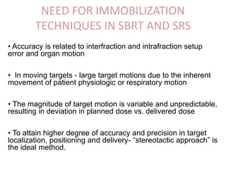
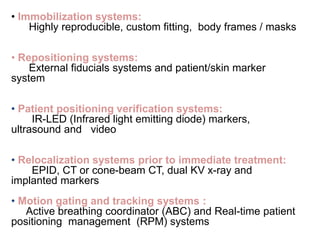
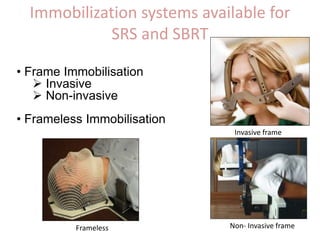
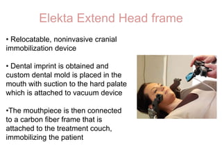
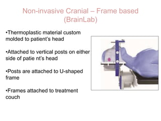
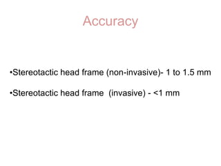
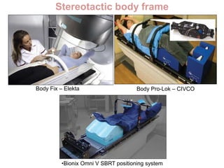
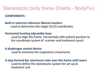
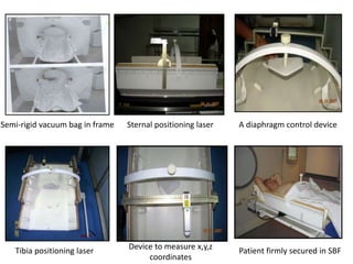
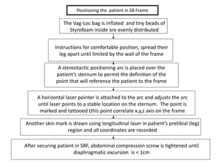
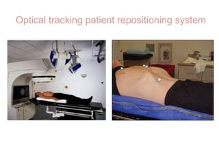
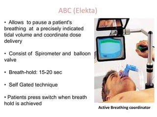
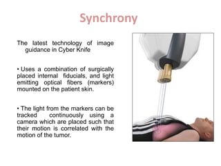
IMMOBILIZATION TECHNIQUES
IN SRS AND SRT
Dr. Shreya Singh
JR-III
Department of Radiation Oncology
IMS, BHU
NEED FOR IMMOBILIZATION
TECHNIQUES IN SBRT AND SRS
• Accuracy is related to interfraction and intrafraction setup
error and organ motion
• In moving targets - large target motions due to the inherent
movement of patient physiologic or respiratory motion
• The magnitude of target motion is variable and unpredictable,
resulting in deviation in planned dose vs. delivered dose
• To attain higher degree of accuracy and precision in target
localization, positioning and delivery- “stereotactic approach” is
the ideal method.
• Immobilization systems:
Highly reproducible, custom fitting, body frames / masks
• Repositioning systems:
External fiducials systems and patient/skin marker
system
• Patient positioning verification systems:
IR-LED (Infrared light emitting diode) markers,
ultrasound and video
• Relocalization systems prior to immediate treatment:
EPID, CT or cone-beam CT, dual KV x-ray and
implanted markers
• Motion gating and tracking systems :
Active breathing coordinator (ABC) and Real-time patient
positioning management (RPM) systems
IMMOBILZATION
AND
REPOSITIONING SYSTEMS
Immobilization systems available for
SRS and SBRT
• Frame Immobilisation
 Invasive
 Non-invasive
• Frameless Immobilisation
Invasive frame
Non- Invasive frameFrameless
Stereotactic head frame
The head frame serves three main purposes:
• It enables accurate reproducibility of the treatment
position for each planning and treatment session.
• It prevents movement of your head during the
treatment
• It removes the need for ink marks on your head or
face.
Invasive Stereotactic head frame
• Leksell system
• BRW system
• CRW system
• TALON system
Leksell system (Elekta)
•Compatible with gamma knife
or LINAC platforms
•The arc employs the center-of-
arc principle for encompassing
the surgical target in three
dimensions, enabling full access
to any intracranial area
•Used for diagnostic and
therapeutic stereotactic
neurosurgery
BRW system (Integra)
• Has set of vertical and diagonal
rods ( 6 and 3) creating ‘N’ shape
alignment for stereotactic
localization and alignment
• 4 pins into the skull
• Arc guidance frame – creates
infinite pathways but for each
trajectory, the computation must
include entry coordinates.
CRW system
•Simplified and improved form of BRW
• MRI compatible
•Its target-centered arc principle
means that all arc trajectories pass to
the target, thus obviating the need for
fixed entry point
•Frame is affixed to cranium via four
screws placed through graphite posts
(red arrows). Outer cage (blue arrow)
serves as a CT localizer for image
registration
TALON (Best Namos)
•Removable head frame system
for fractionated SRT
•Utilises two self-tapping titanium
screws inserted into patient’s
skull at vertex
•TALON assembly is then
attached to the screws on
treatment table
•Frame can be removed after
each fraction with two screws
remanining in the patient’s skull
Non-invasive Cranial Immobilization
•Elekta Extend Head frame
•BrainLab Mask System
•Stereoadapter 5000
•Gill-Thomas-Cosman (GTC) Relocatable Head
Ring (Integra)
Elekta Extend Head frame
• Relocatable, noninvasive cranial
immobilization device
• Dental imprint is obtained and
custom dental mold is placed in the
mouth with suction to the hard palate
which is attached to vacuum device
•The mouthpiece is then connected
to a carbon fiber frame that is
attached to the treatment couch,
immobilizing the patient
Non-invasive Cranial – Frame based
(BrainLab)
•Thermoplastic material custom
molded to patient’s head
•Attached to vertical posts on either
side of patie nt’s head
•Posts are attached to U-shaped
frame
•Frames attached to treatment
couch
Accuracy
•Stereotactic head frame (non-invasive)- 1 to 1.5 mm
•Stereotactic head frame (invasive) - <1 mm
Stereotactic body frame
Body Fix – Elekta Body Pro-Lok – CIVCO
•Bionix Omni V SBRT positioning system
Stereotactic body frame (Elekta - BodyFix)
COMPONENTS :
Built-in external reference fiducial markers
used to determine the target (X,Y,Z) coordinates
Horizontal leveling adjustable base
used to align the frame horizontally with patient position to
the coordinate system of scanner and treatment couch
A diaphragm control device
used to minimize the respiratory movements
A step formed-bar aluminum ruler over the frame with lasers
used to define the stereotactic system for set up at
treatment unit
A diaphragm control deviceSemi-rigid vacuum bag in frame Sternal positioning laser
Tibia positioning laser Patient firmly secured in SBFDevice to measure x,y,z
coordinates
Positioning the patient in SB Frame
The Vag-Loc bag is inflated and tiny beads of
Styrofoam inside are evenly distributed
Instructions for comfortable position, spread their
leg apart until limited by the wall of the frame
A stereotactic positioning arc is placed over the
patient’s sternum to permit the definition of the
point that will reference the patient to the frame
A horizontal laser pointer is attached to the arc and adjusts the arc
until laser points to a stable location on the sternum. The point is
marked and tattooed (this point correlate x,y,z axis on the frame
Another skin mark is drawn using longitudinal laser in patient’s pretibial (leg)
region and all coordinates are recorded
After securing patient in SBF, abdominal compression screw is tightened until
diaphragmatic excursion is < 1cm
Stereotactic Fiducial System
•Central to stereotactic method is requirement for reference
markers to be imaged when attached to the stereotactic
immobilisation system fixed to the patient.
•These markers are vital in providing accurate geometrical
information on the coordinates of the planned isocentres.
•They are commonly in the form of
oVarious rod configurations attached to support rings
oEtchings / steel balls on the sides
oWires stretched between rigid spacers
Patient Positioning Verification
Systems
• Optical tracking is a means of determining real-time position
of an object by tracking the positions of either active or passive
infrared markers attached to the object.
• Optical tracking systems uses infrared light to determine a
target’s position via active or passive markers.
• Common active marker is infrared light-emitting diodes
(IRLED).
• The position of the point of reflection is determined using a
camera system
Optical Tracking system
Optical Tracking systems
Active markers Passive markers
Camera array
gold marker implant
fiducial
Optical tracking technology in SBRT
Target (lesion)
Stereo cameras
y
z
x
center of rotation
a)
c)
b)
d)
The stereo cameras locate the exact
coordinates of passive or active IR marker in
treatment room
The marker is in the same location as it was
in fig a, but because of the patient is
rotated around the marker point, the lesion
has moved undetected by the cameras
An array of marker is used now, fixed to the
patient surface. The collection of points in the
array of markers is tracked
Using an array of markers allow the
rotational movement to be detected
Optical tracking patient repositioning system
Relocalization Systems Prior to
Immediate Treatment
Methods
•Radiographic imaging of fiducial markers
• Video-based surface tracking
• In-room CT imaging
• EPID
•Cone Beam CT imaging
Motion Gating and Tracking
System
Methods for tracking motion
• Skull and spine tracking-
bony landmarks are tracked.
• Fiducial tracking-
radio-opaque marker are placed near soft tissue targets
and tracked.
• Respiratory tracking -
LED’s on the exterior of the patient are correlated with
the movement of the target/tumor and fiducials.
Management of respiratory motion
Methods to account for respiratory motion
• Breath-hold technique
• Forced shallow breathing
• Respiratory Gated technique
Respiratory management devices
• Active breathing coordinator (ABC) (Elekta)
• Real-time Position Management (RPM) system (Varian)
Abdominal Compression Device
•For forced shallow
breathing
•A plate compresses the
abdominal wall few
centimeters below the
xiphoid
•This plate is fixed to the
arch
•Arch is attached to the
body frame
RPM System (Varian)
• External gating system
• System consists of
Infra-red camera
Marker block containing 2 reflectors
• The marker block is placed on the patient’s
skin in the abdominal region
• Surrogate signal - abdominal surface cmotion
correlates to tumor motion
• The x-ray signal from the CT scanner
is recorded synchronously with the respiration
signal.
Real-time position
Management system (RPM)
Video Camera
ABC (Elekta)
• Allows to pause a patient's
breathing at a precisely indicated
tidal volume and coordinate dose
delivery
• Consist of Spirometer and balloon
valve
• Breath-hold: 15-20 sec
• Self Gated technique
• Patients press switch when breath
hold is achieved
Active Breathing coordinator
Shortcomings of Respiration techniques
Internal and external gating techniques, do not localize the
tumor (volume, shape) itself. Thus, they do not detect
• Deformation
• Migration
• Anatomical changes
Also, patient breath coaching is important
Real - time Tumor Tracking
• Monitor tumor motion and synchronize the radiation beam
with tumor motion
• Real-time tracking using (i) dynamic MLC (ii) Robotic couch
• Interrupt and restart the treatment when target motion differs
from the simulated pattern
Advantages
• Accuracy is increased with tracking because residual motion
is lowered compared to gating technique
• Deformation can be considered
Synchrony
The latest technology of image
guidance in Cyber Knife
• Uses a combination of surgically
placed internal fiducials, and light
emitting optical fibers (markers)
mounted on the patient skin.
• The light from the markers can be
tracked continuously using a
camera which are placed such that
their motion is correlated with the
motion of the tumor.
Calypso
• Used to monitor motion during
radiosurgery treatment
• Uses electro-magnetic transponder
fiducial implanted in or near the target that
is monitored by electromagnetic detector
•Detector’s position is tracked by optical
cameras
•Allows near real-time continuous tracking
without using ionizing radiation
•Requires invasive procedure for
implantation
THANK YOU

Immobilization techniques in SRS and SBRT

  • 1.
    IMMOBILIZATION TECHNIQUES IN SRSAND SRT Dr. Shreya Singh JR-III Department of Radiation Oncology IMS, BHU
  • 2.
    NEED FOR IMMOBILIZATION TECHNIQUESIN SBRT AND SRS • Accuracy is related to interfraction and intrafraction setup error and organ motion • In moving targets - large target motions due to the inherent movement of patient physiologic or respiratory motion • The magnitude of target motion is variable and unpredictable, resulting in deviation in planned dose vs. delivered dose • To attain higher degree of accuracy and precision in target localization, positioning and delivery- “stereotactic approach” is the ideal method.
  • 3.
    • Immobilization systems: Highlyreproducible, custom fitting, body frames / masks • Repositioning systems: External fiducials systems and patient/skin marker system • Patient positioning verification systems: IR-LED (Infrared light emitting diode) markers, ultrasound and video • Relocalization systems prior to immediate treatment: EPID, CT or cone-beam CT, dual KV x-ray and implanted markers • Motion gating and tracking systems : Active breathing coordinator (ABC) and Real-time patient positioning management (RPM) systems
  • 4.
  • 5.
    Immobilization systems availablefor SRS and SBRT • Frame Immobilisation  Invasive  Non-invasive • Frameless Immobilisation Invasive frame Non- Invasive frameFrameless
  • 6.
    Stereotactic head frame Thehead frame serves three main purposes: • It enables accurate reproducibility of the treatment position for each planning and treatment session. • It prevents movement of your head during the treatment • It removes the need for ink marks on your head or face.
  • 7.
    Invasive Stereotactic headframe • Leksell system • BRW system • CRW system • TALON system
  • 8.
    Leksell system (Elekta) •Compatiblewith gamma knife or LINAC platforms •The arc employs the center-of- arc principle for encompassing the surgical target in three dimensions, enabling full access to any intracranial area •Used for diagnostic and therapeutic stereotactic neurosurgery
  • 9.
    BRW system (Integra) •Has set of vertical and diagonal rods ( 6 and 3) creating ‘N’ shape alignment for stereotactic localization and alignment • 4 pins into the skull • Arc guidance frame – creates infinite pathways but for each trajectory, the computation must include entry coordinates.
  • 10.
    CRW system •Simplified andimproved form of BRW • MRI compatible •Its target-centered arc principle means that all arc trajectories pass to the target, thus obviating the need for fixed entry point •Frame is affixed to cranium via four screws placed through graphite posts (red arrows). Outer cage (blue arrow) serves as a CT localizer for image registration
  • 11.
    TALON (Best Namos) •Removablehead frame system for fractionated SRT •Utilises two self-tapping titanium screws inserted into patient’s skull at vertex •TALON assembly is then attached to the screws on treatment table •Frame can be removed after each fraction with two screws remanining in the patient’s skull
  • 12.
    Non-invasive Cranial Immobilization •ElektaExtend Head frame •BrainLab Mask System •Stereoadapter 5000 •Gill-Thomas-Cosman (GTC) Relocatable Head Ring (Integra)
  • 13.
    Elekta Extend Headframe • Relocatable, noninvasive cranial immobilization device • Dental imprint is obtained and custom dental mold is placed in the mouth with suction to the hard palate which is attached to vacuum device •The mouthpiece is then connected to a carbon fiber frame that is attached to the treatment couch, immobilizing the patient
  • 14.
    Non-invasive Cranial –Frame based (BrainLab) •Thermoplastic material custom molded to patient’s head •Attached to vertical posts on either side of patie nt’s head •Posts are attached to U-shaped frame •Frames attached to treatment couch
  • 16.
    Accuracy •Stereotactic head frame(non-invasive)- 1 to 1.5 mm •Stereotactic head frame (invasive) - <1 mm
  • 17.
    Stereotactic body frame BodyFix – Elekta Body Pro-Lok – CIVCO •Bionix Omni V SBRT positioning system
  • 18.
    Stereotactic body frame(Elekta - BodyFix) COMPONENTS : Built-in external reference fiducial markers used to determine the target (X,Y,Z) coordinates Horizontal leveling adjustable base used to align the frame horizontally with patient position to the coordinate system of scanner and treatment couch A diaphragm control device used to minimize the respiratory movements A step formed-bar aluminum ruler over the frame with lasers used to define the stereotactic system for set up at treatment unit
  • 19.
    A diaphragm controldeviceSemi-rigid vacuum bag in frame Sternal positioning laser Tibia positioning laser Patient firmly secured in SBFDevice to measure x,y,z coordinates
  • 20.
    Positioning the patientin SB Frame The Vag-Loc bag is inflated and tiny beads of Styrofoam inside are evenly distributed Instructions for comfortable position, spread their leg apart until limited by the wall of the frame A stereotactic positioning arc is placed over the patient’s sternum to permit the definition of the point that will reference the patient to the frame A horizontal laser pointer is attached to the arc and adjusts the arc until laser points to a stable location on the sternum. The point is marked and tattooed (this point correlate x,y,z axis on the frame Another skin mark is drawn using longitudinal laser in patient’s pretibial (leg) region and all coordinates are recorded After securing patient in SBF, abdominal compression screw is tightened until diaphragmatic excursion is < 1cm
  • 21.
    Stereotactic Fiducial System •Centralto stereotactic method is requirement for reference markers to be imaged when attached to the stereotactic immobilisation system fixed to the patient. •These markers are vital in providing accurate geometrical information on the coordinates of the planned isocentres. •They are commonly in the form of oVarious rod configurations attached to support rings oEtchings / steel balls on the sides oWires stretched between rigid spacers
  • 22.
  • 23.
    • Optical trackingis a means of determining real-time position of an object by tracking the positions of either active or passive infrared markers attached to the object. • Optical tracking systems uses infrared light to determine a target’s position via active or passive markers. • Common active marker is infrared light-emitting diodes (IRLED). • The position of the point of reflection is determined using a camera system Optical Tracking system
  • 24.
    Optical Tracking systems Activemarkers Passive markers Camera array gold marker implant fiducial
  • 25.
    Optical tracking technologyin SBRT Target (lesion) Stereo cameras y z x center of rotation a) c) b) d) The stereo cameras locate the exact coordinates of passive or active IR marker in treatment room The marker is in the same location as it was in fig a, but because of the patient is rotated around the marker point, the lesion has moved undetected by the cameras An array of marker is used now, fixed to the patient surface. The collection of points in the array of markers is tracked Using an array of markers allow the rotational movement to be detected
  • 26.
    Optical tracking patientrepositioning system
  • 27.
    Relocalization Systems Priorto Immediate Treatment
  • 28.
    Methods •Radiographic imaging offiducial markers • Video-based surface tracking • In-room CT imaging • EPID •Cone Beam CT imaging
  • 29.
    Motion Gating andTracking System
  • 30.
    Methods for trackingmotion • Skull and spine tracking- bony landmarks are tracked. • Fiducial tracking- radio-opaque marker are placed near soft tissue targets and tracked. • Respiratory tracking - LED’s on the exterior of the patient are correlated with the movement of the target/tumor and fiducials.
  • 31.
    Management of respiratorymotion Methods to account for respiratory motion • Breath-hold technique • Forced shallow breathing • Respiratory Gated technique Respiratory management devices • Active breathing coordinator (ABC) (Elekta) • Real-time Position Management (RPM) system (Varian)
  • 32.
    Abdominal Compression Device •Forforced shallow breathing •A plate compresses the abdominal wall few centimeters below the xiphoid •This plate is fixed to the arch •Arch is attached to the body frame
  • 33.
    RPM System (Varian) •External gating system • System consists of Infra-red camera Marker block containing 2 reflectors • The marker block is placed on the patient’s skin in the abdominal region • Surrogate signal - abdominal surface cmotion correlates to tumor motion • The x-ray signal from the CT scanner is recorded synchronously with the respiration signal. Real-time position Management system (RPM) Video Camera
  • 34.
    ABC (Elekta) • Allowsto pause a patient's breathing at a precisely indicated tidal volume and coordinate dose delivery • Consist of Spirometer and balloon valve • Breath-hold: 15-20 sec • Self Gated technique • Patients press switch when breath hold is achieved Active Breathing coordinator
  • 35.
    Shortcomings of Respirationtechniques Internal and external gating techniques, do not localize the tumor (volume, shape) itself. Thus, they do not detect • Deformation • Migration • Anatomical changes Also, patient breath coaching is important
  • 36.
    Real - timeTumor Tracking • Monitor tumor motion and synchronize the radiation beam with tumor motion • Real-time tracking using (i) dynamic MLC (ii) Robotic couch • Interrupt and restart the treatment when target motion differs from the simulated pattern Advantages • Accuracy is increased with tracking because residual motion is lowered compared to gating technique • Deformation can be considered
  • 37.
    Synchrony The latest technologyof image guidance in Cyber Knife • Uses a combination of surgically placed internal fiducials, and light emitting optical fibers (markers) mounted on the patient skin. • The light from the markers can be tracked continuously using a camera which are placed such that their motion is correlated with the motion of the tumor.
  • 38.
    Calypso • Used tomonitor motion during radiosurgery treatment • Uses electro-magnetic transponder fiducial implanted in or near the target that is monitored by electromagnetic detector •Detector’s position is tracked by optical cameras •Allows near real-time continuous tracking without using ionizing radiation •Requires invasive procedure for implantation
  • 39.