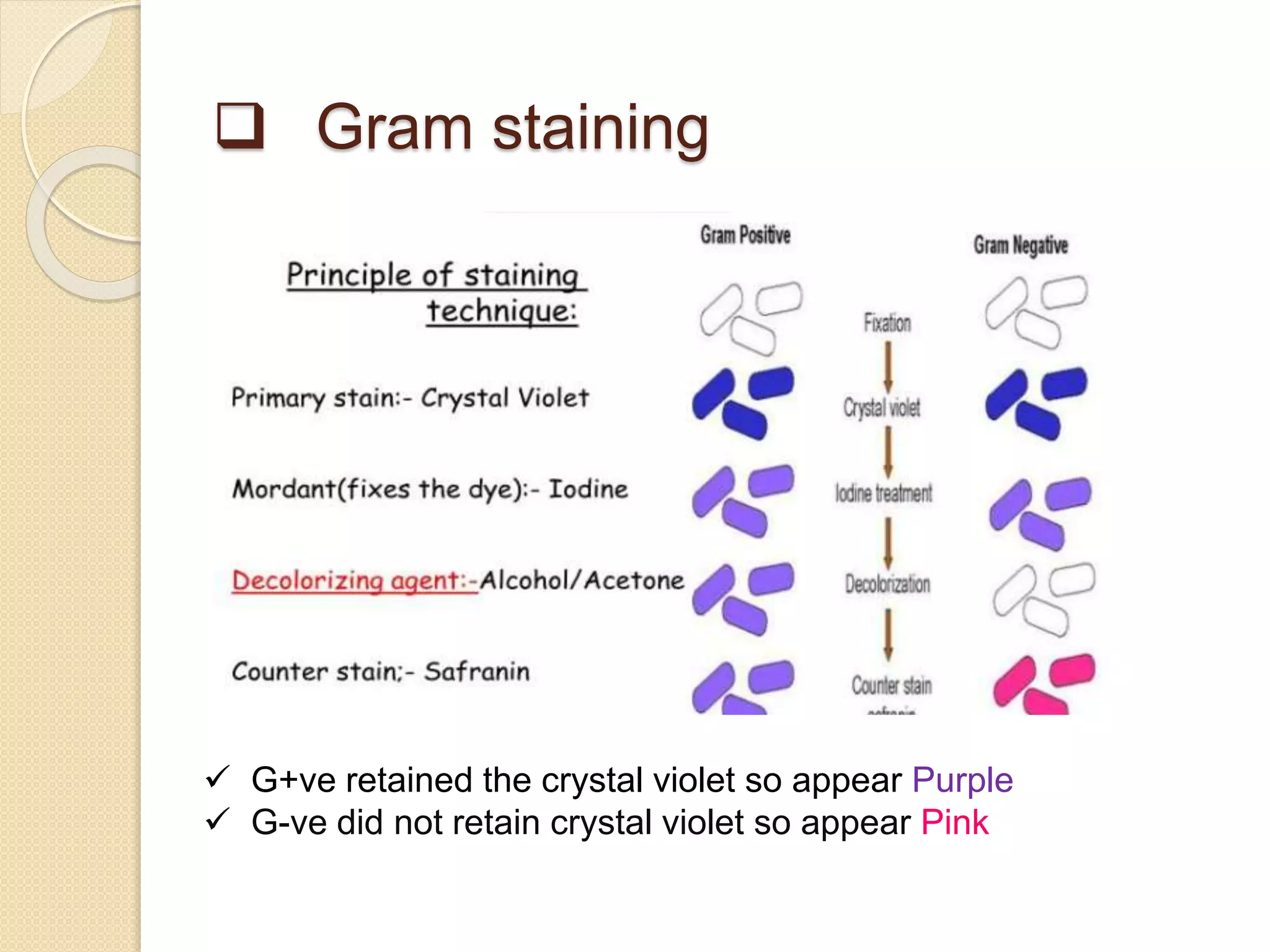
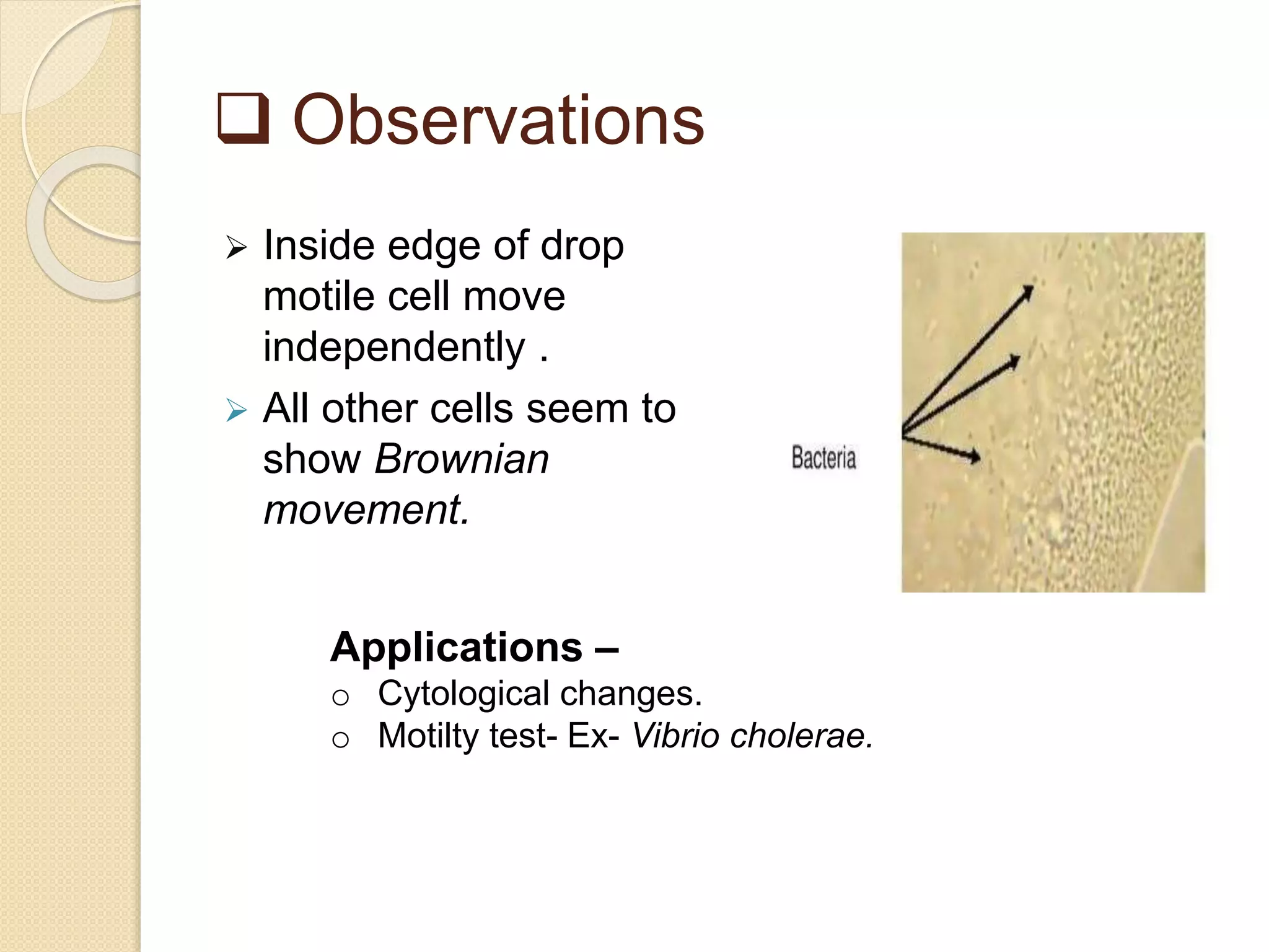
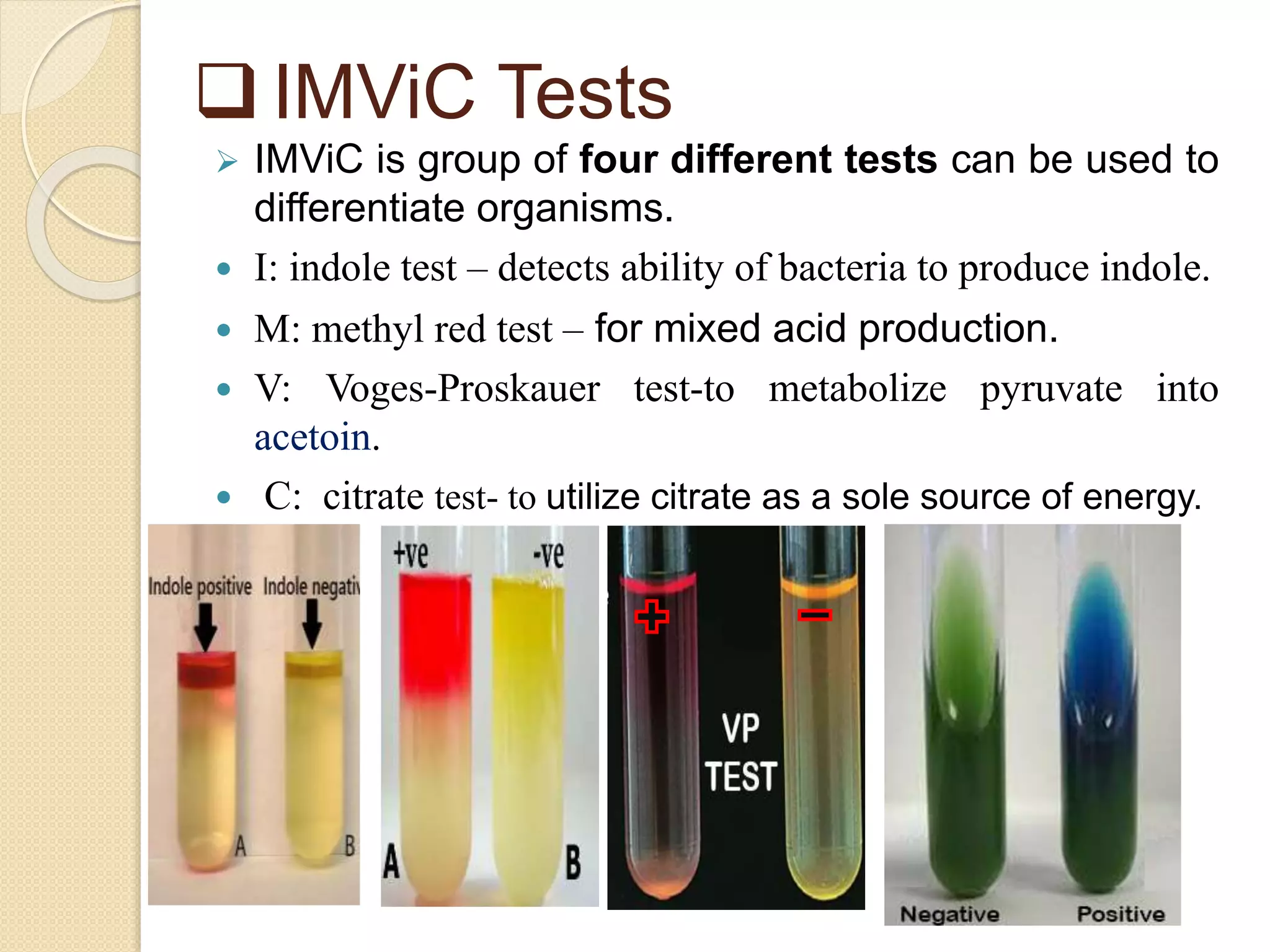
.
Submitted by – Ankur Kumar
M.Sc. Microbiology
Central University Of Haryana
 Classification and identification?
 Classification- Placing an organism in groups of
related species. List of characteristics of known
organisms.
 Identification- Matching characteristics of an
unknown to list of known organisms.
 Identification and characterization
 Morphological approach
 Physiological and Biochemical approach
 Immunologial approach
 Molecular approach
 Staining:-
 Biochemical technique of coloring specimen.
 Use to increase visibility of microorganisms being
studied and to identify the shape of bacteria.
 Use to determine the morphological features of
microorganisms.
 Use to differentiate and classify
microorganisms.
 Use to detect bacterial parts such as capsule,
spores, flagella or inclusion bodies.
 Types of staining
 Simple Staining Technique:- Only one stain is
used. Crystal Violet, and Methylene Blue are
examples of basic dyes used in simple staining
technique.
 Differential Staining:- Used to classify microbes
into separate groups based on their distinct
staining properties.
 Use of two contrasting stains seperated by
decolurizing agents
 Examples- Gram Stain, Acid-Fast Stain and Spore-
Staining .
 Gram staining
 G+ve retained the crystal violet so appear Purple
 G-ve did not retain crystal violet so appear Pink
 Acid-Fast Stain
 Acid-fast stain is a differential stain used to identify
acid fast organisms.
 First introduced by Dr. Paul Ehrlich.
 Also known as the Ziehl-Neelson’s staining.
 Acid fast bacteria appear as bright red bacilli against a
light blue background.
 Used to differentiate Mycobacteria (M. tuberculosis)
from other bacteria.
 EX- Corynebacterium ,Nocardia spp.,
Rhodococcus spp.
 Acid fast bacteria do not decolourize on applying
acid alcohol so appear Red
 Non acid fast bacteria appear blue after applying
methylene blue. (counter stain)
 Special Staining
 Negative Staining - India Ink or Nigrosine
is used to stain capsule.
 Schaefer-Fulton- Malachite Green and
Safranin is used to stain endospores.
 Hanging Drop Method
.
 To examine the cell motility and morphology by
taking the living microorganism from the media.
 Observations
 Inside edge of drop
motile cell move
independently .
 All other cells seem to
show Brownian
movement.
Applications –
o Cytological changes.
o Motilty test- Ex- Vibrio cholerae.
 Cultural charateristics
Characteristics of growth in broth.
• Clear – no growth
• surface
• subsurface
• sediments
• Amount of Growth
 Characteristics of colonies on plates
 size, color, form, elevation, and margin
Characteristics of growth on slants
 amount of growth
 colour
 Opacity
 form
 Biochemical Tests
 Each species of bacteria has specific metabolic
needs and relies on different enzymes to fuel those
unique needs.
 The presence of catalase, gelatinase, oxidase,
urease can be used to identify the species of
bacteria.
 Biochemical characterization relies on the
characteristic patterns of substrate utilization,
metabolic product formation and sugar formation,
etc.
.
1.Fermentation tests- whether the microbe is
capable to carry out various fermentation reactions or
not.
Ex., Mixed-acid fermentation (methyl red test)
2.Hydrolytic Reactions:- starch , casein, fat,
tryptophan, urea, phenylalanine.
Starch Hydrolysis
Positive test: A clear zone
around the line of growth
indicates that the organism has
hydrolyzed starch.
Negative test: A blue, purple, or
black coloration of the medium
(depending on the concentration of
iodine).
.
3.oxidative tests- for respiratory metabolism.
 Eg., Catalase, and Nitrate tests.
The presence of the enzyme in a bacterial isolate is evident when
a small inoculum is introduced into hydrogen peroxide,
and the rapid elaboration of oxygen bubbles occurs.
 IMViC Tests
 IMViC is group of four different tests can be used to
differentiate organisms.
 I: indole test – detects ability of bacteria to produce indole.
 M: methyl red test – for mixed acid production.
 V: Voges-Proskauer test-to metabolize pyruvate into
acetoin.
 C: citrate test- to utilize citrate as a sole source of energy.
.
Name of
Bacteria
Indole Test MR Test VP Test Citrate Test
E. coli + ve + ve – ve – ve
Klebsiella
pneumoniae
– ve – ve + ve + ve
Klebsiella
oxytoca
+ ve – ve + ve + ve
Pseudomona
s aeruginosa
– ve – ve – ve + ve
Salmonella
Paratyphi A
– ve + ve – ve – ve
Salmonella
Typhimurium
– ve + ve – ve + ve
 Molecular approaches
Ribotyping- rRna sequencing.
 Polymerase Chain Reaction (PCR)- can be
used to amplify a small amount of microbial DNA in
a sample.
o The presence or identification of an organism is
indicated by amplified DNA
 Nucleic acid hybridization
 Single strands of DNA or RNA, from related organisms
will hydrogen bond to form a double stranded molecule;
this bonding is called nucleic acid hybridization.
Taxonomical
characterization
of newly
isolated strains.
 Referance:
 Microbiology, a loboratory manual ,12th Edition
James G. Cappuccino
 Prescott's Microbiology 9th Edition, by Willey.
 https://www.sciencedirect.com
 https://www.ncbi.nlm.nih.gov/.
 https://microbenotes.com

Identification Of Unknown Bacteria And Biochemical Tests.pptx

  • 1.
    . Submitted by –Ankur Kumar M.Sc. Microbiology Central University Of Haryana
  • 2.
     Classification andidentification?  Classification- Placing an organism in groups of related species. List of characteristics of known organisms.  Identification- Matching characteristics of an unknown to list of known organisms.
  • 3.
     Identification andcharacterization  Morphological approach  Physiological and Biochemical approach  Immunologial approach  Molecular approach
  • 5.
     Staining:-  Biochemicaltechnique of coloring specimen.  Use to increase visibility of microorganisms being studied and to identify the shape of bacteria.  Use to determine the morphological features of microorganisms.  Use to differentiate and classify microorganisms.  Use to detect bacterial parts such as capsule, spores, flagella or inclusion bodies.
  • 6.
     Types ofstaining  Simple Staining Technique:- Only one stain is used. Crystal Violet, and Methylene Blue are examples of basic dyes used in simple staining technique.  Differential Staining:- Used to classify microbes into separate groups based on their distinct staining properties.  Use of two contrasting stains seperated by decolurizing agents  Examples- Gram Stain, Acid-Fast Stain and Spore- Staining .
  • 7.
     Gram staining G+ve retained the crystal violet so appear Purple  G-ve did not retain crystal violet so appear Pink
  • 9.
     Acid-Fast Stain Acid-fast stain is a differential stain used to identify acid fast organisms.  First introduced by Dr. Paul Ehrlich.  Also known as the Ziehl-Neelson’s staining.  Acid fast bacteria appear as bright red bacilli against a light blue background.  Used to differentiate Mycobacteria (M. tuberculosis) from other bacteria.  EX- Corynebacterium ,Nocardia spp., Rhodococcus spp.
  • 10.
     Acid fastbacteria do not decolourize on applying acid alcohol so appear Red  Non acid fast bacteria appear blue after applying methylene blue. (counter stain)
  • 11.
     Special Staining Negative Staining - India Ink or Nigrosine is used to stain capsule.  Schaefer-Fulton- Malachite Green and Safranin is used to stain endospores.
  • 12.
     Hanging DropMethod .  To examine the cell motility and morphology by taking the living microorganism from the media.
  • 13.
     Observations  Insideedge of drop motile cell move independently .  All other cells seem to show Brownian movement. Applications – o Cytological changes. o Motilty test- Ex- Vibrio cholerae.
  • 14.
     Cultural charateristics Characteristicsof growth in broth. • Clear – no growth • surface • subsurface • sediments • Amount of Growth
  • 15.
     Characteristics ofcolonies on plates  size, color, form, elevation, and margin
  • 16.
    Characteristics of growthon slants  amount of growth  colour  Opacity  form
  • 17.
     Biochemical Tests Each species of bacteria has specific metabolic needs and relies on different enzymes to fuel those unique needs.  The presence of catalase, gelatinase, oxidase, urease can be used to identify the species of bacteria.  Biochemical characterization relies on the characteristic patterns of substrate utilization, metabolic product formation and sugar formation, etc.
  • 18.
    . 1.Fermentation tests- whetherthe microbe is capable to carry out various fermentation reactions or not. Ex., Mixed-acid fermentation (methyl red test) 2.Hydrolytic Reactions:- starch , casein, fat, tryptophan, urea, phenylalanine.
  • 19.
    Starch Hydrolysis Positive test:A clear zone around the line of growth indicates that the organism has hydrolyzed starch. Negative test: A blue, purple, or black coloration of the medium (depending on the concentration of iodine).
  • 20.
    . 3.oxidative tests- forrespiratory metabolism.  Eg., Catalase, and Nitrate tests. The presence of the enzyme in a bacterial isolate is evident when a small inoculum is introduced into hydrogen peroxide, and the rapid elaboration of oxygen bubbles occurs.
  • 21.
     IMViC Tests IMViC is group of four different tests can be used to differentiate organisms.  I: indole test – detects ability of bacteria to produce indole.  M: methyl red test – for mixed acid production.  V: Voges-Proskauer test-to metabolize pyruvate into acetoin.  C: citrate test- to utilize citrate as a sole source of energy.
  • 22.
    . Name of Bacteria Indole TestMR Test VP Test Citrate Test E. coli + ve + ve – ve – ve Klebsiella pneumoniae – ve – ve + ve + ve Klebsiella oxytoca + ve – ve + ve + ve Pseudomona s aeruginosa – ve – ve – ve + ve Salmonella Paratyphi A – ve + ve – ve – ve Salmonella Typhimurium – ve + ve – ve + ve
  • 23.
     Molecular approaches Ribotyping-rRna sequencing.  Polymerase Chain Reaction (PCR)- can be used to amplify a small amount of microbial DNA in a sample. o The presence or identification of an organism is indicated by amplified DNA
  • 24.
     Nucleic acidhybridization  Single strands of DNA or RNA, from related organisms will hydrogen bond to form a double stranded molecule; this bonding is called nucleic acid hybridization.
  • 25.
  • 26.
     Referance:  Microbiology,a loboratory manual ,12th Edition James G. Cappuccino  Prescott's Microbiology 9th Edition, by Willey.  https://www.sciencedirect.com  https://www.ncbi.nlm.nih.gov/.  https://microbenotes.com