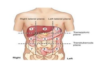
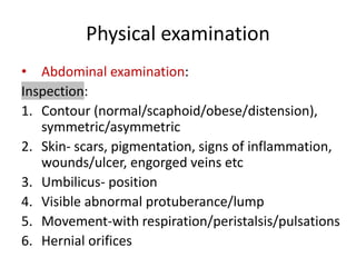
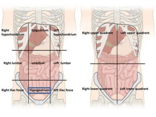
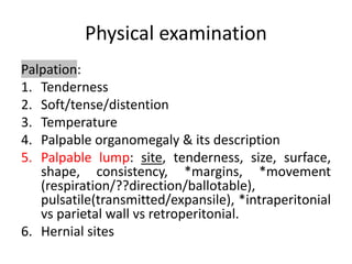
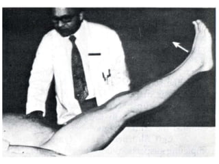
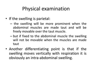
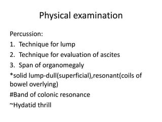
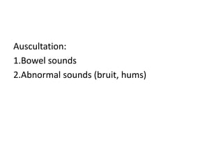
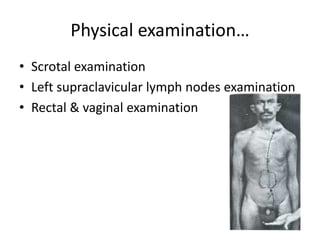
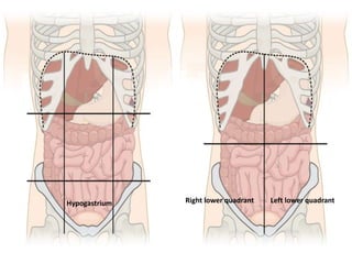
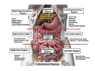
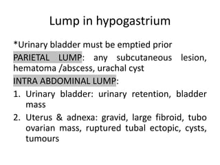
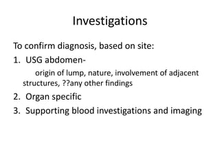
Approach to a patient with
hypogastric lump
Dr.Ankita Singh
Assistant Professor
General Surgery
L.H.M.C.
Objectives
Adopt sequential approach to a patient
complaining about abdominal lump by
• Extracting relevant history
• Thorough clinical examination
• Clue to right diagnosis by conducting
investigations guided by above history &
examination
• Offering adequate treatment
history
Demography:
• Age
• Gender
• Occupation
• Address
• Socio economic status
History…
Presenting complaints:
• Abdominal lump: appearance, duration,
site, approx. Size, progression/regression, any
association with posture/ straining/
trauma/*pain, disappears spontaneously
• Pain: site, onset, duration, character,
radiation/migration/referred, any relieving
/aggravating factors
History …
other associated complaints-
• Nausea/vomiting: character, amount,
frequency, content (hemetemesis),
relieving/aggrevating factors,
• Altered bowel habit: diarrhea, constipation,
obstipation, flatulent dyspepsia etc
• Melaena/hematochezia
• Jaundice: onset, duration,progression etc.
History…
• Associated fever episodes
• Appetite
• Weight loss
• Any other relevant history
History…
Past & personal history:
• Comorbidity/ treatments- medical or
surgical
• Sleep-wake cycle/ eating habits/ appetite/
bladder & bowel habits/ substance abuse
Family/ menstrual & obstetric history
Physical examination
Prerequisites: Consent, environment, chaperon
General survey:
• built, nutrition, mentation
• vitals
• signs-pallor, icterus, cyanosis, pedal edema,
lymphadenopathy, clubbing.
Systemic examination:
• CNS/Respiratory/Cardiovascular
examination
Physical examination
• Abdominal examination:
Inspection:
1. Contour (normal/scaphoid/obese/distension),
symmetric/asymmetric
2. Skin- scars, pigmentation, signs of inflammation,
wounds/ulcer, engorged veins etc
3. Umbilicus- position
4. Visible abnormal protuberance/lump
5. Movement-with respiration/peristalsis/pulsations
6. Hernial orifices
Right
hypochondrium
Epigastrium Left
hypochondrium
Right lumbar Left lumbar
umbilical
Right iliac fossa left iliac fossa
Hypogastrium
Right upper quadrant Left upper quadrant
Right lower quadrant Left lower quadrant
Physical examination
Palpation:
1. Tenderness
2. Soft/tense/distention
3. Temperature
4. Palpable organomegaly & its description
5. Palpable lump: site, tenderness, size, surface,
shape, consistency, *margins, *movement
(respiration/??direction/ballotable),
pulsatile(transmitted/expansile), *intraperitonial
vs parietal wall vs retroperitonial.
6. Hernial sites
Physical Examination…
*The abdominal muscles made taut by asking
patient
(i) to raise his shoulders from the bed with
the arms folded over the chest —
the 'rising-test' or
(ii) to raise both the extended legs from the
bed — the 'leg lifting test’(Carnett's test) or
(iii) to try to blow out with his nose and
mouth shut.
Physical examination
• If the swelling is parietal:
– the swelling will be more prominent when the
abdominal muscles are made taut and will be
freely movable over the taut muscle.
– but if fixed to the abdominal muscle the swelling
will not be movable when the muscles are made
taut
• Another differentiating point is that if the
swelling moves vertically with respiration it is
obviously an intra-abdominal swelling.
Physical examination
Percussion:
1. Technique for lump
2. Technique for evaluation of ascites
3. Span of organomegaly
*solid lump-dull(superficial),resonant(coils of
bowel overlying)
#Band of colonic resonance
~Hydatid thrill
Auscultation:
1.Bowel sounds
2.Abnormal sounds (bruit, hums)
Physical examination…
• Scrotal examination
• Left supraclavicular lymph nodes examination
• Rectal & vaginal examination
Differential diagnosis of hypogastric
lump
Hypogastrium Right lower quadrant Left lower quadrant
Lump in hypogastrium
*Urinary bladder must be emptied prior
PARIETAL LUMP: any subcutaneous lesion,
hematoma /abscess, urachal cyst
INTRA ABDOMINAL LUMP:
1. Urinary bladder: urinary retention, bladder
mass
2. Uterus & adnexa: gravid, large fibroid, tubo
ovarian mass, ruptured tubal ectopic, cysts,
tumours
Investigations
To confirm diagnosis, based on site:
1. USG abdomen-
origin of lump, nature, involvement of adjacent
structures, ??any other findings
2. Organ specific
3. Supporting blood investigations and imaging
Thank you
Any queries?

hypogastric lump abdomen.pptx

  • 1.
    Approach to apatient with hypogastric lump Dr.Ankita Singh Assistant Professor General Surgery L.H.M.C.
  • 2.
    Objectives Adopt sequential approachto a patient complaining about abdominal lump by • Extracting relevant history • Thorough clinical examination • Clue to right diagnosis by conducting investigations guided by above history & examination • Offering adequate treatment
  • 4.
    history Demography: • Age • Gender •Occupation • Address • Socio economic status
  • 5.
    History… Presenting complaints: • Abdominallump: appearance, duration, site, approx. Size, progression/regression, any association with posture/ straining/ trauma/*pain, disappears spontaneously • Pain: site, onset, duration, character, radiation/migration/referred, any relieving /aggravating factors
  • 6.
    History … other associatedcomplaints- • Nausea/vomiting: character, amount, frequency, content (hemetemesis), relieving/aggrevating factors, • Altered bowel habit: diarrhea, constipation, obstipation, flatulent dyspepsia etc • Melaena/hematochezia • Jaundice: onset, duration,progression etc.
  • 7.
    History… • Associated feverepisodes • Appetite • Weight loss • Any other relevant history
  • 8.
    History… Past & personalhistory: • Comorbidity/ treatments- medical or surgical • Sleep-wake cycle/ eating habits/ appetite/ bladder & bowel habits/ substance abuse Family/ menstrual & obstetric history
  • 9.
    Physical examination Prerequisites: Consent,environment, chaperon General survey: • built, nutrition, mentation • vitals • signs-pallor, icterus, cyanosis, pedal edema, lymphadenopathy, clubbing. Systemic examination: • CNS/Respiratory/Cardiovascular examination
  • 10.
    Physical examination • Abdominalexamination: Inspection: 1. Contour (normal/scaphoid/obese/distension), symmetric/asymmetric 2. Skin- scars, pigmentation, signs of inflammation, wounds/ulcer, engorged veins etc 3. Umbilicus- position 4. Visible abnormal protuberance/lump 5. Movement-with respiration/peristalsis/pulsations 6. Hernial orifices
  • 11.
    Right hypochondrium Epigastrium Left hypochondrium Right lumbarLeft lumbar umbilical Right iliac fossa left iliac fossa Hypogastrium Right upper quadrant Left upper quadrant Right lower quadrant Left lower quadrant
  • 12.
    Physical examination Palpation: 1. Tenderness 2.Soft/tense/distention 3. Temperature 4. Palpable organomegaly & its description 5. Palpable lump: site, tenderness, size, surface, shape, consistency, *margins, *movement (respiration/??direction/ballotable), pulsatile(transmitted/expansile), *intraperitonial vs parietal wall vs retroperitonial. 6. Hernial sites
  • 13.
    Physical Examination… *The abdominalmuscles made taut by asking patient (i) to raise his shoulders from the bed with the arms folded over the chest — the 'rising-test' or (ii) to raise both the extended legs from the bed — the 'leg lifting test’(Carnett's test) or (iii) to try to blow out with his nose and mouth shut.
  • 14.
    Physical examination • Ifthe swelling is parietal: – the swelling will be more prominent when the abdominal muscles are made taut and will be freely movable over the taut muscle. – but if fixed to the abdominal muscle the swelling will not be movable when the muscles are made taut • Another differentiating point is that if the swelling moves vertically with respiration it is obviously an intra-abdominal swelling.
  • 15.
    Physical examination Percussion: 1. Techniquefor lump 2. Technique for evaluation of ascites 3. Span of organomegaly *solid lump-dull(superficial),resonant(coils of bowel overlying) #Band of colonic resonance ~Hydatid thrill
  • 16.
  • 17.
    Physical examination… • Scrotalexamination • Left supraclavicular lymph nodes examination • Rectal & vaginal examination
  • 18.
    Differential diagnosis ofhypogastric lump
  • 19.
    Hypogastrium Right lowerquadrant Left lower quadrant
  • 21.
    Lump in hypogastrium *Urinarybladder must be emptied prior PARIETAL LUMP: any subcutaneous lesion, hematoma /abscess, urachal cyst INTRA ABDOMINAL LUMP: 1. Urinary bladder: urinary retention, bladder mass 2. Uterus & adnexa: gravid, large fibroid, tubo ovarian mass, ruptured tubal ectopic, cysts, tumours
  • 22.
    Investigations To confirm diagnosis,based on site: 1. USG abdomen- origin of lump, nature, involvement of adjacent structures, ??any other findings 2. Organ specific 3. Supporting blood investigations and imaging
  • 23.

Editor's Notes

  • #16 Solid lump- dull if superficial, but not dull if deep and overlapped by bowel loops- resonant
  • #18 In appendicitis(pelvic),