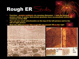
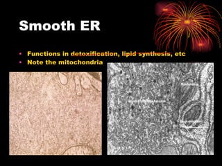
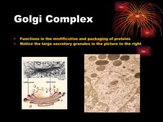
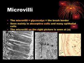
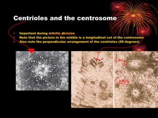
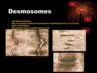
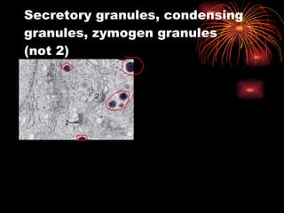
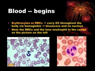
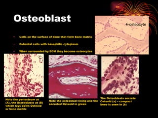
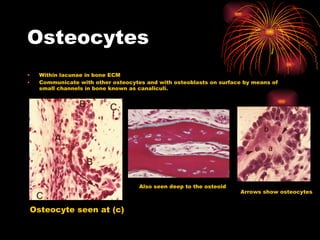
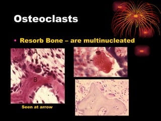
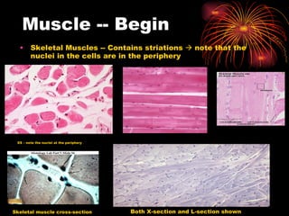
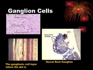
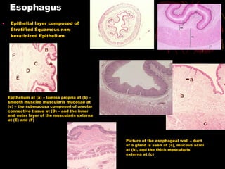
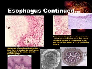
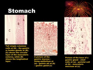
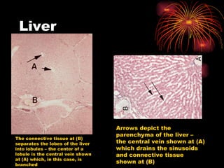
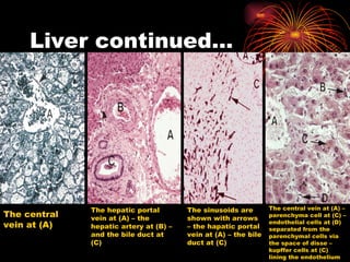
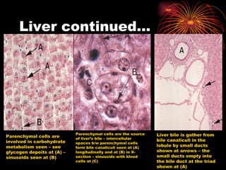
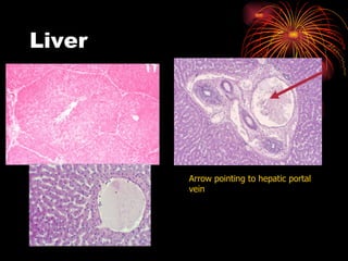
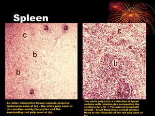
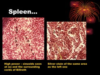
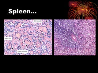
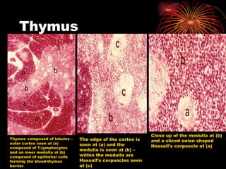
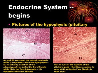
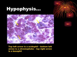
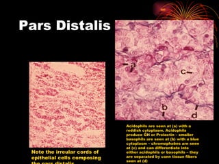
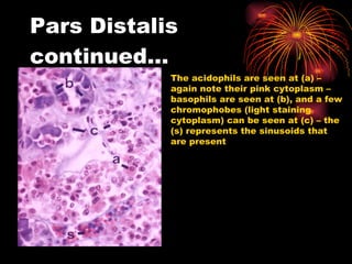
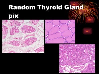
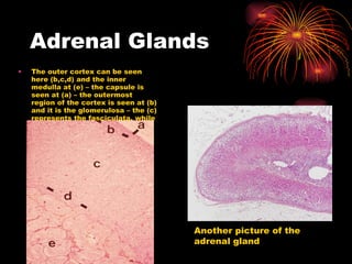
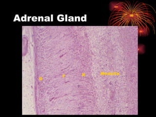
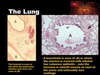
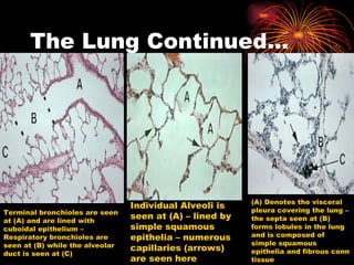
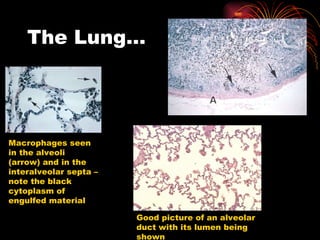
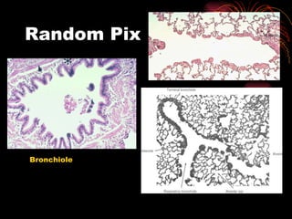
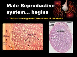
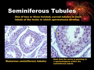
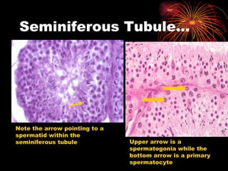
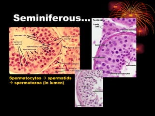
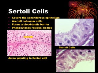
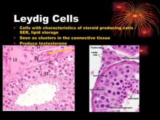
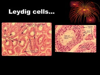
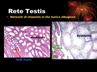
This document provides a histology slide list that summarizes key structures and functions seen across various tissue and cell types, including the nucleus, chromatin, mitochondria, rough ER, smooth ER, Golgi complex, microvilli, centrioles, desmosomes, blood cells, epithelial cells, connective tissue, glands, integumentary system, cartilage, bone, and muscle. Key structures are highlighted on photomicrographs with labels indicating their location.

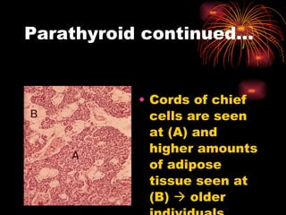
![Parathyroid Gland Picture of thyroid [(C) and (B)] and parathyroid glands (A) – note the parathyroid gland is composed of cords of cells and is separated from the thyroid by a capsule Note the cells are lined up along capillaries – chief cells are seen at (a) and comprise most of the cells – some chief cells are large and contain lots of glycogen (b) – note they are more clear – these chief cells release PTH – chief cells are also seen at (c) and are in contact with capillaries](https://image.slidesharecdn.com/slidelist-awesome-110913221922-phpapp01/85/Slide-list-awesome-131-320.jpg)


![2. epithelial-t[1]](https://cdn.slidesharecdn.com/ss_thumbnails/c55mbqopt3axovrntgld-signature-4c28f0f13a30c4ea316a9d58353990586de4897ab085203d01a9b7b7228e72f9-poli-180213061217-thumbnail.jpg?width=640&height=640&fit=bounds)


