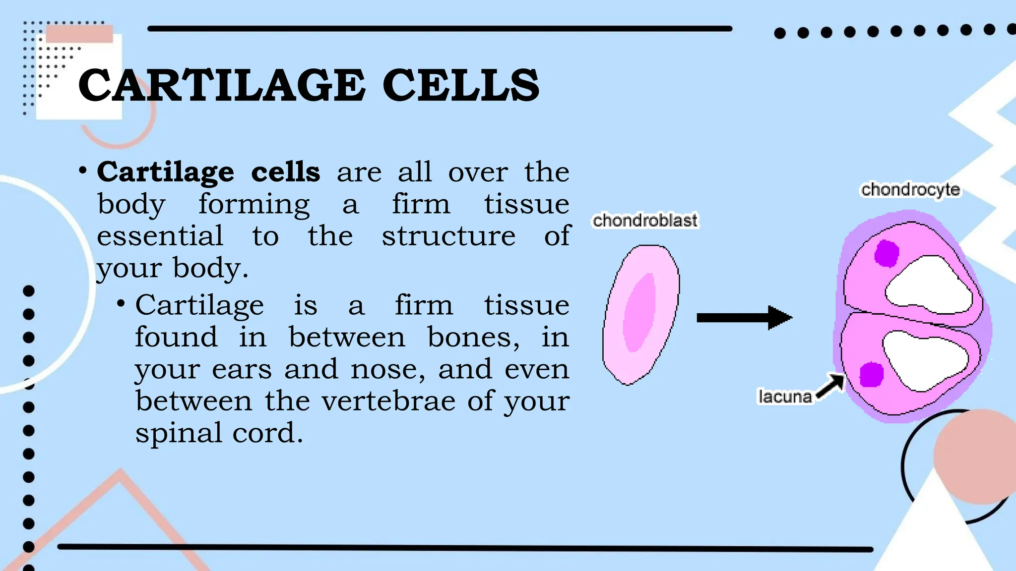
CELL CLASSIFICATION
AND TYPES
GENERAL BIOLOGY 1
Somatic cells (body cells) and
Gametes (sex cells)
• Somatic cells are a regular type of body cell that is not
involved in any way in sexual reproduction. In humans, such
cells are diploid and reproduce using the process
of mitosis to create identical diploid copies of themselves
when they split.
• Gamete or sex cells are cells that can produce and
reproduce. These produce gametes in living entities through
sexual reproduction. In humans, the gametes are called the
sperm (in the male) and the egg (in the female).
• These are formed by the process of meiosis, which can turn a
diploid cell into four haploid gametes.
VARIOUS TYPES OF
CELLS IN HUMANS
Nerve Cells- also known as neurons, these
are the basic unit of the nervous system
transmitting information throughout your
body.
• transmit signals as little electrical
signals and sent through the brain,
spinal cord, and organs.
• warn you or inform you of what is
happening throughout your body.
NERVE CELLS
CARTILAGE CELLS
• Cartilage cells are all over the
body forming a firm tissue
essential to the structure of
your body.
• Cartilage is a firm tissue
found in between bones, in
your ears and nose, and even
between the vertebrae of your
spinal cord.
BONE CELLS
• Bone cells- One of the strongest cells in the body. These gives
body strength and support in its skeletal system. There are
many different types of bone cells in the body:
• Osteoclasts- aid in bone resorption releasing enzymes and
acids to help the process of breaking down bones.
• Osteoblasts- help form new bones by managing bone
mineralization.
• Osteocytes- osteoblast form osteocytes which are found in
the bone. These cells produce growth on the bone in
response to strain and help keep calcium balanced. Without
bone cells you wouldn’t have your skeletal system.
MUSCLE CELLS
Muscle cells- These cells form muscle tissue which is necessary
for body movements. These are multiple types of muscle such as
skeletal muscle tissue, smooth and cardiac muscle tissue.
• Skeletal muscle tissue- enables voluntary movement by
attaching to bones and tendons.
• Smooth muscle tissue- responsible for contractions in the
bladder, lungs, and other parts of the body such as the digestive
system.
• Cardiac muscle tissue- part of the most important muscle
tissue in the body-heart. These cells contract in harmony to
create the contractions of the heart.
BLOOD CELLS
Blood cells- Blood cells they transport oxygen through the
body, fight infections, and are critical to our body. There are
different types of blood cells in the body such as red blood
cells, white blood cells, and platelets. Each of these cells
perform a different task in the bloodstream.
• Red blood cells- carry oxygen through the body.
• White blood cells- fight off diseases and illnesses.
• Platelet- cloth the blood to prevent too much blood loss. For
example, when you cut your finger platelets come together to
stop the cut from bleeding. Each of these three cells are
produced through bone marrow.
FAT CELLS
Fat cells- Fat cells are also known as adipocytes.
• Adipocytes contain triglycerides (stored fat) used for
energy. When storing fat these cells swell and
become round; however, when the fat is being used
the cells shrink down.
• These fat cells also have an endocrine function to
them. They influence multiple hormones for your
metabolism, sex drive, blood pressure and clotting,
and cell signaling.
SKIN CELLS
Skin Cells- Your skin is made of
multiple layers of skin cells
including epithelial and
connective tissue, along with a
subcutaneous layer. Composed
of a layer of epithelial tissue
that is supported by a layer of
connective tissue and an and
an underlying subcutaneous
layer.
The outermost layer is made of many different types of
cells packed tightly together. These cells include:
• Keratinocytes- produce keratin proteins which block your body
from toxins.
• Merkel cells- give you the ability to feel when you touch an
item.
• Langerhans cells- are antigens for the skin. If there is a cut that
becomes infected these cells fight the infection for your body.
• Melanocytes- produce melanin giving your skin its color. Your
skin is the largest organ in your body. It protects you from
dehydration, germs, damage, and stores fats and vitamins
within.
SEX CELLS
Sex cells- also called gametes, reproductive cells produced in
the male and female reproductive organs.
• Male sex cells (sperm) are long and mobile with tail-like
extension. They have a head, a midsection, and a tail that
propels them. The head contains the DNA and are covered
with enzymes to penetrate the ova.
• Female sex cells (eggs or ova/ovum) are non-moving cells
and are large in comparison to sperm. Ova is produced in
the body during embryonic stages. During fertilization ova
and sperm cells unite to form a new being.
STEM CELLS
Stem cells- these cells are unique to the body as they begin
unspecialized and can develop into specialized cells.
• Once stem cells become specialized, they work with specific
organs and tissues- often repairing or replacing old tissue in
the body.
• Stem cells repair tissue by dividing and duplicating multiple
times.
• Stem cells can treat multiple diseases, tissues repairs, and
transplants.
ANIMAL TISSUES
GENERAL BIOLOGY 1
Animal Tissues
Tissues result from the differentiation of several groups of
cells that will form a group of cells having the same
function.
In animals, the tissues are either epithelial, connective,
muscular, or nervous tissue. Different tissues arise from a
particular germ layer during embryonic development.
• Histology- the study of different tissues.
Epithelial tissue
1. Epithelial tissue- made up of polygonal cells nearly
placed together with very few or no extracellular
matrix.
• Their function includes shielding the organism against
harmful infective agents (e.g., skin), uptake and pre-
metabolism of nutrients/drugs (e.g., intestine), and sensation
(e.eg. neuroepithelium).
Types of Epithelial Tissue
1. Simple epithelium-.has only one layer of cells classified as squamous,
columnar, and cuboidal, where the shape and structure are bases.
a. Simple squamous epithelium- flat cells with a non-regular outline,
closely fitted together to form a sheet. This type of cell is prevalent in the
alveoli, endothelium of blood vessels, and covering mesothelium of body
cavities.
b. Simple cuboidal epithelium- it has a box-shaped structure with
brush border in its cell lining, usually found in kidneys.
c. Simple columnar epithelium- cylindrical with a noticeable height
plus straited border. It has goblet cells that secrete mucus, normally
found in the stomach.
2. The stratified epithelium -comprises of more than one layer of
cells, classified according to structure of the topmost tissue layer.
a. Stratified squamous epithelium- multilayer of cell groups,
often bound in the skin that protects the organism from bacterial
invasion. It can be keratinized or non-keratinized.
b. Transition epithelium- are overlapping layers of cells that
line the ureters and urinary bladder.
c. Stratified cuboidal epithelium- are multilayered cube-
shaped cells fused together in some glands.
d. Stratified columnar epithelium- stratified, elongated cell
structure often found in the conjunctiva and human salivary
glands.
3. Glandular epithelium is distinct due to cells that function for
molecular secretion.
Types of Glandular epithelium, classified according to:
a. Number of cells- monocellular or multicellular.
b. Morphology or shape- simple, compound, or saccular.
c. Types of secretion- serous, mucous, or mixed serous-
mucous.
d. Presence or absence of secretory ducts- endocrine or
exocrine; and
e. The integrity of the secretory cells- holocrine, apocrine,
and merocrine.
The Connective Tissues and
Special Connective Tissues
• Connective tissues are groups of tissue that structurally
differ according to their localization.
• They can be fibrous, elastic, reticular, or collagenous.
They are important for support, protection, transport,
insulation, and repair.
• They are also responsible for blood cell differentiation or
formation (hematopoiesis) and immunological responses.
Types of Connective Tissue
1. Collagenous connective tissue- structurally made up
of type II collagen. Examples of these are the fasciae
containing loose connective tissue, tensons containing
dense regular connective tissue and the periosteum
containing dense irregular connective tissue.
2. Reticular connective tissues-structurally made up of
type III collagen and usually found in the supporting
structure of the hematopoietic and lymphoid organs.
Examples are the basal lamina and stroma of glandular
organs.
Types of Connective Tissue
3. Elastic connective tissue- structurally made up
of type II collagen containing retractile fibers with
elastin, usually found in the trachea, bronchi, and
blood vessels.
4. Connective tissues with special properties-
these are the adipose tissue which acts as insulator
via fat storage and does mechanical support and
the mucous connective tissue, found in the
umbilical cord and young tooth pulp.
Specialized Connective tissue- specialized tissues
that are found in blood, cartilage, and bone.
1. Blood and lymph- structurally fiber in dry form that functions
primarily for gas exchange, immunity, hormonal transport and much
more.
2. Cartilage- structurally made up of elastic fibers and collagen. Its
function is for support and flexibility.
3. Bones- structurally a rigid connective tissue composed of
osteoblast, osteocytes, and osteoclast plus a calcified matrix of organic
(collagen and polysaccharides) and inorganic materials (e.g., calcium).
Bones can be cancellous/spongy (irregular branching bony spicules) or
compact (concentric layer surrounding the Haversian system).
The Muscular Tissue
The muscular tissue function for movement and locomotion,
hence, are structurally elongated. Their function varies
according to its location and type.
1. Smooth muscles- have no cross-striations and are fusiform-
shaped. Functionally, they are slow contracting and not
voluntary. Examples are smooth muscle of the visceral organs.
2. Skeletal muscles- are structurally elongated, multinucleated,
and cylindrical in shape. They are functionally capable of
voluntary contractions and are the major structures that
constitute the muscular system.
The Muscular Tissue
3. Cardiac muscle- composed of elongated branched cells that
adhere parallel to each other. They are functionally capable of
involuntary rhythmic contractions and are distinguishable from the
other muscle types because of the presence of an intercalated disc
that connects one cardiac cell from the other. The heart and major
blood vessels are the only organs that have cardiac muscles.
4. Nervous tissue- receives stimuli from the environment and
internal signals, which produce relay functionality. The functionality
is dependent on synapses, which serves as the site for the
physiological relay of signals. It is composed of a variety of cells
including the neuron, the functional unit of the nervous system.
Classification of Neurons based on:
1. Function- Sensory neurons are responsible for
transmission of signals from the environment or other
parts of the body to the Central Nervous System (CNS).
Interneurons are for are the integration and processing
of signals. Motor neurons command the effector.
2. A number of processes-Neurons can be categorized
into unipolar (one process), bipolar (two processes),
and multipolar (multiple processes).
3. The length of signal relay- Interneurons is small
neurons(connector) whose axons are contained within specific
area of the brain whereas the projection neurons (excitatory) have
long axons projecting to distant targets or the brain regions.
4. Chemical Messenger-Neurons can be differentiated regarding
chemicals used to transfer impulses from one part of the body to
another such as cholinger (e.g., acetylcholine as a
neurotransmitter), adrenergic (e.g., epinephrine and
norepinephrine), dopaminergic (e.g., dopamine), and serotonergic
(e.g., serotonin).
PLANT CELL TYPES
GENERAL BIOLOGY 1
Parenchyma tissue
1. Parenchyma tissue-represents most of the cells in a plant.
• They are found in leaves and carry out photosynthesis and
cellular respiration, along with other metabolic processes.
• They also store substances like starches and proteins and
have a role in plant wound repair.
• They are live thin-walled cells with permeable walls that are
undifferentiated.
• They do not have a specialized structure hence they easily
adapt and differentiate into a variety of cells performing
different functions.
TWO TYPES OF PARENCHYMA
CELLS
• Palisade parenchyma cells are columnar elongated
structured cells found in a variety of leaves, lying below
the epidermal tissue. Palisades are closely linked cells in
layers of mesophyll cells found in leaf cells.
• Ray parenchyma cells has both radial and horizontal
arrangement majorly found within the stem wood of the
plant.
Collenchyma tissue
2. Collenchyma tissue- provides support to growing parts
of a plant. They are elongated, have thick cell walls, and
can grow and change shape as a plant grows. There are
four types of collenchyma based on the thickness of the
wall and the cell arrangement.
• Annular collenchyma- The walls are uniformly thickened. The
cells appear to be circular in shape.
• Lamellar collenchyma- The cells are thickened on the periphery
making them appear tangentially arranged in rows. They are
closely packed together and therefore they don’t have intracellular
spaces. They are commonly formed and found in the leaf’s
petioles.
• Lacunar Collenchyma- These are cells are formed spaciously
leaving intracellular spaces between each other. The cell wall
thickens around the intracellular spaces. They appear spherically
shaped. They are formed and found in the walls of fruits.
Sclerenchyma tissue
3. Sclerenchyma tissue- contains hard cells that are
the main supporting cells in the areas of plant that
have ceased growing. Sclerenchyma cells are dead
and have very thick cell walls. There are two types of
sclerenchyma cells.
• Fiber sclerenchyma cells
• Sclereid sclerenchyma cells
4.Xylem cells - transport mostly water and few
nutrients throughout a plant, from the roots to the
stem and leaves.
5.Phloem cells- transport nutrients made during
photosynthesis to all parts of a plant. They transport
sap, which is a watery solution high in sugars.
6.Meristematic cells - They are also known as the
meristems. These are the cells in a plant that divide
continuously throughout the life of a plant. They
have a self-renewal ability and high metabolisms to
control the cell.
There are three types of meristematic cells
classified according to the tissue they exist in.
• Apical meristems – they are found at the tips of
roots and stems that have started growing and they
contribute to the length of the plant.
• Lateral meristems – They are found in the radial
part of the stem and roots, and they contribute to
the plant thickness.
• Intercalary meristems – they are found at the base
of the leaves and the contribution to the size
variance of the leaves.
Epidermal cell
• Epidermal cell -These are
the external cells of the
plants offering protection
from water loss, pathogenic
invaders such as fungi. They
are placed closely together
with no intracellular spaces.
They are covered with a waxy
cuticle layer to reduce water
loss. These cells cover the
plant stems, leaves, roots,
and plant seeds.

gen bio CELL CLASSIFICATION AND TYPES.pptx

  • 1.
  • 2.
    Somatic cells (bodycells) and Gametes (sex cells) • Somatic cells are a regular type of body cell that is not involved in any way in sexual reproduction. In humans, such cells are diploid and reproduce using the process of mitosis to create identical diploid copies of themselves when they split. • Gamete or sex cells are cells that can produce and reproduce. These produce gametes in living entities through sexual reproduction. In humans, the gametes are called the sperm (in the male) and the egg (in the female). • These are formed by the process of meiosis, which can turn a diploid cell into four haploid gametes.
  • 3.
    VARIOUS TYPES OF CELLSIN HUMANS Nerve Cells- also known as neurons, these are the basic unit of the nervous system transmitting information throughout your body. • transmit signals as little electrical signals and sent through the brain, spinal cord, and organs. • warn you or inform you of what is happening throughout your body. NERVE CELLS
  • 4.
    CARTILAGE CELLS • Cartilagecells are all over the body forming a firm tissue essential to the structure of your body. • Cartilage is a firm tissue found in between bones, in your ears and nose, and even between the vertebrae of your spinal cord.
  • 6.
    BONE CELLS • Bonecells- One of the strongest cells in the body. These gives body strength and support in its skeletal system. There are many different types of bone cells in the body: • Osteoclasts- aid in bone resorption releasing enzymes and acids to help the process of breaking down bones. • Osteoblasts- help form new bones by managing bone mineralization. • Osteocytes- osteoblast form osteocytes which are found in the bone. These cells produce growth on the bone in response to strain and help keep calcium balanced. Without bone cells you wouldn’t have your skeletal system.
  • 8.
    MUSCLE CELLS Muscle cells-These cells form muscle tissue which is necessary for body movements. These are multiple types of muscle such as skeletal muscle tissue, smooth and cardiac muscle tissue. • Skeletal muscle tissue- enables voluntary movement by attaching to bones and tendons. • Smooth muscle tissue- responsible for contractions in the bladder, lungs, and other parts of the body such as the digestive system. • Cardiac muscle tissue- part of the most important muscle tissue in the body-heart. These cells contract in harmony to create the contractions of the heart.
  • 10.
    BLOOD CELLS Blood cells-Blood cells they transport oxygen through the body, fight infections, and are critical to our body. There are different types of blood cells in the body such as red blood cells, white blood cells, and platelets. Each of these cells perform a different task in the bloodstream. • Red blood cells- carry oxygen through the body. • White blood cells- fight off diseases and illnesses. • Platelet- cloth the blood to prevent too much blood loss. For example, when you cut your finger platelets come together to stop the cut from bleeding. Each of these three cells are produced through bone marrow.
  • 12.
    FAT CELLS Fat cells-Fat cells are also known as adipocytes. • Adipocytes contain triglycerides (stored fat) used for energy. When storing fat these cells swell and become round; however, when the fat is being used the cells shrink down. • These fat cells also have an endocrine function to them. They influence multiple hormones for your metabolism, sex drive, blood pressure and clotting, and cell signaling.
  • 13.
    SKIN CELLS Skin Cells-Your skin is made of multiple layers of skin cells including epithelial and connective tissue, along with a subcutaneous layer. Composed of a layer of epithelial tissue that is supported by a layer of connective tissue and an and an underlying subcutaneous layer.
  • 14.
    The outermost layeris made of many different types of cells packed tightly together. These cells include: • Keratinocytes- produce keratin proteins which block your body from toxins. • Merkel cells- give you the ability to feel when you touch an item. • Langerhans cells- are antigens for the skin. If there is a cut that becomes infected these cells fight the infection for your body. • Melanocytes- produce melanin giving your skin its color. Your skin is the largest organ in your body. It protects you from dehydration, germs, damage, and stores fats and vitamins within.
  • 16.
    SEX CELLS Sex cells-also called gametes, reproductive cells produced in the male and female reproductive organs. • Male sex cells (sperm) are long and mobile with tail-like extension. They have a head, a midsection, and a tail that propels them. The head contains the DNA and are covered with enzymes to penetrate the ova. • Female sex cells (eggs or ova/ovum) are non-moving cells and are large in comparison to sperm. Ova is produced in the body during embryonic stages. During fertilization ova and sperm cells unite to form a new being.
  • 18.
    STEM CELLS Stem cells-these cells are unique to the body as they begin unspecialized and can develop into specialized cells. • Once stem cells become specialized, they work with specific organs and tissues- often repairing or replacing old tissue in the body. • Stem cells repair tissue by dividing and duplicating multiple times. • Stem cells can treat multiple diseases, tissues repairs, and transplants.
  • 20.
  • 21.
    Animal Tissues Tissues resultfrom the differentiation of several groups of cells that will form a group of cells having the same function. In animals, the tissues are either epithelial, connective, muscular, or nervous tissue. Different tissues arise from a particular germ layer during embryonic development. • Histology- the study of different tissues.
  • 22.
    Epithelial tissue 1. Epithelialtissue- made up of polygonal cells nearly placed together with very few or no extracellular matrix. • Their function includes shielding the organism against harmful infective agents (e.g., skin), uptake and pre- metabolism of nutrients/drugs (e.g., intestine), and sensation (e.eg. neuroepithelium).
  • 23.
    Types of EpithelialTissue 1. Simple epithelium-.has only one layer of cells classified as squamous, columnar, and cuboidal, where the shape and structure are bases. a. Simple squamous epithelium- flat cells with a non-regular outline, closely fitted together to form a sheet. This type of cell is prevalent in the alveoli, endothelium of blood vessels, and covering mesothelium of body cavities. b. Simple cuboidal epithelium- it has a box-shaped structure with brush border in its cell lining, usually found in kidneys. c. Simple columnar epithelium- cylindrical with a noticeable height plus straited border. It has goblet cells that secrete mucus, normally found in the stomach.
  • 24.
    2. The stratifiedepithelium -comprises of more than one layer of cells, classified according to structure of the topmost tissue layer. a. Stratified squamous epithelium- multilayer of cell groups, often bound in the skin that protects the organism from bacterial invasion. It can be keratinized or non-keratinized. b. Transition epithelium- are overlapping layers of cells that line the ureters and urinary bladder. c. Stratified cuboidal epithelium- are multilayered cube- shaped cells fused together in some glands. d. Stratified columnar epithelium- stratified, elongated cell structure often found in the conjunctiva and human salivary glands.
  • 25.
    3. Glandular epitheliumis distinct due to cells that function for molecular secretion. Types of Glandular epithelium, classified according to: a. Number of cells- monocellular or multicellular. b. Morphology or shape- simple, compound, or saccular. c. Types of secretion- serous, mucous, or mixed serous- mucous. d. Presence or absence of secretory ducts- endocrine or exocrine; and e. The integrity of the secretory cells- holocrine, apocrine, and merocrine.
  • 26.
    The Connective Tissuesand Special Connective Tissues • Connective tissues are groups of tissue that structurally differ according to their localization. • They can be fibrous, elastic, reticular, or collagenous. They are important for support, protection, transport, insulation, and repair. • They are also responsible for blood cell differentiation or formation (hematopoiesis) and immunological responses.
  • 27.
    Types of ConnectiveTissue 1. Collagenous connective tissue- structurally made up of type II collagen. Examples of these are the fasciae containing loose connective tissue, tensons containing dense regular connective tissue and the periosteum containing dense irregular connective tissue. 2. Reticular connective tissues-structurally made up of type III collagen and usually found in the supporting structure of the hematopoietic and lymphoid organs. Examples are the basal lamina and stroma of glandular organs.
  • 28.
    Types of ConnectiveTissue 3. Elastic connective tissue- structurally made up of type II collagen containing retractile fibers with elastin, usually found in the trachea, bronchi, and blood vessels. 4. Connective tissues with special properties- these are the adipose tissue which acts as insulator via fat storage and does mechanical support and the mucous connective tissue, found in the umbilical cord and young tooth pulp.
  • 29.
    Specialized Connective tissue-specialized tissues that are found in blood, cartilage, and bone. 1. Blood and lymph- structurally fiber in dry form that functions primarily for gas exchange, immunity, hormonal transport and much more. 2. Cartilage- structurally made up of elastic fibers and collagen. Its function is for support and flexibility. 3. Bones- structurally a rigid connective tissue composed of osteoblast, osteocytes, and osteoclast plus a calcified matrix of organic (collagen and polysaccharides) and inorganic materials (e.g., calcium). Bones can be cancellous/spongy (irregular branching bony spicules) or compact (concentric layer surrounding the Haversian system).
  • 30.
    The Muscular Tissue Themuscular tissue function for movement and locomotion, hence, are structurally elongated. Their function varies according to its location and type. 1. Smooth muscles- have no cross-striations and are fusiform- shaped. Functionally, they are slow contracting and not voluntary. Examples are smooth muscle of the visceral organs. 2. Skeletal muscles- are structurally elongated, multinucleated, and cylindrical in shape. They are functionally capable of voluntary contractions and are the major structures that constitute the muscular system.
  • 31.
    The Muscular Tissue 3.Cardiac muscle- composed of elongated branched cells that adhere parallel to each other. They are functionally capable of involuntary rhythmic contractions and are distinguishable from the other muscle types because of the presence of an intercalated disc that connects one cardiac cell from the other. The heart and major blood vessels are the only organs that have cardiac muscles. 4. Nervous tissue- receives stimuli from the environment and internal signals, which produce relay functionality. The functionality is dependent on synapses, which serves as the site for the physiological relay of signals. It is composed of a variety of cells including the neuron, the functional unit of the nervous system.
  • 32.
    Classification of Neuronsbased on: 1. Function- Sensory neurons are responsible for transmission of signals from the environment or other parts of the body to the Central Nervous System (CNS). Interneurons are for are the integration and processing of signals. Motor neurons command the effector. 2. A number of processes-Neurons can be categorized into unipolar (one process), bipolar (two processes), and multipolar (multiple processes).
  • 33.
    3. The lengthof signal relay- Interneurons is small neurons(connector) whose axons are contained within specific area of the brain whereas the projection neurons (excitatory) have long axons projecting to distant targets or the brain regions. 4. Chemical Messenger-Neurons can be differentiated regarding chemicals used to transfer impulses from one part of the body to another such as cholinger (e.g., acetylcholine as a neurotransmitter), adrenergic (e.g., epinephrine and norepinephrine), dopaminergic (e.g., dopamine), and serotonergic (e.g., serotonin).
  • 34.
  • 36.
    Parenchyma tissue 1. Parenchymatissue-represents most of the cells in a plant. • They are found in leaves and carry out photosynthesis and cellular respiration, along with other metabolic processes. • They also store substances like starches and proteins and have a role in plant wound repair. • They are live thin-walled cells with permeable walls that are undifferentiated. • They do not have a specialized structure hence they easily adapt and differentiate into a variety of cells performing different functions.
  • 37.
    TWO TYPES OFPARENCHYMA CELLS • Palisade parenchyma cells are columnar elongated structured cells found in a variety of leaves, lying below the epidermal tissue. Palisades are closely linked cells in layers of mesophyll cells found in leaf cells. • Ray parenchyma cells has both radial and horizontal arrangement majorly found within the stem wood of the plant.
  • 38.
    Collenchyma tissue 2. Collenchymatissue- provides support to growing parts of a plant. They are elongated, have thick cell walls, and can grow and change shape as a plant grows. There are four types of collenchyma based on the thickness of the wall and the cell arrangement.
  • 39.
    • Annular collenchyma-The walls are uniformly thickened. The cells appear to be circular in shape. • Lamellar collenchyma- The cells are thickened on the periphery making them appear tangentially arranged in rows. They are closely packed together and therefore they don’t have intracellular spaces. They are commonly formed and found in the leaf’s petioles. • Lacunar Collenchyma- These are cells are formed spaciously leaving intracellular spaces between each other. The cell wall thickens around the intracellular spaces. They appear spherically shaped. They are formed and found in the walls of fruits.
  • 40.
    Sclerenchyma tissue 3. Sclerenchymatissue- contains hard cells that are the main supporting cells in the areas of plant that have ceased growing. Sclerenchyma cells are dead and have very thick cell walls. There are two types of sclerenchyma cells. • Fiber sclerenchyma cells • Sclereid sclerenchyma cells
  • 41.
    4.Xylem cells -transport mostly water and few nutrients throughout a plant, from the roots to the stem and leaves. 5.Phloem cells- transport nutrients made during photosynthesis to all parts of a plant. They transport sap, which is a watery solution high in sugars. 6.Meristematic cells - They are also known as the meristems. These are the cells in a plant that divide continuously throughout the life of a plant. They have a self-renewal ability and high metabolisms to control the cell.
  • 42.
    There are threetypes of meristematic cells classified according to the tissue they exist in. • Apical meristems – they are found at the tips of roots and stems that have started growing and they contribute to the length of the plant. • Lateral meristems – They are found in the radial part of the stem and roots, and they contribute to the plant thickness. • Intercalary meristems – they are found at the base of the leaves and the contribution to the size variance of the leaves.
  • 45.
    Epidermal cell • Epidermalcell -These are the external cells of the plants offering protection from water loss, pathogenic invaders such as fungi. They are placed closely together with no intracellular spaces. They are covered with a waxy cuticle layer to reduce water loss. These cells cover the plant stems, leaves, roots, and plant seeds.

Editor's Notes

  • #2 In higher animals, such as fishes, birds, reptiles, amphibians, mammals, and other vertebrates, there are major types of cells found in their body. These are somatic cells (body cells) and reproductive cells (sex cells). Almost all multicellular eukaryotic organisms that undergo sexual reproduction use gametes, or sex cells, to create offspring. Since two parents are necessary to create individuals for the next generation of the species, gametes are typically haploid cells. That way, each parent can contribute half of the total DNA to the offspring. When two haploid gametes fuse during fertilization, they each contribute one set of chromosomes to make a single diploid zygote.
  • #4 However, because there are no blood vessels in cartilage this tissue repairs much slower than most and nutrients are diffused from surrounding tissue.
  • #9 https://www.labtestsguide.com/muscle-cells-myocytes
  • #34 Plants are also multicellular organisms composed of different types of cells. There are five types of tissue formed by plant cells, each with different functions.