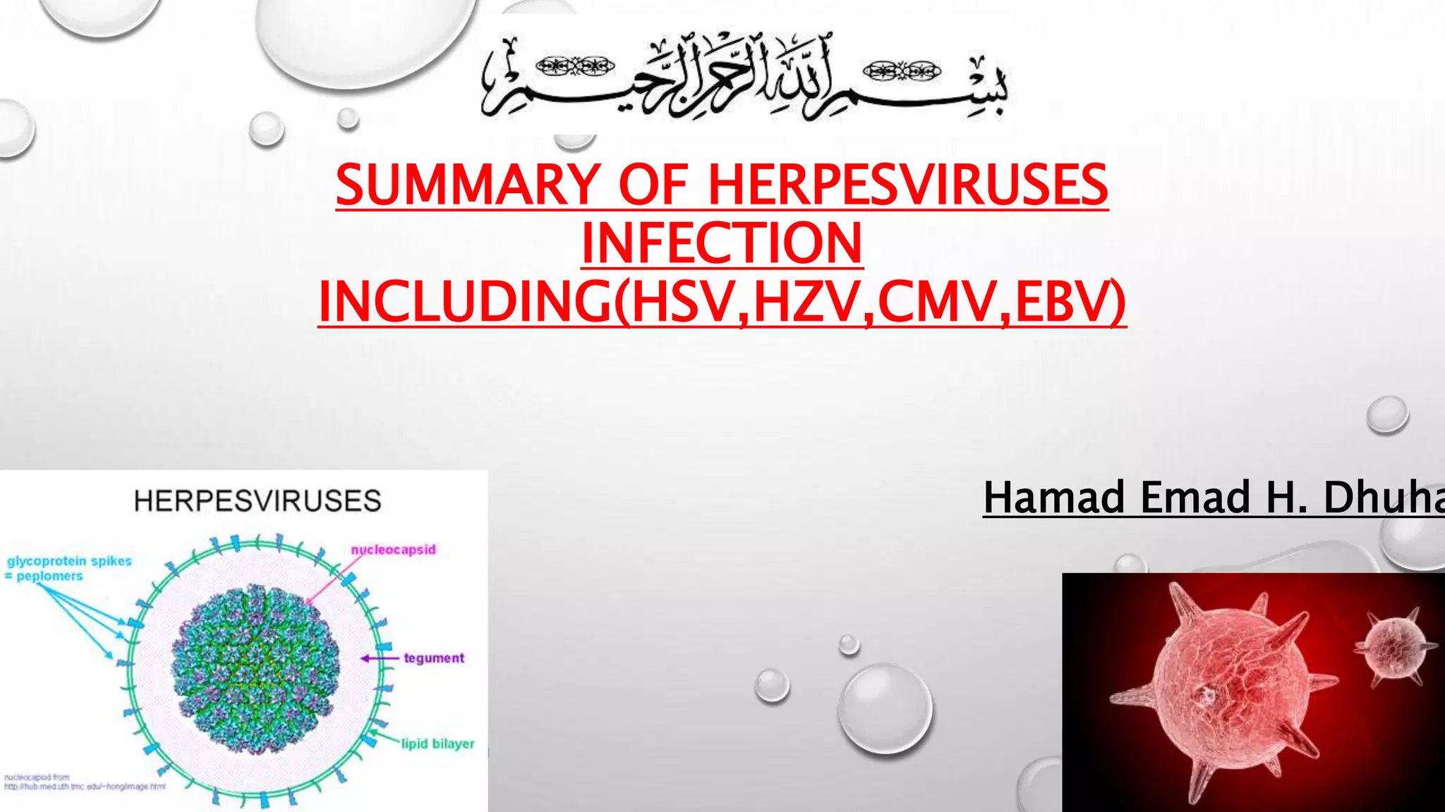
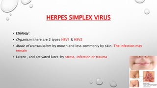
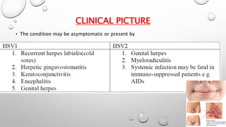
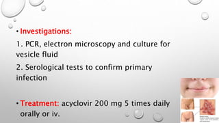
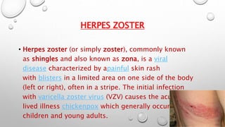
SUMMARY OF HERPESVIRUSES 
INFECTION 
INCLUDING(HSV,HZV,CMV,EBV) 
Hamad Emad H. Dhuhayr
CONTENTS 
• HSV 
• HZV 
• CMV 
• EBV
HERPES SIMPLEX VIRUS 
• Etiology: 
• Organism: there are 2 types HSV1 & HSV2 
• Mode of transmission: by mouth and less commonly by skin. The infection may 
remain 
• Latent , and activated later by stress, infection or trauma
CLINICAL PICTURE 
• The condition may be asymptomatic or present by
• Investigations: 
1. PCR, electron microscopy and culture for 
vesicle fluid 
2. Serological tests to confirm primary 
infection 
• Treatment: acyclovir 200 mg 5 times daily 
orally or iv.
HERPES ZOSTER 
• Herpes zoster (or simply zoster), commonly known 
as shingles and also known as zona, is a viral 
disease characterized by apainful skin rash 
with blisters in a limited area on one side of the body 
(left or right), often in a stripe. The initial infection 
with varicella zoster virus (VZV) causes the acute, short-lived 
illness chickenpox which generally occurs in 
children and young adults.
• Clinical picture: 
1. results from reactivation of latent VZV in ganglia 
2. Occurs most common in the elderly 
3. Skin lesion are unilateral and are of dermatome distribution 
4. Usually proceeded by sever pain of dermatome distribution (thoracic 
dermatomes, ophthalmic division of trigeminal nerve) 
complication 
1. Post herpetic neuralgia 
2. Encephalitis, myelitis
• Dx 
Pcr or culture of fluid of vesicles 
Treatment 
Acyclovir 800mg 5 times daily orally or iv 
Analgesics carbamazepine
CYTOMEGALOVIRUS 
Etiology: 
· Virus: DNA herpes virus 
· Mode of transmission: transplacental, passage through 
infected birth canal, Blood transfusion, sexual intercourse, 
or organ transplantation.
Pathogenesis: 
· The infected cells are 2-4 times larger than normal. 
· There is severe lymphocytic reaction which is 
characterized by atypical 
Lymphocytes in the peripheral blood. 
· Once infected, the host will have CMV indefinitely. 
· The manifestations of the virus will appear when the host 
is 
Immunosupressed.
Clinical picture: 
1. Congenital CMV infection 
· CNS: microcephaly+ cerebral calcification & mental retardation 
· GIT: hepatosplenomegaly, jaundice 
· Petichea 
2. Perinatal CMV infection 
Ø mainly asymptomatic 
Ø interstitial pneumonitis 
Ø lymphadenopathy 
Ø hepatitis 
Ø rash 
Ø anemia
Investigations: 
1. Blood: 
· Rbcs: may show hemolyytic anemia 
· WBS: relative lymphocytosis(> 10% atypical lymphocytes) 
· Pancytopenia 
· Detection of virus by PCR 
2. Liver enzymes: mild elevation in SGOT, SGPT & ALP 
3. Serology: four fold increase in antibodies detected by ELISA or IF 
Treatment: 
Prophylaxis: screening blood donors & checking donors and recipients of organ 
Transplantation for CMV 
Curative: gancyclovir (the best treatment) · Acyclovir or interferon
EPSTEIN BARR VIRUS 
Etiology: 
· Organism: epstein-barr virus 
· Mode of transmission: droplet infection 
Presentations: 
1. Infectious mononucleosis 
2. Nasopharyngeal carcinoma 
3. Burkitt’s lymphoma and other B cell lymphomas
Clinical picture: 
· Incubation period: 4-8 weeks 
· Clinical triad: fever, pharyngitis, lymphadenopathy: 
O fever: higher in the afternoon and may reach 40 C 
O pharyngitis: with sore throat & red spots 
O lymphadenopathy: usually cervical but may be generalized 
O others: splenomegaly (50-75% of cases), rash (which may 
represent drug 
Eruption when treated by ampicillin).
· Other presentations: 
O neurological: peripheral neuritis(guillan-barre syndrome), bell’s palsy 
O cardiac: pericarditis, myocarditis 
O GIT type: 
1. Hepatomegaly + jaundice(dd viral hepatitis) 
2. Fever + splenomegaly (typhoid picture) 
3. Oropharyngeal type: sore throat + hyperplasia of 
Pharyngeal lymphoid tissue (DD follicular tonsillitis) 
O cutanouse eruption : may be precipitated by ampicillin 
O latent form: laboratory diagnosis only.
Investigations: 
1. Cbc: 
O lymphocytosis with atypical lymphocytes (larger, stains deeper & with 
Vacuolated cytoplasm). 
O thrombocytopenia 
O auto-immune hemolytic aneamia 
2. Serology: 
O paul-bunnel test: hetereophil antibodies in the serum that agglutinate sheep 
Rbcs 
O monospot test: serum of the patient + guinea big kidney + ox rbcs on slide 
→ Agglutination 
O anti- EBV antibodies in the serum 
3.Others: 
O BM aspiration to exclude leukemia 
O LN biopsy to exclude lymphoma 
O throat swab to exclude other causes of sore throat
REFERENCES 
• CECIEL 
• WEBSITE

Hsv,ebs,cmv and hzv

  • 1.
    SUMMARY OF HERPESVIRUSES INFECTION INCLUDING(HSV,HZV,CMV,EBV) Hamad Emad H. Dhuhayr
  • 2.
    CONTENTS • HSV • HZV • CMV • EBV
  • 3.
    HERPES SIMPLEX VIRUS • Etiology: • Organism: there are 2 types HSV1 & HSV2 • Mode of transmission: by mouth and less commonly by skin. The infection may remain • Latent , and activated later by stress, infection or trauma
  • 4.
    CLINICAL PICTURE •The condition may be asymptomatic or present by
  • 5.
    • Investigations: 1.PCR, electron microscopy and culture for vesicle fluid 2. Serological tests to confirm primary infection • Treatment: acyclovir 200 mg 5 times daily orally or iv.
  • 6.
    HERPES ZOSTER •Herpes zoster (or simply zoster), commonly known as shingles and also known as zona, is a viral disease characterized by apainful skin rash with blisters in a limited area on one side of the body (left or right), often in a stripe. The initial infection with varicella zoster virus (VZV) causes the acute, short-lived illness chickenpox which generally occurs in children and young adults.
  • 7.
    • Clinical picture: 1. results from reactivation of latent VZV in ganglia 2. Occurs most common in the elderly 3. Skin lesion are unilateral and are of dermatome distribution 4. Usually proceeded by sever pain of dermatome distribution (thoracic dermatomes, ophthalmic division of trigeminal nerve) complication 1. Post herpetic neuralgia 2. Encephalitis, myelitis
  • 8.
    • Dx Pcror culture of fluid of vesicles Treatment Acyclovir 800mg 5 times daily orally or iv Analgesics carbamazepine
  • 9.
    CYTOMEGALOVIRUS Etiology: ·Virus: DNA herpes virus · Mode of transmission: transplacental, passage through infected birth canal, Blood transfusion, sexual intercourse, or organ transplantation.
  • 10.
    Pathogenesis: · Theinfected cells are 2-4 times larger than normal. · There is severe lymphocytic reaction which is characterized by atypical Lymphocytes in the peripheral blood. · Once infected, the host will have CMV indefinitely. · The manifestations of the virus will appear when the host is Immunosupressed.
  • 11.
    Clinical picture: 1.Congenital CMV infection · CNS: microcephaly+ cerebral calcification & mental retardation · GIT: hepatosplenomegaly, jaundice · Petichea 2. Perinatal CMV infection Ø mainly asymptomatic Ø interstitial pneumonitis Ø lymphadenopathy Ø hepatitis Ø rash Ø anemia
  • 12.
    Investigations: 1. Blood: · Rbcs: may show hemolyytic anemia · WBS: relative lymphocytosis(> 10% atypical lymphocytes) · Pancytopenia · Detection of virus by PCR 2. Liver enzymes: mild elevation in SGOT, SGPT & ALP 3. Serology: four fold increase in antibodies detected by ELISA or IF Treatment: Prophylaxis: screening blood donors & checking donors and recipients of organ Transplantation for CMV Curative: gancyclovir (the best treatment) · Acyclovir or interferon
  • 13.
    EPSTEIN BARR VIRUS Etiology: · Organism: epstein-barr virus · Mode of transmission: droplet infection Presentations: 1. Infectious mononucleosis 2. Nasopharyngeal carcinoma 3. Burkitt’s lymphoma and other B cell lymphomas
  • 14.
    Clinical picture: ·Incubation period: 4-8 weeks · Clinical triad: fever, pharyngitis, lymphadenopathy: O fever: higher in the afternoon and may reach 40 C O pharyngitis: with sore throat & red spots O lymphadenopathy: usually cervical but may be generalized O others: splenomegaly (50-75% of cases), rash (which may represent drug Eruption when treated by ampicillin).
  • 15.
    · Other presentations: O neurological: peripheral neuritis(guillan-barre syndrome), bell’s palsy O cardiac: pericarditis, myocarditis O GIT type: 1. Hepatomegaly + jaundice(dd viral hepatitis) 2. Fever + splenomegaly (typhoid picture) 3. Oropharyngeal type: sore throat + hyperplasia of Pharyngeal lymphoid tissue (DD follicular tonsillitis) O cutanouse eruption : may be precipitated by ampicillin O latent form: laboratory diagnosis only.
  • 16.
    Investigations: 1. Cbc: O lymphocytosis with atypical lymphocytes (larger, stains deeper & with Vacuolated cytoplasm). O thrombocytopenia O auto-immune hemolytic aneamia 2. Serology: O paul-bunnel test: hetereophil antibodies in the serum that agglutinate sheep Rbcs O monospot test: serum of the patient + guinea big kidney + ox rbcs on slide → Agglutination O anti- EBV antibodies in the serum 3.Others: O BM aspiration to exclude leukemia O LN biopsy to exclude lymphoma O throat swab to exclude other causes of sore throat
  • 17.