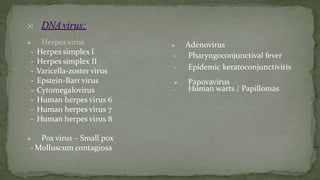
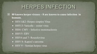
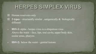
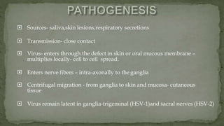
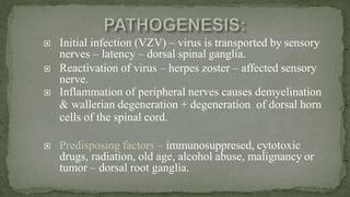
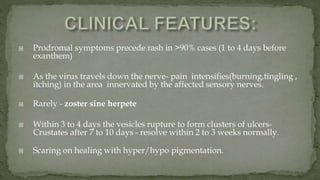
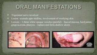
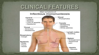
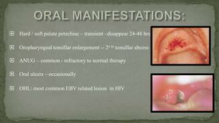
4. Found in almost every ecosystem on Earth - most abundant type of biological entity.
The study of viruses - virology.
5. They are small (10 -200 millimicrons)
They are totally dependent on living cells, either eukaryotic
or prokaryotic for replication and existence.
They possess only one species of nucleic acid, either DNA
or RNA.
They have a receptor binding protein for attaching to cells.
6.
7. Enters the skin
Multiplies initially then spread to
regional nodes through lymphatics
Enters the bloodstream and
transported to central foci for
viral multiplication
Massive spillover to blood stream–
appearance of prodromal symptoms
Reaches the target –
multiplication
produces lesion
9. Herpesvirus
- Herpes simplex I
- Herpes simplex II
- Varicella-zoster virus
- Epstein-Barr virus
- Cytomegalovirus
- Human herpes virus 6
- Human herpes virus 7
- Human herpes virus 8
Pox virus – Small pox
- Molluscum contagiosa
Adenovirus
- Pharyngoconjunctival fever
- Epidemic keratoconjunctivitis
Papovavirus
- Human warts / Papillomas
10. 80 known herpes viruses – 8 are known to cause infection in
humans.
HSV-1&2- Herpes simplex Virus
HHV-3- Varicella – zoster virus
HH4- CMV – Infective mononucleosis
HHV5- EBV
HHV6 and 7- Roseolovirus
HHV 8- Kaposi’s sarcoma
HSV 9 – Semian herpes virus
11. Human reservoirs only
2 types – structurally similar , antigenically & biologically
different
HSV-1: alpha - herpes virus is a ubiquitous virus
Above the waist – face, lips, oral cavity, upper body skin,
ocular areas, pharynx
HSV-2: below the waist – genital lesions
12. Sources- saliva,skin lesions,respiratory secretions
Transmission- close contact
Virus- enters through the defect in skin or oral mucous membrane –
multiplies locally- cell to cell spread.
Enters nerve fibers – intra-axonally to the ganglia
Centrifugal migration - from ganglia to skin and mucosa- cutaneous
tissue
Virus remain latent in ganglia-trigeminal (HSV-1)and sacral nerves (HSV-2)
14. Most common pattern of symptomatic primary HSV
infection
Incidence of primary HSV-1 increases after 6 months
Peak of incidence – b/w 2 and 3 years of age
Incidence of HSV-2 does not increase until sexual
activity begins
15. Many primary infections are subclinical
Prodromal- fever, headache, malaise , nausea,
vomiting
Oral lesions appear –1-2 days after prodromals
Numerous 1-2mm pinhead vesicles – collapse –
enlarge – central area of ulceration with yellow fibrin
– larger shallow irregular ulcers – heal 10-14 days
Satellite vesicles – perioral skin – common
Clinical Features:
16. Self innoculation – fingers, eyes, genital areas
Children – gen. initial – macular, later – purpuric cutaneous rash
Adults - pharyngotonsillitis – sore throat, fever, malaise, headache.
Numerous small vesicles – tonsils & posterior pharynx
Diffuse grayish yellow exudate over the ulcers.
Mild cases resolve within 5 to 7 days.
Rare complications- keratocoinjunctivitis, oesophagitis,meningitis and
encephalitis
17. Recurrence of HSV1 –lips – recurrent herpes labialis
Prodromal signs – 6 to 24 hrs before lesions develop –
pain, burning, itching, tingling, localized warmth,
erythema of involved epithellium
Multiple, small erythematous papules – clusters of fluid-filled
vesicles – rupture – crust (2days) – healing (7-10 days)
Variable intervals – months - years
18. Observe cytopathic effects of the cells inoculated with HSV
Rate of CPE depends on type of host cell, type of virus , conc. Of virus
Cytology: scrapings – base of lesion smeared onto glass slides.
Stained – Wright, Giemsa stain (Tzanck preparation) -
Multinucleated giant cells / intranuclear inclusions like Cowdry typeA
PCR: most sensitive method-detection of viral DNA
Does not require viable virus or infected cells for detection
Used to distinguish HSV typess
19. Pain Control:
2% viscous lidocaine (swish and spit out 5ml 4-5times/day)
Systemic analgesics
Supportive care:
Hydration
Soft bland diet
Antipyretics
20. Within 24-48hrs, antiviral medication help in reducing the
healing time:
Acyclovir ( Tab. Aciherpin) –inhibits viral replication
Dosage: 200mg 5 times daily for 7 to 10 days or
400mg TID for 7-10 days
Valacyclovir (Tab. valcivir) and famciclovi(Tab. famtrex)
–Better bioavailability, hence fewer daily doses.
Valacyclovir 1000mg BID for 7-10 days
Famcilcovir 250 mg TID for 7-10 days
21. Topical antiviral medication:
5% acyclovir cream - every 4hr for 5 days
3% penciclovir cream - 3-6 times/day for 5 days
Systemic therapy:
Tab.Zovirax (Acyclovir): 200mg 5times/day for 7 days
Tab.Valcivir (Valaciclovir):500-1000mg three times/day for 5 days.
Tab.Famtrex (Famciclovir):500-1000mg three times/day for 5-7 days
22. Enveloped DNA virus – HHV-3
Primary infection – Varicella (chicken pox)
Latent – dorsal root ganglia/ganglia of cranial nerves
Reactivation – Herpes zoster infection (shingles)
Transmission: Air droplets/ direct contact withlesions.
Children < 13 years
Incubation period: 10-20 days.
23. Chicken pox is an acute, ubiquitous, extremely contagious disease usually
occuring in children
Characterized by an exanthematous vesicular rash.
Source of infection-chicken pox or zoster patients
Portal of entry- conjunctiva or respiratory tract
Incubation period of about 2 weeks
24. Clinical features:
First 2 decades of life
Begins with prodromals
Maculopapular rash appear –followed by vesicles-”dewdrop-like“
Burst and scab with crust falling off after 1 to 2 weeks
Centrifugal spread
Typically continue to erupt for 4 - 7 days
Contagious from 2 days before eruptions until all the lesions crust
25. Commonly precede skin lesions
Most common sites - vermillion border, palate, buccal mucosa
Gingival lesions may resemble Primary herpes.
Initial raised vesicles (3-4mm) with surrounding erythema
Rupture and form eroded ulcers (1-3mm)
26. History and clinical examination
Confirmation : demonstration of viral cytopathic effects in
cells harvested from the vesicular fluid
Further confirmatory tests:
Viral isolation in cell culture- rapid diagnosis.
Direct fluorescent antibody testing: VZV monoclonal antibodies
PCR: VZV DNA in CSF
27. Warm bath with soap (lipid envelope destroyed)
Calamine lotion and systemic diphenhydramine -relieve pruritis
Antipyretics other than aspirin: Acetaminophen.
Systemic:
To be administered within first 24 hours of the rash
For pts at high risk or more severity
1. Acyclovir (Tab Zovirax) 800 mg 5 times/day for 7-10 days.
2. Valacyclovir (Tab Valtrex) 1000 mg tid for 7-10 days
3. Famciclovir (Tab Famvir) 500 mg tid for 7-10 days
Immunocompromised:
Purified VZV Ig can be given to modify the clinical manifestations of
the infection.
Within 96 hours of initial exposure
28. Herpes zoster is an acute infectious viral
disease of an extremely painful &
incapacitating nature - characterized by
inflammation of dorsal root ganglia or
extramedullary cranial nerve ganglia.
Associated with vesicular eruptions of skin or
mucous membranes in areas supplied by the
affected sensory nerves.
29. Initial infection (VZV) – virus is transported by sensory
nerves – latency – dorsal spinal ganglia.
Reactivation of virus – herpes zoster – affected sensory
nerve.
Inflammation of peripheral nerves causes demyelination
& wallerian degeneration + degeneration of dorsal horn
cells of the spinal cord.
Predisposing factors – immunosuppresed, cytotoxic
drugs, radiation, old age, alcohol abuse, malignancy or
tumor – dorsal root ganglia.
30. Prodromal symptoms precede rash in >90% cases (1 to 4 days before
exanthem)
As the virus travels down the nerve- pain intensifies(burning,tingling ,
itching) in the area innervated by the affected sensory nerves.
Rarely - zoster sine herpete
Within 3 to 4 days the vesicles rupture to form clusters of ulcers-
Crustates after 7 to 10 days - resolve within 2 to 3 weeks normally.
Scaring on healing with hyper/hypo pigmentation.
31.
Trigeminal nerve involved
Lesion –extends upto midline, involvement of overlying skin
Lesions – 1-4mm white opaque vesicles (painful) – buccal mucosa, hard palate,
gingiva,uvula, pharynx – rupture and form shallow ulcers
33. Commonly affects paients olderthan 60 yrs
Pain- burning, throbbing, aching, itching orstabbing
Neuralgias – resolve within 1 year
Pain subsides after initiation of long- term (Tab. famciclovir)
Facial paralysis associated with herpes zoster of faceor external
auditory meatus
34. Special form of zoster infection of geniculate ganglion
with ipsilateral involvement – facial & auditory nerves
Facial paralysis (Bell’s palsy) + pain + unilateral vesicles over
External auditory meatus & pinna of ear, Oral cavity & pharynx
Hoarseness of voice, loss of taste sensation – ant 2/3rd of tongue
35. Supportive care – hydration, soft bland diet.
Specific treatment
Antiviral drugs
Acyclovir (Tab. Zovirax): 800 mg 5 times/day for 7-10 days
Valacyclovir (Tab. Valtrex):1000 mg tid for 7-10 days
Famciclovir (Tab. Famvir): 500 mg tid for 7-10 days
Management of Post herpetic neuralgia
Gabapentin & 5% lidocaine patch –as first line of treatment
36. Mono Glandular Fever, “Kissing disease”
Exposure to Epstein-Barr virus (EBV) – HHV-4
Burkitt’s lymphoma, Hodgkin’s disease, Oral hairy leukoplakia
Transmission:
Intimate contact –once exposed –EBV
remains in host for life
Children- contaminated saliva – finger , toys , other objects
Adults – direct salivary transfer – shared straws , kissing
37. Multiplies locally - invades the blood stream and infects B-lymphocytes
Virus either become latent inside the B-lymphocytes
cause cell death and lead to release of mature virions
In response to these b-cells, abnormal CD8+ T-lymphocytes are released by
host immune - Downey cells
38.
39. Hard / soft palate petechiae – transient –disappear 24-48 hrs
Oropharyngeal tonsillar enlargement -- 2o to tonsillar abcess
ANUG – common - refractory to normal therapy
Oral ulcers – occasionally
OHL: most common EBV related lesion in HIV
40. Classic serologic test-
Paul bunnel test – detect heterophile antiodies
Titre above 100 suggestive of infection
Positive only in young adults
For children younger than 4: ELISA can confirm EBV
infections
41. No specific treatment
Self limiting, 2-4 weeks
Bed rest , adequate diet
Antipyretics (non-aspirin)
NSAIDs
Antibiotics – avoided – may cause skin rashes
Corticosteroids – recommended in life threatening cases
43. Entry into the host cell is achieved by attachment
of the viral glycoproteins to host receptors
The hallmark of such infection appearance of atypical
lymphocytes in the peripheral blood; these cells are
predominantly activated CD8+ T lymphocytes
44. 90% - asymptomatic
Neonatal infection:
Hepatosplenomegaly , extra-medullary cutaneous erythropoiesis,
mental retardation & motor retardation , thrombocytopenia
Acute adult infection:
Symptoms ranges from influenza -like to lethal multiorgan involvement
Only 1/3rd of pts demonstarte pharyngitis and lymphadenopathy
Rarely, acute sialadenitis-xerostomia and enlargement of affected glands
45. Salivary gland involvement-all major and minor glands
Chronic mucosal ulcerations-mostly single large ulcers –
Deep penetrating –lips , tongue , pharynx
Gingivitis , gingival hyperplasia
Co-infection – CMV + HSV(1/3rd cases)
Neonatal CMV – developmental tooth defects – diffuse enamel
hypoplasia , enamel hypomaturation , yellow discoloration –
underlying dentin
46. Biopsy :
Characteristic “owl eye” appearance of cellular inclusions
Specific verification by detection of viral antigens:
By immunohistochemiostry and PCR
Demonstrate viral antibody titers
ELISA of CMV is recommended for any pts with an
unexplianed fever
47. Prevention : By passive immunization with hyperimmune
gammaglobulin can be successfully prevented
Pain control : 2% viscous lidocaine, Systemic analgesics.
Supportive care: Hydration , Soft bland diet
Definitive treatment:
Cidofovir -5 mg/kg once a week for the initial 2 weeks and then
followed by maintenance regimens of 3–5 mg/kg every 2 weeks.
Ganciclovir –500mg t.i.d for 7 days..
48. An acute, contagious, dermatrophic viral infection affecting
children produced by paramyxovirus family of genus
morbilli virus.
RNA virus-linear single stranded RNA
TRANSMISSION:
Direct contact with respiratory secretions and aerosols
Outbreak cycle: 2-3 years
49. Infects skin, respiratory tract, viral replication in local
lymph nodes
Spread of virus within leukocytes- RE cells
Host cell necrosis
Suppresion of cellular immunity.
Secondary – Viremia, coryza and cough
50. Incubation period 10-12 days
Infectious from 2 days before becoming symptomatic
until 4 days after appearance of rash.
Lymphoid hyperplasia involving lymph nodes, tonsils,
adenoids, peyers patches
3 STAGES: 9 DAYMEASLES
51. Cough
Coryza
C onjunctivitis
Accompanied by fever
Koplik’s Spots – buccal mucosa
“Grains Of Salt In A Red Background”-
Pathognomic
52. Fever continues
Koplik’s spots fade
Morbiliform rash begins - blanches on pressure
Face is involved 1st then spread to trunk andextremities
Abdominal pain – secondary to lymphatic involvement
53. Fever ends
Rash begins to fade
Downward progression -Replacement by brown pigments
Complications:
Young children: otitis media, pneumonia,persistent bronchitis and diarrhea
Immunocompromised pts: more atypical rashes
54. -
Apart from koplik spots-
Candidiasis, ANUG and necrotising
stomatitis can occur
Severe measles in children –affect
odontogenesis-pitted enamel hypoplasia.
Enlargement of tonsils.
55. Desmonstration of rising serologic antibody titers-
appear within 1 to 3 days after exanthem- peaks in 3
to 4 weeks
56. No specific drugs
Antiviral drug –Ribavirin is effective against measles.
Cap. Ribavin 200mg QID
Syrup Ribavin 5ml daily –2 divided doses.
VitaminA should be given - VitaminA Chewable TAB – 50000 IU.
.
57. MMR vaccine -Introduced in 1963
Incidence decreased by 99%
MMR vaccine:
MMR 1 - 1year
MMR 2 - 4-6yrs.
Passive immunization: Human immunoglobulin (im)
<1 yr – 250mg ;
>1 yr – 500mg.
58. Primarily in winter
Toga virus
Transmission: droplet infection, congenital
rubella syndrome
Infection of the mother by Rubella virus
during pregnancy can be serious
Spontaneous abortion occurs in up to 20%
of cases
"Blueberry muffin lesions."
59. Transmitted by the respiratory route and replicates
in the nasopharynx and lymph nodes.
The virus is found in the blood 5 to 7 days after
infection and spreads throughout the body.
Teratogenic properties.
60. Incubation Period 14-21 Days
Contagious - from 1 week before exanthem to about
5 days after development of rashes
Mostly asymptomatic
Prodromal symptoms-1 to 5 days before exanthem
Lymphadenopathy-persists for weeks - cervical, postauricular
Exanthematous rash- entire body within 3 days : 1st sign-pink macule-
papules-flaky desquamtion-resolves by day 3
Most common complication is arthritis
61. Forehheimer’s sign : 20 % cases 6 hrs after 1st symptom
Dark red papules on soft palate, hard
palate along with rash
Palatal petechiae
1st month of pregnancy- hypoplasia,
caries, delayed eruption of decidous teeth
62. A four fold rise in rubella IgG antibody titre between acute
and convalescent serum specimens.
A positive serologic test for rubella-specific IgMantibody.
A positive rubella culture (isolation of rubellavirus).
Serologic studies are best performed within 7 to 10 days afterthe
onset of the rash and should be repeated two to three weekslater
63. No specific Rx- non aspirin antipyretics and antipruritics
Prevention: MMR vaccine, sub cutaneous inj.
1st around 1 year
2nd: 4-5 years.
Passive immunity : human rubella immunoglobulin.
64. Caused by paramyxovirus family , genus:Rubulavirus
Epidemiology markedly affected by MMR vaccine
Before vaccination, epidemics were seen every 2 to 5 years- 90% before age 15
Post vaccination, incidence reached an all time low in 1985 (98% decrease)
Resurgence in 1986-mainly among 10 to 19 yr olds –due to loss of diligence
65. Transmitted through urine, saliva, respiratory droplets
Incubation period – 15 to 18 days
Contagious from 1 day before the clinical appearance to 14 days
after itsclinical resolution
Site of involvement- salivary glands, pancreas, choroid plexus,mature
ovaries and testis.
One attack confers life long immunity
66. Infection and Proliferation of virus in upper Resp tract
Replication in respiratory epithelium
Localisation in glandular & neural tissues
Perivascular and interstitial mononuclear infiltrate,
necrosis- acinar cells
67. 30% subclinical
5-15 YRS
Incubation Period: 2-4weeks
Prodromal symptoms: pain below ear, headache,malaise,myalgia
Parotid gland is most commonly affected-Swelling occurs within 24-48 hrs
accompanied by pain on mastication.
Elevation of ear lobe
25% unilateral. Starts off on one side and followed by contralateral involvemnt
within a few days
Oral manifestation is redness and enlargemnt of Whartons and stensons ductal
openings
68. Most frequently used confirmatory measure is demonstration of
mumps- specific IgM or a 4-fold rise in IgG titers when measured
during acute phase and about 2 weeks later respectively
Viral isolation from swabs obtained from salivary secretion of parotid
Reverse-transcriptase – polymerase chain reaction testing- to detect viral
RNA
69. Palliative in nature
Avoidance of sour foods and more liquid helps to decrease the
salivary gland discomfort
Application of intermittent ice
Warm saline gargles, soft foods, and extra fluids
Prevention:
M-M-R (Measles, Mumps, and Rubella Virus Vaccine
Live)
70. First identified in 1937, in birds that had the potential to devastate
poultry stocks.
Evidence of human coronaviruses found in the 1960s, in the noses of
people with the common cold.
Human coronaviruses that are particularly prevalent include 229E,
NL63, OC43, and HKU1.
The name “coronavirus” comes from the crown-like projections on their
surfaces.
“Corona” in Latin means “halo” or “crown.”
71. Common in animals worldwide, rarely affects humans.
WHO used the term 2019 novel coronavirus to refer to a coronavirus that
affected the lower respiratory tract of patients with pneumonia in Wuhan,
China on 29 December 2019.
WHO named 2019 novel coronavirus as coronavirus disease (COVID-19)
Current reference name for the virus is severe acute respiratory syndrome
coronavirus 2 (SARS-CoV-2).
Highly transmissible than the SARS-CoV.
Incubation duration ranges from 7 to 14 days
Most people who get COVID-19 will have a mild form of the disease.
According to WHO, around 80% of people who get COVID-19 will recover
without needing hospitalization.
Remaining 20% become seriously ill and develop difficulty breathing.
72. Most common symptoms: Fever, dry cough. tiredness.
Less common symptoms:
Aches and pains, sore throat, diarrhea, Conjunctivitis,
headache, loss of taste and smell, a rash on skin.
Serious symptoms: Difficulty breathing or shortness of
breath.
Chest pain or pressure.
73. Multiple ulcers with an erythematous halo and symmetric distribution
on the right hard palate
Blisters on the inner lip mucosa
75. The most common reason for oral ulcerations and blisters
is viral infections.
Major challenge in the diagnosis of oral viral infections is
due to complex clinical presentations
Early recognition of oral viral infections will reduce the
morbidity, comorbidity, and the clinical care cost.
Hence, diagnosing oral lesions with presence of systemic
manifestations will serve as a useful tool in clinical
situation.
76. Oral & maxillofacial pathology: Neville, 3rd
edition.
Text book of oral medicine: Burkitts, 11th edition
Shafer’s text book of oral pathology, 6th edition