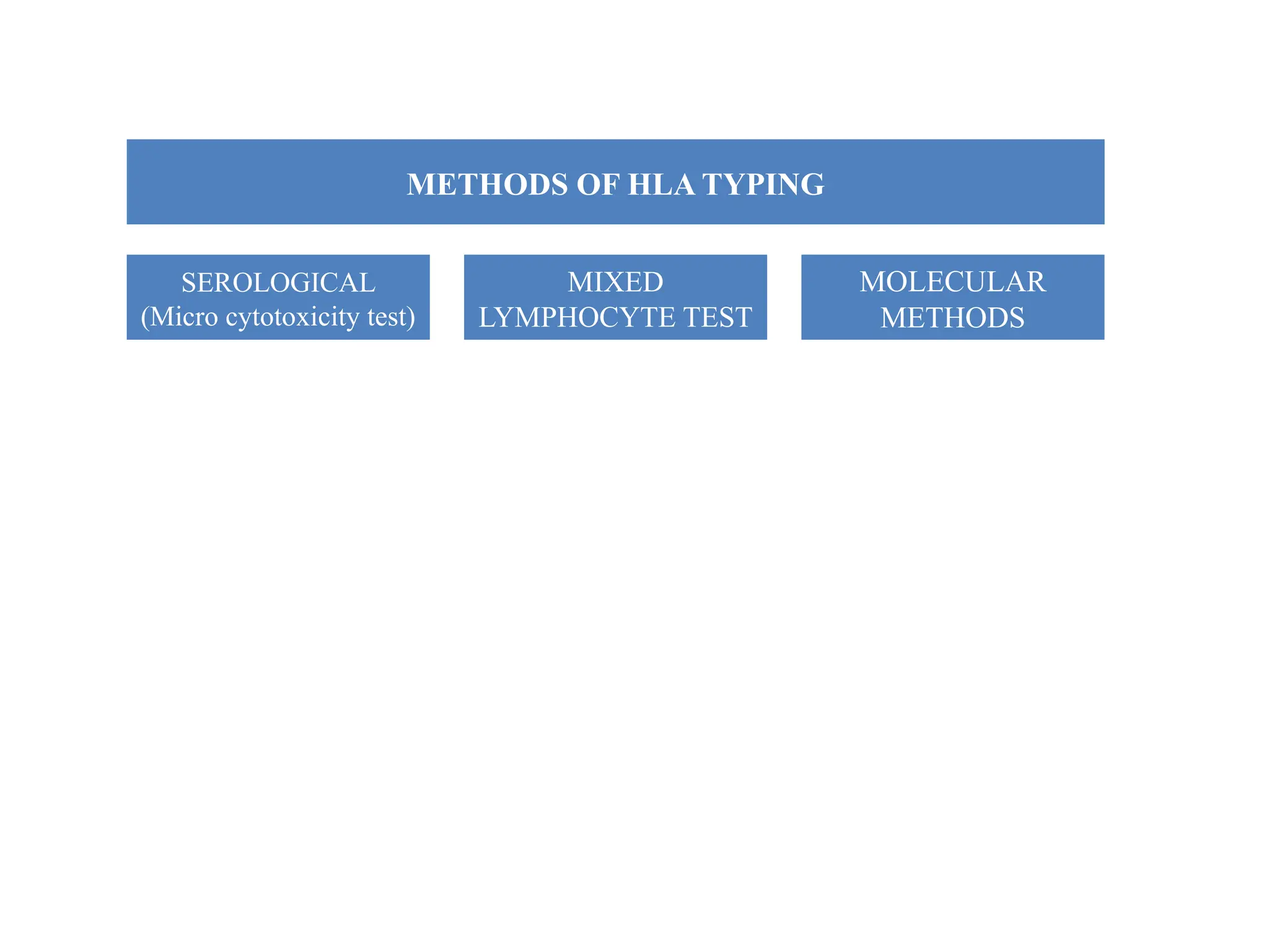
What Is the HLA System?
The HLA system refers to a group of related genes that play an
important role in the immune system.
Together, the proteins made from these genes form something
called the major histocompatibility complex (MHC). These
proteins are attached to almost all of the cells of our body
(excluding red blood cells).
HLA stands for human leukocyte antigen
HLA typing is a kind of genetic test used to identify certain
individual variations in a person’s immune system.
The process is critical for identifying which people can safely
donate bone marrow, cord blood or an organ to a person who
needs a transplant.
HLA typing is also sometimes called HLA matching.
PURPOSE OF HLA TYPING
• The most common reason for HLA typing is to help determine
which people can provide the safest tissue transplants (
solid organ or hematopoietic stem cell transplantation).
• Potential tissue recipients must have the typing, as must anyone
who might potentially want to donate tissue.
• HLA type included in a bone marrow registry, for stem cell
transplantation.
• HLA typing may also be performed on terminally ill or recently
deceased people who will be serving as organ donors.
• The best possible donors have HLAs that closely match the
HLA patterns of the recipient.
CONDITIONS THAT REQUIRING TRANSPLANTATION
 There are many different health conditions that may need to be
treated through a transplant.
 For example, various types of blood cancers and genetic blood
disorders are treated through stem cell transplantation (taken
either from the bone marrow or from the peripheral blood).
 A solid organ transplant might be necessary for any essential
organ that has become severely damaged.
 This might happen through trauma, infection, autoimmune
disease, genetic illness, toxins, or many other disease processes.
 For example, one might need a kidney, liver or lung transplant if
one’s own organs are very functioning very poorly.
THE PROCESS OF HLA TYPING
HLA typing assesses the particular HLA genes that have inherited
Because there are a number of different HLA genes, as well as different
variations of these genes, there are very many different possible color
combinations that together make up your specific HLA type.
HLA typing also usually includes testing for antibodies targeted to specific
HLA proteins. Antibodies are made by part of the immune system.
If a person already has an antibody against an HLA protein it may attack that
protein if it is transplanted. This may cause the transplant to fail.
So generally, shouldn’t receive a transplant from someone if already have an
antibody against one of their HLA proteins.
CONT…….
HLA typing also often includes something called lymphocyte
cross matching. Lymphocytes are a type of immune cell.
Lymphocyte cross matching checks to see if the recipient has an
antibody against a protein on the donor’s lymphocytes.
If so, that person generally shouldn’t receive a transplant from
that particular person. These people are at high risk of a
transplant that won’t be successful.
Is HLA Typing the Same as Blood Typing?
No. HLA is much more complicated than blood typing because
there are many more HLA markers that make a person’s cells
unique.
There are only eight basic blood types, and many people can
safely receive more than one type of blood (depending on their
type).
To receive only blood from a person, do not need to be an HLA
match, because HLA is not present on red blood cells.
To receive a solid organ transplant, the recipient must have a
compatible blood type with the donor, as well as the best HLA
match possible.
For stem cell donations, one needs a very strong HLA match, but
blood type is not as important as it is for solid organ transplants.
How Are HLA Genes Inherited?
Because the HLA genes are located close together on DNA, they are usually
inherited as a group.
HLA type is composed of the set of HLA genes you inherited from mother
and the HLA genes you inherited from father.
Biological parents always share half of their HLA proteins with their children.
This is also called a “half match.” Conversely, a child always is a half match
with their parents.
The set of HLA alleles found on one chromosome is called haplotype.
Determination of haplotype is important for identification of HLA identical
siblings because sharing of antigen from different haplotypes is common.
HLA TYPING IMPORTANT ROLE
The primary target of immune responses to allogeneic transplants
Critical for response to antigenic stimuli
Implicated in genetic susceptibility to auto immune disease.
METHODS OF HLA TYPING
SEROLOGICAL
(Micro cytotoxicity test)
MIXED
LYMPHOCYTE TEST
MOLECULAR
METHODS
SEROLOGICAL TEST
Potential donor′s and recipient's WBC′s added to the different
wells of microtitre plates
Antibodies specific for HLA class I and II added
After incubation complement is added
Cytotoxicity is assessed by uptake or exclusion of dye
SEROLOGY
HLA Typing is done serologically by MICROCYTOTOXICITY
(microlymphocytotoxicity) which tests for complement
mediated lysis of peripheral blood lymphocytes with a
standard set of typing sera.
Viable peripheral blood lymphocyte are obtained by
discontinuous density gradient centrifugation using Ficol/
Tryosil or Ficol / Sodium Metrizoate at a density of 1.077 at
19⁰C-22⁰C
Microlymphocytotoxic test
3 stages
• Viable lymphocyte are incubated with HLA specific antibodies. If
the specific antigen is present on the cell the antibody is bound.
• Rabbit serum as a source of complement is added, incubate. If
antibody is bound HLA antigen on the cell surface it activates the
complement which damages the cell membrane making it
permeable to vital stains.
• Results are visualized by adding dye usually a flurochrome eg.
Ethidium Bromide although both trypan blue and Eosin Y have
been used .
• If the reaction has takes place the EB enters the cell and binds to
the DNA
• For ease double staining is normally used. A cocktail of Ethidium
Bromide and Acridine Orange, quenched using Bovine
Hemoglobin to allow simultaneous visualization of both living
and dead cells.
• White blood cells from potential donors and the recipient are
added to separate wells of a microtiter plate. The example depicts
the reaction of donor and recipient cells with a single antibody
directed against an HLA-A antigen.
• The reaction sequence shows that if the antigen is present on the
lymphocytes, addition of complement will cause them to become
porous and unable to exclude the added dye.
Presence or absence of
various MHC alleles
determined by antibody
mediated cytotoxicity
Cont…….
• Test is left for 10 minutes and then read using an inverted
fluorescent microscope.
• A mixture of T and B lymphocytes can be used for HLA class I
typing.
• B lymphocytes are required for HLA class II typing by
serology. (Normal population 85-90% to T and 10-15% B
cells)
• This can be achieve using a number of methods
In the past neuraminidase treated sheep red blood cell resetting
and nylon wool have been used.
Immunomagnetic bead separation is the current method of choice.
It utilizes polystyrene microspheres with a magnet able core
coated in monoclonal antibody for a HLA class II b chain
monomorphic epitope. Positive selection
N
MIXED LYMPHOCYTE REACTION
o It has been observed the lymphocytes from one donor, when
cultured with lymphocytes from an unrelated donor, are
stimulated to proliferate.
o It has been established that this proliferation is primarily due to
a disparity in the Class II MHC (DR) antigens and T cells of
one individual interact with allogeneic class-II MHC antigens
bearding cells (B cells, dendritic cells, Langerhans cells, etc…)
this reactivity was termed with mixed leukocyte reaction
(MLR) and has been used for studying the degree of
histocompatibility.
In this test, the lymphocyte (responder cells) are mixed with
irradiated or mitomycin C treated leukocytes from the recipient,
containing B-lymphocytes and monocytes (stimulator cells).
The responder T cells will recognize the foreign class II antigens
found on the donor and undergo transformation (DNA synthesis and
enlargement: blastogenesis) and proliferation (mitogenesis) occurs
The T cells that respond to foreign Class II antigens are typically
CD4+ TH-1 type cells
These changes are recorded by the addition of radioactive (tritiated,
3H)
NN
Molecular HLA typing
DNA based procedures has increased the accuracy of HLA typing
and lead to the identification of serologically undetected alleles and
many subtypes of serological specificities.
DNA based methods of type for HLA alleles have therefore
focused in the analysis of nucleotide variation occurring in both
exon 2 and 3 of class I genes and exon-2 of class II genes.
PCR BASED METHODS/ THREE CATEGORIES
 Polymorphism is identified directly as part of the PCR process,
although there are post amplification steps e. g (SSP)
 Product containing internally located polymorphism that can be
identified by a second technique e. g PCR sequence specific
oligonucleotide probing (SSOP), PCR- RFLP,PCR followed by
sequencing.
 Conformational analysis
STEPS OF
MOLECULAR
CLONING
DNA EXTRACTION
Genomic DNA extracted from nucleated cells; the purity of the
DNA extracted may be an important factor for successful results
GENE AMPLIFICATION/PCR
The specificity of the amplification can be locus specific e. g (HLA-
A, HLA-B, HLA-DRB1), group specific… e.g. (DRB1-01, DRB1-
02) Or allele specific DRB1-0401, DRB1-0402
The primers are selected to amplify a single allele or a group of
alleles
For PCR, the specificity is determined by the sequence of the
primers and amplification condition. Most typing scheme require
conditions the avoid the co-amplification of pseudogene.
Amplification of exon 2 (approximately 270bp ) of HLA class II
is sufficient to achieve the highest resolution level.
For typing HLA class I, both exon 2 and 3 and the intervening
intron (fragment longer than 900bp ) are amplified by single pair
of primers.
Detection of sequence polymorphism that define the alleles
• Sequence specific primers (SSP) group and alleles specific
primers
• Hybridization with sequence specific oligonucleotide probes
(SSOP)
• Sequence based typing (SSP)
Sequence specific priming
• The procedure relies on the
specificity of primer extension that
is matched or mismatched with the
template at its 3’end.
• A combination of 2 primers
designed for each of 2 polymorphic
sequence motifsin cis allows the
identification of an allele or a group
of alleles that are characterized by
these 2 motifs.
• The method is rapid and ideally suited
for small numbers of samples. Because very few sequences are
absolutely allele specific, SSP combine several primer to
discriminate a particular allele unambiguously.
Hybridization with sequence specific oligonucleotide probes (SSOP)
The procedure relies on the locus-specific
amplification of the genomic DNA segment
comprising the polymorphic sites of HLA alleles.
Amplified DNA is then immobilized on a
solid support, usually a nylon membrane,
and then hybridized with a battery of
(SSOP) (direct hybridization).
Fluorochromes are linked with the probes to
allow their detection by chemiluminescence.
Alternatively, SSO probes can be immobilized
on a solid support, for example color-coded microspheres,
and hybridized with labeled PCR product (reverse hybridization).
The higher the number of probes the better the resolution level. Usually 50-
100 probes per locus are used for intermediate/high resolution typing,
however, only the probe completely matched with the target sequence
amplified will hybridize and give a positive signal.
SEQUENCE BASED HLA TYPING
• Sequence based HLA typing involves determining the nucleotide
sequence of an amplified segment of HLA gene
• SBT presents the advantages over the other procedure because of
the relatively fast (24-48 hours), high level resolution
• More reliable and specific method
CONFORMATIONALANALYSIS
Different mutation generates specific conformational changes in
PCR products, these are identified by electrophoresis analysis.
E.g.….. heteroduplex analysis, single strand conformation
polymorphism (SSCP), denaturing gradient gel electrophoresis
(DGGE), temperature gradient gel electrophoresis (TGGE)
CLINICAL SIGNIFICANCE OF HLA TYPING
In organ transplantation
In transfusion therapy
Disease association
Disputed paternity
In cancer prevention
Anthropological studies
What is Transplant rejection?
• Transplant rejection is process in which the
transplanted tissue (of donor) is rejected by
the recipient's immune system and may result
in fatal illness if not treated/removed early.
Based on genetic relationship between donor and
recipient there are four types of Grafting methods:
1. Autografts are grafts in which the donor and
recipient is the same individual.
2. Isografts are grafts between the donor and
recipient of the same genotype.
3. Allografts are those in which the donor is of the
same species but of a different genotype.
4. Xenografts are those in which the donor is of a
different species from that of the recipient.
• For any successful tissue transplant without
immunological rejection, matched Major
Histocompatibility Locus Antigens (HLA)
between the donor and recipient are of
paramount importance.
• The greater the genetic disparity between
donor and recipient in HLA system, the
stronger and more rapid will be the rejection
reaction.
• All types of grafts have been performed in
human beings but xenografts have been found
to be rejected invariably due to genetic
disparity.
• Most commonly practiced grafting are:
- Skin grafting
- Kidney
- Bone marrow transplantation.
Exceptions in Rejections:
• Cornea transplants are rarely rejected because
the cornea has no blood supply.
• Also, transplants from one identical twin to
another are almost never rejected.
Graft versus Host Reaction
• In some cases esp. when the transplanted
tissue is bone marrow, a peculiar illness arises
besides rejection of the transplanted bone
marrow called GRAFT Vs HOST REACTION.
• The intensity of GVH reaction depends upon
the extent of genetic disparity between the
donor and recipient.
• The clinical features of Graft Vs Host reaction
include:
- Fever,
- Weight Loss,
- Anaemia,
- Dermatitis,
- Diarrhoea,
- Intestinal Malabsorption,
- Pneumonia,
- Hepatosplenomegaly.
MECHANISMS OF GRAFT REJECTION
• Except for autografts and isografts, an immune
response against allografts is
inevitable/unavoidable.
• The development of immunosuppressive
drugs has made the survival of allografts in
recipients possible.
• Rejection of allografts involves both cell-
mediated and humoral immunity.
1. CELL-MEDIATED IMMUNE REACTIONS
• Mainly responsible for graft rejection and are
mediated by T cells (mainly by cytotoxic T
cells).
• T cells attack the graft and destroy it.
2. HUMORAL IMMUNE REACTIONS
• In addition to the cell-mediated immune
reactions, humoral antibodies cause certain
rejection reactions.
TYPES OF REJECTION REACTIONS
• Based on the underlying mechanism and time
period, rejection reactions are classified into 3
types:
1- Hyperacute Rejection
2- Acute Rejection
3- Chronic Rejection
1. HYPERACUTE REJECTION
• Hyperacute rejection appears within minutes
to hours of placing the transplant and destroys
it.
• It is mediated by Humoral system antibodies.
• This type of rejection is seen when a recipient
is given the wrong type of blood.
• For example, when a person is given type A
blood when he or she is type B.
2. ACUTE REJECTION
• This usually becomes evident within a few
days to a few months of transplantation.
• Acute graft rejection may be mediated by
cellular or humoral mechanisms.
3. CHRONIC REJECTION
• Chronic rejection may develop slowly over a
period of months to a year or so.
• The underlying mechanisms of chronic
rejection may be immunologic or ischaemic.
• Patients with chronic rejection of renal
transplant show progressive deterioration in
renal function as seen by rising serum
creatinine levels.
How to overcome transplant rejection?
• Tissue typing is done to prevent transplant rejection
• Here both the organ donor and the person who is
receiving the organ tissue typing is done to ensure that
the organ or tissue is as similar as possible to the tissues
of the recipient.
• The more similar the antigens are between the donor and
recipient, the less likely that the organ will be rejected.
• No match is usually 100 percent identical.
• No two people, except identical twins, have identical
tissue antigens.
AIDS
(Acquired Immunodeficiency
Syndrome )
08/29/2024
Definition
Definition of AIDS is a syndrome caused by
infection with the human immunodificiency
virus (HIV) with ensuing compromise of the
body's immune system.
Mode of transmission
• AIDS can transmit by body fluid,
vaginal secretions, amniotic fluid and
breast milk.
• Blood product also can transmit to
recipient by blood transfusion.
IV injection drug
use
Sexual relationship
with HIV infected
Person who
received HIV
infected blood
Baby breastfeed
with HIV-infected
mother
Frequency exposure
to blood
Frequency exposure
to body fluid
Etiology
Clinical Manifestations
Fever Cough Chest pain
Skin rashed
Loss of
appetite
Diarrhea
Fatigue Tubercolusis Weight loss
Laboratory Investigation
1. Blood test such as
CBC and WBC.
2. ELISA ( Enzyme-
linked immunosorbent
assay ) test used to
detect HIV infection.
Complications
1. Kaposi's sarcoma.
2. Lymphomas.
3. Wasting syndrome.
4. Neurological
complications.
5. Kidney disease.
Management
Medical management
1. Antiretroviral drugs. (trx to fight the HIV
infection and slow down the spread of the
virus)
2. Antifungal cream Ciclopirox may help
eradicate HIV.
3. Antiemetics drug to treat the symptoms.
4. Antipyretics drug to treat the symptoms such
as PCM
Nursing management.
1. Notification.
2. Encourage patient to have protected sex such
as use a condom. HIV can spread by having
unprotected sex.
3. Encourage patient to do proper hygiene.
4. Advice patient to do not give blood donation.
5. Educate patient for risk factors for acquiring
AIDS diseases.
Health Education
1. Avoid from sharing needles and
toothbrushes.
2. Do not reuse condom.
3. Reduce the number of sexual partner to one.
4. Avoid donating blood, plasma, body organs
or sperm.
5. Encourage patient to have fluid intake to
avoid dehydration.
Hypersensitivity
• Exaggerated or Inappropriate
immune response that is
harmful to the host
Atopy:
• Atopy is the term for the genetic trait to have a
predisposition for localized anaphylaxis
• Atopic individuals have higher levels of IgE and Eosinophills
Allergens
• Allergens are nonparasite antigens that can stimulate a
type I hypersensitivity response.
• Allergens bind to IgE and trigger degranulation of
chemical mediators.
Hypersensitivity Reactions
• Immediate hypersensitivity refers to antibody
mediated reactions – symptoms develop within
minutes to hours
• Delayed hypersensitivity refers to cell mediated
immunity, symptoms not observed for 24 to 48
hours.
Four Classifications
• Type I (Immediate) Hypersensitivity
• Type II (cytotoxic) hypersensitivity
• Type III (immune complex mediated) hypersensitivity
• Type IV (delayed) hypersensitivity
Type I Hypersensitivity
(Anaphylactic) Reactions/Allergy
– Occur within minutes of exposure to antigen
– Antigens combine with IgE antibodies
– IgE binds to mast cells and basophils, causing
them to undergo degranulation and release
several mediators:
• Histamine
• Prostaglandins
• Leukotrienes
• Anaphylactic shock
Type I (Immediate) Hypersensitivity
Mast Cells and the Allergic Response
Type I (Immediate) Hypersensitivity
Examples of type 1
• Allergic asthma
• Allergic conjunctivitis
• Allergic rhinitis
• Anaphylaxis
• Angioedema
• Urticaria (Hives)
Type II Hypersensitivity (Cytotoxic)
Reactions/antibody-dependent
– Involve activation of complement by IgG or IgM
binding to an antigenic cell.
– Antigenic cell is lysed
– Transfusion reactions:
• ABO Blood group system: Type O is universal
donor. Incompatible donor cells are lysed as
they enter bloodstream.
Hemolytic disease of the newborn is caused by type II
hypersensitivity reactions
When an Rh- mother carries an Rh+ fetus
Examples of type II
• Autoimmune hemolytic anemia
• Thrombocytopenia
• Erythroblastosis fetalis
Type III Hypersensitivity (Immune Complex)
Reactions
– Involve reactions against soluble antigens circulating in
serum.
– Antibody-Antigen immune complexes are deposited in
organs, activate complement, and cause inflammatory
damage.
• Glomerulonephritis: Inflammatory kidney damage.
– Occurs with slightly high antigen-antibody ratio is
present.
Type III (immune complex mediated) Hypersensitivity
Immune Complex Mediated Hypersensitivity
Examples of type III
• Serum sickness
• Arthus reaction
• Systemic lupus erythematosus (SLE)
• Post streptococcal Glomerulonephritis
• IgA nephropathy
• Rheumatoid arthritis
Hypersensitivity Type III Reactions
Systemic Reactions
Local Reactions
 Arthus Reaction:
Inflammation caused by
the deposition of immune
complexes at a localized
site.
Clinical Manifestation is :
Hypersensitivity
Pneumonitis
 Serum Sickness:
Systemic inflammatory
response to deposited
immune complexes at many
areas of body.
Clinical Manifestation is :
Fever, Urticaria, Artheralgia,
Eosinophila, Spleenomegally,
and Lymph adenopathy
Type IV (delayed) Hypersensitivity
• Used to describe the signs and symptoms associated with a
cell mediated immune response.
• Results from reactions involving T lymphocytes.
• Characteristics of this phenomenon are:
– Delayed, taking 12 hours to develop.
– Causes accumulation of lymphs and macrophages.
– Reaction is not mediated by histamine.
– Antibodies are not involved in the reaction.
Type IV Mechanism
APC resident in the skin
process antigen and
migrate to regional lymph
nodes where they activate
T cells
Sensitised T cells migrate
back to the the skin where
they produce cytokines
which attract macrophages
which cause tissue damage
Examples of Type IV
• Contact dermatitis
• Mantoux test
• Chronic transplant rejection
• An injection of tuberculin beneath the skin causes
reaction in individual exposed to tuberculosis or
tuberculosis vaccine
• Used to diagnose contact with antigens of
M. tuberculosis
» No response when individual not infected or
vaccinated
» Red, hard swelling develops in individuals
previously infected or immunized
The tuberculin response:
85
A positive tuberculin test
• Cell-mediated immune response
• Results in an intensely irritating skin rash
• Triggered by chemically modified skin proteins that the
body regards as foreign
• Acellular, fluid-filled blisters develop in severe cases
Contact Dermatitis
87
Contact dermatitis
Classification of Hypersensitivity
90
Thank you

The HLA system , Immunolgy, Graft versus Host Reaction

  • 1.
    What Is theHLA System? The HLA system refers to a group of related genes that play an important role in the immune system. Together, the proteins made from these genes form something called the major histocompatibility complex (MHC). These proteins are attached to almost all of the cells of our body (excluding red blood cells).
  • 2.
    HLA stands forhuman leukocyte antigen HLA typing is a kind of genetic test used to identify certain individual variations in a person’s immune system. The process is critical for identifying which people can safely donate bone marrow, cord blood or an organ to a person who needs a transplant. HLA typing is also sometimes called HLA matching.
  • 3.
    PURPOSE OF HLATYPING • The most common reason for HLA typing is to help determine which people can provide the safest tissue transplants ( solid organ or hematopoietic stem cell transplantation). • Potential tissue recipients must have the typing, as must anyone who might potentially want to donate tissue. • HLA type included in a bone marrow registry, for stem cell transplantation. • HLA typing may also be performed on terminally ill or recently deceased people who will be serving as organ donors. • The best possible donors have HLAs that closely match the HLA patterns of the recipient.
  • 4.
    CONDITIONS THAT REQUIRINGTRANSPLANTATION  There are many different health conditions that may need to be treated through a transplant.  For example, various types of blood cancers and genetic blood disorders are treated through stem cell transplantation (taken either from the bone marrow or from the peripheral blood).  A solid organ transplant might be necessary for any essential organ that has become severely damaged.  This might happen through trauma, infection, autoimmune disease, genetic illness, toxins, or many other disease processes.  For example, one might need a kidney, liver or lung transplant if one’s own organs are very functioning very poorly.
  • 5.
    THE PROCESS OFHLA TYPING HLA typing assesses the particular HLA genes that have inherited Because there are a number of different HLA genes, as well as different variations of these genes, there are very many different possible color combinations that together make up your specific HLA type. HLA typing also usually includes testing for antibodies targeted to specific HLA proteins. Antibodies are made by part of the immune system. If a person already has an antibody against an HLA protein it may attack that protein if it is transplanted. This may cause the transplant to fail. So generally, shouldn’t receive a transplant from someone if already have an antibody against one of their HLA proteins.
  • 6.
    CONT……. HLA typing alsooften includes something called lymphocyte cross matching. Lymphocytes are a type of immune cell. Lymphocyte cross matching checks to see if the recipient has an antibody against a protein on the donor’s lymphocytes. If so, that person generally shouldn’t receive a transplant from that particular person. These people are at high risk of a transplant that won’t be successful.
  • 7.
    Is HLA Typingthe Same as Blood Typing? No. HLA is much more complicated than blood typing because there are many more HLA markers that make a person’s cells unique. There are only eight basic blood types, and many people can safely receive more than one type of blood (depending on their type). To receive only blood from a person, do not need to be an HLA match, because HLA is not present on red blood cells. To receive a solid organ transplant, the recipient must have a compatible blood type with the donor, as well as the best HLA match possible. For stem cell donations, one needs a very strong HLA match, but blood type is not as important as it is for solid organ transplants.
  • 8.
    How Are HLAGenes Inherited? Because the HLA genes are located close together on DNA, they are usually inherited as a group. HLA type is composed of the set of HLA genes you inherited from mother and the HLA genes you inherited from father. Biological parents always share half of their HLA proteins with their children. This is also called a “half match.” Conversely, a child always is a half match with their parents. The set of HLA alleles found on one chromosome is called haplotype. Determination of haplotype is important for identification of HLA identical siblings because sharing of antigen from different haplotypes is common.
  • 9.
    HLA TYPING IMPORTANTROLE The primary target of immune responses to allogeneic transplants Critical for response to antigenic stimuli Implicated in genetic susceptibility to auto immune disease.
  • 10.
    METHODS OF HLATYPING SEROLOGICAL (Micro cytotoxicity test) MIXED LYMPHOCYTE TEST MOLECULAR METHODS
  • 11.
    SEROLOGICAL TEST Potential donor′sand recipient's WBC′s added to the different wells of microtitre plates Antibodies specific for HLA class I and II added After incubation complement is added Cytotoxicity is assessed by uptake or exclusion of dye
  • 12.
    SEROLOGY HLA Typing isdone serologically by MICROCYTOTOXICITY (microlymphocytotoxicity) which tests for complement mediated lysis of peripheral blood lymphocytes with a standard set of typing sera. Viable peripheral blood lymphocyte are obtained by discontinuous density gradient centrifugation using Ficol/ Tryosil or Ficol / Sodium Metrizoate at a density of 1.077 at 19⁰C-22⁰C
  • 13.
    Microlymphocytotoxic test 3 stages •Viable lymphocyte are incubated with HLA specific antibodies. If the specific antigen is present on the cell the antibody is bound. • Rabbit serum as a source of complement is added, incubate. If antibody is bound HLA antigen on the cell surface it activates the complement which damages the cell membrane making it permeable to vital stains. • Results are visualized by adding dye usually a flurochrome eg. Ethidium Bromide although both trypan blue and Eosin Y have been used .
  • 14.
    • If thereaction has takes place the EB enters the cell and binds to the DNA • For ease double staining is normally used. A cocktail of Ethidium Bromide and Acridine Orange, quenched using Bovine Hemoglobin to allow simultaneous visualization of both living and dead cells. • White blood cells from potential donors and the recipient are added to separate wells of a microtiter plate. The example depicts the reaction of donor and recipient cells with a single antibody directed against an HLA-A antigen. • The reaction sequence shows that if the antigen is present on the lymphocytes, addition of complement will cause them to become porous and unable to exclude the added dye.
  • 15.
    Presence or absenceof various MHC alleles determined by antibody mediated cytotoxicity
  • 17.
    Cont……. • Test isleft for 10 minutes and then read using an inverted fluorescent microscope. • A mixture of T and B lymphocytes can be used for HLA class I typing. • B lymphocytes are required for HLA class II typing by serology. (Normal population 85-90% to T and 10-15% B cells) • This can be achieve using a number of methods
  • 18.
    In the pastneuraminidase treated sheep red blood cell resetting and nylon wool have been used. Immunomagnetic bead separation is the current method of choice. It utilizes polystyrene microspheres with a magnet able core coated in monoclonal antibody for a HLA class II b chain monomorphic epitope. Positive selection
  • 19.
  • 21.
    MIXED LYMPHOCYTE REACTION oIt has been observed the lymphocytes from one donor, when cultured with lymphocytes from an unrelated donor, are stimulated to proliferate. o It has been established that this proliferation is primarily due to a disparity in the Class II MHC (DR) antigens and T cells of one individual interact with allogeneic class-II MHC antigens bearding cells (B cells, dendritic cells, Langerhans cells, etc…) this reactivity was termed with mixed leukocyte reaction (MLR) and has been used for studying the degree of histocompatibility.
  • 22.
    In this test,the lymphocyte (responder cells) are mixed with irradiated or mitomycin C treated leukocytes from the recipient, containing B-lymphocytes and monocytes (stimulator cells). The responder T cells will recognize the foreign class II antigens found on the donor and undergo transformation (DNA synthesis and enlargement: blastogenesis) and proliferation (mitogenesis) occurs The T cells that respond to foreign Class II antigens are typically CD4+ TH-1 type cells These changes are recorded by the addition of radioactive (tritiated, 3H)
  • 24.
  • 25.
    Molecular HLA typing DNAbased procedures has increased the accuracy of HLA typing and lead to the identification of serologically undetected alleles and many subtypes of serological specificities. DNA based methods of type for HLA alleles have therefore focused in the analysis of nucleotide variation occurring in both exon 2 and 3 of class I genes and exon-2 of class II genes.
  • 26.
    PCR BASED METHODS/THREE CATEGORIES  Polymorphism is identified directly as part of the PCR process, although there are post amplification steps e. g (SSP)  Product containing internally located polymorphism that can be identified by a second technique e. g PCR sequence specific oligonucleotide probing (SSOP), PCR- RFLP,PCR followed by sequencing.  Conformational analysis
  • 27.
  • 28.
    DNA EXTRACTION Genomic DNAextracted from nucleated cells; the purity of the DNA extracted may be an important factor for successful results GENE AMPLIFICATION/PCR The specificity of the amplification can be locus specific e. g (HLA- A, HLA-B, HLA-DRB1), group specific… e.g. (DRB1-01, DRB1- 02) Or allele specific DRB1-0401, DRB1-0402 The primers are selected to amplify a single allele or a group of alleles For PCR, the specificity is determined by the sequence of the primers and amplification condition. Most typing scheme require conditions the avoid the co-amplification of pseudogene.
  • 29.
    Amplification of exon2 (approximately 270bp ) of HLA class II is sufficient to achieve the highest resolution level. For typing HLA class I, both exon 2 and 3 and the intervening intron (fragment longer than 900bp ) are amplified by single pair of primers. Detection of sequence polymorphism that define the alleles • Sequence specific primers (SSP) group and alleles specific primers • Hybridization with sequence specific oligonucleotide probes (SSOP) • Sequence based typing (SSP)
  • 30.
    Sequence specific priming •The procedure relies on the specificity of primer extension that is matched or mismatched with the template at its 3’end. • A combination of 2 primers designed for each of 2 polymorphic sequence motifsin cis allows the identification of an allele or a group of alleles that are characterized by these 2 motifs. • The method is rapid and ideally suited for small numbers of samples. Because very few sequences are absolutely allele specific, SSP combine several primer to discriminate a particular allele unambiguously.
  • 31.
    Hybridization with sequencespecific oligonucleotide probes (SSOP) The procedure relies on the locus-specific amplification of the genomic DNA segment comprising the polymorphic sites of HLA alleles. Amplified DNA is then immobilized on a solid support, usually a nylon membrane, and then hybridized with a battery of (SSOP) (direct hybridization). Fluorochromes are linked with the probes to allow their detection by chemiluminescence. Alternatively, SSO probes can be immobilized on a solid support, for example color-coded microspheres, and hybridized with labeled PCR product (reverse hybridization). The higher the number of probes the better the resolution level. Usually 50- 100 probes per locus are used for intermediate/high resolution typing, however, only the probe completely matched with the target sequence amplified will hybridize and give a positive signal.
  • 32.
    SEQUENCE BASED HLATYPING • Sequence based HLA typing involves determining the nucleotide sequence of an amplified segment of HLA gene • SBT presents the advantages over the other procedure because of the relatively fast (24-48 hours), high level resolution • More reliable and specific method
  • 33.
    CONFORMATIONALANALYSIS Different mutation generatesspecific conformational changes in PCR products, these are identified by electrophoresis analysis. E.g.….. heteroduplex analysis, single strand conformation polymorphism (SSCP), denaturing gradient gel electrophoresis (DGGE), temperature gradient gel electrophoresis (TGGE)
  • 35.
    CLINICAL SIGNIFICANCE OFHLA TYPING In organ transplantation In transfusion therapy Disease association Disputed paternity In cancer prevention Anthropological studies
  • 36.
    What is Transplantrejection? • Transplant rejection is process in which the transplanted tissue (of donor) is rejected by the recipient's immune system and may result in fatal illness if not treated/removed early.
  • 37.
    Based on geneticrelationship between donor and recipient there are four types of Grafting methods: 1. Autografts are grafts in which the donor and recipient is the same individual. 2. Isografts are grafts between the donor and recipient of the same genotype. 3. Allografts are those in which the donor is of the same species but of a different genotype. 4. Xenografts are those in which the donor is of a different species from that of the recipient.
  • 39.
    • For anysuccessful tissue transplant without immunological rejection, matched Major Histocompatibility Locus Antigens (HLA) between the donor and recipient are of paramount importance. • The greater the genetic disparity between donor and recipient in HLA system, the stronger and more rapid will be the rejection reaction.
  • 40.
    • All typesof grafts have been performed in human beings but xenografts have been found to be rejected invariably due to genetic disparity. • Most commonly practiced grafting are: - Skin grafting - Kidney - Bone marrow transplantation.
  • 41.
    Exceptions in Rejections: •Cornea transplants are rarely rejected because the cornea has no blood supply. • Also, transplants from one identical twin to another are almost never rejected.
  • 42.
    Graft versus HostReaction • In some cases esp. when the transplanted tissue is bone marrow, a peculiar illness arises besides rejection of the transplanted bone marrow called GRAFT Vs HOST REACTION. • The intensity of GVH reaction depends upon the extent of genetic disparity between the donor and recipient.
  • 43.
    • The clinicalfeatures of Graft Vs Host reaction include: - Fever, - Weight Loss, - Anaemia, - Dermatitis, - Diarrhoea, - Intestinal Malabsorption, - Pneumonia, - Hepatosplenomegaly.
  • 44.
    MECHANISMS OF GRAFTREJECTION • Except for autografts and isografts, an immune response against allografts is inevitable/unavoidable. • The development of immunosuppressive drugs has made the survival of allografts in recipients possible. • Rejection of allografts involves both cell- mediated and humoral immunity.
  • 45.
    1. CELL-MEDIATED IMMUNEREACTIONS • Mainly responsible for graft rejection and are mediated by T cells (mainly by cytotoxic T cells). • T cells attack the graft and destroy it.
  • 46.
    2. HUMORAL IMMUNEREACTIONS • In addition to the cell-mediated immune reactions, humoral antibodies cause certain rejection reactions.
  • 47.
    TYPES OF REJECTIONREACTIONS • Based on the underlying mechanism and time period, rejection reactions are classified into 3 types: 1- Hyperacute Rejection 2- Acute Rejection 3- Chronic Rejection
  • 48.
    1. HYPERACUTE REJECTION •Hyperacute rejection appears within minutes to hours of placing the transplant and destroys it. • It is mediated by Humoral system antibodies. • This type of rejection is seen when a recipient is given the wrong type of blood. • For example, when a person is given type A blood when he or she is type B.
  • 49.
    2. ACUTE REJECTION •This usually becomes evident within a few days to a few months of transplantation. • Acute graft rejection may be mediated by cellular or humoral mechanisms.
  • 50.
    3. CHRONIC REJECTION •Chronic rejection may develop slowly over a period of months to a year or so. • The underlying mechanisms of chronic rejection may be immunologic or ischaemic. • Patients with chronic rejection of renal transplant show progressive deterioration in renal function as seen by rising serum creatinine levels.
  • 51.
    How to overcometransplant rejection? • Tissue typing is done to prevent transplant rejection • Here both the organ donor and the person who is receiving the organ tissue typing is done to ensure that the organ or tissue is as similar as possible to the tissues of the recipient. • The more similar the antigens are between the donor and recipient, the less likely that the organ will be rejected. • No match is usually 100 percent identical. • No two people, except identical twins, have identical tissue antigens.
  • 52.
  • 53.
    08/29/2024 Definition Definition of AIDSis a syndrome caused by infection with the human immunodificiency virus (HIV) with ensuing compromise of the body's immune system.
  • 54.
    Mode of transmission •AIDS can transmit by body fluid, vaginal secretions, amniotic fluid and breast milk. • Blood product also can transmit to recipient by blood transfusion.
  • 55.
    IV injection drug use Sexualrelationship with HIV infected Person who received HIV infected blood Baby breastfeed with HIV-infected mother Frequency exposure to blood Frequency exposure to body fluid Etiology
  • 56.
    Clinical Manifestations Fever CoughChest pain Skin rashed Loss of appetite Diarrhea Fatigue Tubercolusis Weight loss
  • 57.
    Laboratory Investigation 1. Bloodtest such as CBC and WBC. 2. ELISA ( Enzyme- linked immunosorbent assay ) test used to detect HIV infection.
  • 58.
    Complications 1. Kaposi's sarcoma. 2.Lymphomas. 3. Wasting syndrome. 4. Neurological complications. 5. Kidney disease.
  • 59.
    Management Medical management 1. Antiretroviraldrugs. (trx to fight the HIV infection and slow down the spread of the virus) 2. Antifungal cream Ciclopirox may help eradicate HIV. 3. Antiemetics drug to treat the symptoms. 4. Antipyretics drug to treat the symptoms such as PCM
  • 60.
    Nursing management. 1. Notification. 2.Encourage patient to have protected sex such as use a condom. HIV can spread by having unprotected sex. 3. Encourage patient to do proper hygiene. 4. Advice patient to do not give blood donation. 5. Educate patient for risk factors for acquiring AIDS diseases.
  • 61.
    Health Education 1. Avoidfrom sharing needles and toothbrushes. 2. Do not reuse condom. 3. Reduce the number of sexual partner to one. 4. Avoid donating blood, plasma, body organs or sperm. 5. Encourage patient to have fluid intake to avoid dehydration.
  • 62.
    Hypersensitivity • Exaggerated orInappropriate immune response that is harmful to the host
  • 63.
    Atopy: • Atopy isthe term for the genetic trait to have a predisposition for localized anaphylaxis • Atopic individuals have higher levels of IgE and Eosinophills
  • 64.
    Allergens • Allergens arenonparasite antigens that can stimulate a type I hypersensitivity response. • Allergens bind to IgE and trigger degranulation of chemical mediators.
  • 65.
    Hypersensitivity Reactions • Immediatehypersensitivity refers to antibody mediated reactions – symptoms develop within minutes to hours • Delayed hypersensitivity refers to cell mediated immunity, symptoms not observed for 24 to 48 hours.
  • 66.
    Four Classifications • TypeI (Immediate) Hypersensitivity • Type II (cytotoxic) hypersensitivity • Type III (immune complex mediated) hypersensitivity • Type IV (delayed) hypersensitivity
  • 68.
    Type I Hypersensitivity (Anaphylactic)Reactions/Allergy – Occur within minutes of exposure to antigen – Antigens combine with IgE antibodies – IgE binds to mast cells and basophils, causing them to undergo degranulation and release several mediators: • Histamine • Prostaglandins • Leukotrienes • Anaphylactic shock
  • 69.
    Type I (Immediate)Hypersensitivity
  • 70.
    Mast Cells andthe Allergic Response
  • 71.
    Mast Cells andthe Allergic Response
  • 72.
    Type I (Immediate)Hypersensitivity
  • 73.
    Examples of type1 • Allergic asthma • Allergic conjunctivitis • Allergic rhinitis • Anaphylaxis • Angioedema • Urticaria (Hives)
  • 74.
    Type II Hypersensitivity(Cytotoxic) Reactions/antibody-dependent – Involve activation of complement by IgG or IgM binding to an antigenic cell. – Antigenic cell is lysed – Transfusion reactions: • ABO Blood group system: Type O is universal donor. Incompatible donor cells are lysed as they enter bloodstream.
  • 75.
    Hemolytic disease ofthe newborn is caused by type II hypersensitivity reactions When an Rh- mother carries an Rh+ fetus
  • 76.
    Examples of typeII • Autoimmune hemolytic anemia • Thrombocytopenia • Erythroblastosis fetalis
  • 77.
    Type III Hypersensitivity(Immune Complex) Reactions – Involve reactions against soluble antigens circulating in serum. – Antibody-Antigen immune complexes are deposited in organs, activate complement, and cause inflammatory damage. • Glomerulonephritis: Inflammatory kidney damage. – Occurs with slightly high antigen-antibody ratio is present.
  • 78.
    Type III (immunecomplex mediated) Hypersensitivity
  • 79.
    Immune Complex MediatedHypersensitivity
  • 80.
    Examples of typeIII • Serum sickness • Arthus reaction • Systemic lupus erythematosus (SLE) • Post streptococcal Glomerulonephritis • IgA nephropathy • Rheumatoid arthritis
  • 81.
    Hypersensitivity Type IIIReactions Systemic Reactions Local Reactions  Arthus Reaction: Inflammation caused by the deposition of immune complexes at a localized site. Clinical Manifestation is : Hypersensitivity Pneumonitis  Serum Sickness: Systemic inflammatory response to deposited immune complexes at many areas of body. Clinical Manifestation is : Fever, Urticaria, Artheralgia, Eosinophila, Spleenomegally, and Lymph adenopathy
  • 82.
    Type IV (delayed)Hypersensitivity • Used to describe the signs and symptoms associated with a cell mediated immune response. • Results from reactions involving T lymphocytes. • Characteristics of this phenomenon are: – Delayed, taking 12 hours to develop. – Causes accumulation of lymphs and macrophages. – Reaction is not mediated by histamine. – Antibodies are not involved in the reaction.
  • 83.
    Type IV Mechanism APCresident in the skin process antigen and migrate to regional lymph nodes where they activate T cells Sensitised T cells migrate back to the the skin where they produce cytokines which attract macrophages which cause tissue damage
  • 84.
    Examples of TypeIV • Contact dermatitis • Mantoux test • Chronic transplant rejection
  • 85.
    • An injectionof tuberculin beneath the skin causes reaction in individual exposed to tuberculosis or tuberculosis vaccine • Used to diagnose contact with antigens of M. tuberculosis » No response when individual not infected or vaccinated » Red, hard swelling develops in individuals previously infected or immunized The tuberculin response: 85
  • 86.
  • 87.
    • Cell-mediated immuneresponse • Results in an intensely irritating skin rash • Triggered by chemically modified skin proteins that the body regards as foreign • Acellular, fluid-filled blisters develop in severe cases Contact Dermatitis 87
  • 88.
  • 90.
  • 93.