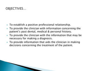
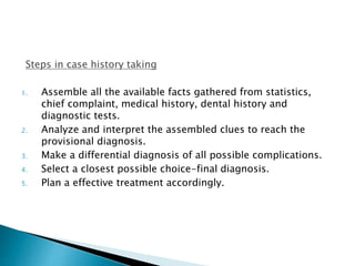
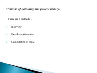
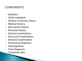
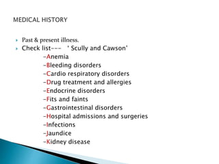
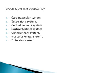
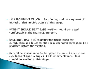
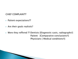
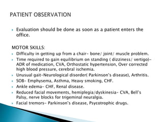
This document provides information on taking a case history for dental patients. It discusses the importance of the case history, outlines the key components that should be covered, and explains the purpose and importance of each component. These include gathering information on the chief complaint, medical history, dental history, social history, and performing an extraoral and intraoral examination. Taking a thorough case history is important for diagnosis, treatment planning, and managing the patient properly.
Gingival Index comes under the chapter of Dental Indices. Gingival Index is used to determine the severity of Gingivits/Gingival Inflammation in a patient.
DEAN’S FLUOROSIS INDEX 1943 (PUBLIC HEALTH DENTISTRY)Jeban Sahu
INTRODUCTION
INDEX:“A NUMERICAL VALUE DESCRIBING THE RELATIVE STATUS OF A POPULATION ON A GRADUATED SCALE WITH DEFINITE UPPER AND LOWER LIMITS, WHICH IS DESIGNED TO PERMIT AND FACILITATE COMPARISION WITH OTHER POPULATIONS CLASSIFIED BY THE SAME CRITERIA AND METHODS.”
-RUSSELL A.L.
INDICES USED TO MEASURE FLUOROSIS
DEAN’S FLUOROSIS INDEX
Introduced by TRENDLEY H. DEAN in 1934.
Devised an index for assessing the presence and severity of mottled enamel.
It is also known as ‘DEAN’S CLASSIFICATION SYSTEM FOR DENTAL FLUOROSIS.’
DEAN’S FLUOROSIS INDEX- Modified Criteria (1942)
SCORING CRITERIA
COMMUNITY FLUOROSIS INDEX (CFI)
SUMMARY
The content narrates about commercially available disclosing agents for the detection of dental plaque. It holds its significance from both clinician and patient viewpoint, especially in reinforcing oral hygiene measures and early detection of inflammatory changes in the gums.
Dental Plaque
Soft deposits that form the biofilm adhering to the tooth surface or other hard surfaces in the oral cavity, including removable & fixed restorations”
Bowen , 1976
Bacterial aggregations on the teeth or other solid oral structures
Lindhe, 2003
Anemia (pronounced /əˈniːmiə/, also spelled anaemia or anæmia; from Ancient Greek ἀναιμία anaimia, meaning "lack of blood") is a decrease in normal number of red blood cells (RBCs) or less than the normal quantity of hemoglobin in the blood.[1][2] However, it can include decreased oxygen-binding ability of each hemoglobin molecule due to deformity or lack in numerical development as in some other types of hemoglobin deficiency.
It is a planned professional conversation that enables the patient to communicate their symptoms , feeling and fear to the clinician, so that the nature of the patient’s real and suspected illness and mental attitudes may be determined.
Definition
Contents of case history Personal Information
General Physical Examination
Extra oral examination Intra oral examination Investigations Diagnosis
List of references
Conclusion
Case history is the most important part when we enter clinics , it has a greater impact on the diagnosis and treatment planning . It also helps to maintain a good rapport with the patient . It is most important with concern to medicolegal point of view . Thus, a Dr should always have a detailed case history.
Diagnosis and treatment planning in conservative dentistry and endodonticsIndian dental academy
Indian Dental Academy: will be one of the most relevant and exciting training center with best faculty and flexible training programs for dental professionals who wish to advance in their dental practice,Offers certified courses in Dental implants,Orthodontics,Endodontics,Cosmetic Dentistry, Prosthetic Dentistry, Periodontics and General Dentistry.
CASE HISTORY AND PHYSICAL EVALUATION OF DENTAL PATIENTS /prosthodontic coursesIndian dental academy
The Indian Dental Academy is the Leader in continuing dental education , training dentists in all aspects of dentistry and
offering a wide range of dental certified courses in different formats.for more details please visit
www.indiandentalacademy.com
CHAPTER 1 SEMESTER V PREVENTIVE-PEDIATRICS.pdfSachin Sharma
This content provides an overview of preventive pediatrics. It defines preventive pediatrics as preventing disease and promoting children's physical, mental, and social well-being to achieve positive health. It discusses antenatal, postnatal, and social preventive pediatrics. It also covers various child health programs like immunization, breastfeeding, ICDS, and the roles of organizations like WHO, UNICEF, and nurses in preventive pediatrics.
Trauma Outpatient Center is a comprehensive facility dedicated to addressing mental health challenges and providing medication-assisted treatment. We offer a diverse range of services aimed at assisting individuals in overcoming addiction, mental health disorders, and related obstacles. Our team consists of seasoned professionals who are both experienced and compassionate, committed to delivering the highest standard of care to our clients. By utilizing evidence-based treatment methods, we strive to help our clients achieve their goals and lead healthier, more fulfilling lives.
Our mission is to provide a safe and supportive environment where our clients can receive the highest quality of care. We are dedicated to assisting our clients in reaching their objectives and improving their overall well-being. We prioritize our clients' needs and individualize treatment plans to ensure they receive tailored care. Our approach is rooted in evidence-based practices proven effective in treating addiction and mental health disorders.
Dr. David Greene R3 stem cell Breakthroughs: Stem Cell Therapy in CardiologyR3 Stem Cell
Dr. David Greene, founder and CEO of R3 Stem Cell, is at the forefront of groundbreaking research in the field of cardiology, focusing on the transformative potential of stem cell therapy. His latest work emphasizes innovative approaches to treating heart disease, aiming to repair damaged heart tissue and improve heart function through the use of advanced stem cell techniques. This research promises not only to enhance the quality of life for patients with chronic heart conditions but also to pave the way for new, more effective treatments. Dr. Greene's work is notable for its focus on safety, efficacy, and the potential to significantly reduce the need for invasive surgeries and long-term medication, positioning stem cell therapy as a key player in the future of cardiac care.
ALKAMAGIC PLAN 1350.pdf plan based of door to door delivery of alkaline water...rowala30
Alka magic plan 1350 -we deliver alkaline water at your door step and you can make handsome money by referral programme
we also help and provide systematic guideline to setup 1000 lph alkaline water plant
TEST BANK For Accounting Information Systems, 3rd Edition by Vernon Richardso...rightmanforbloodline
TEST BANK For Accounting Information Systems, 3rd Edition by Vernon Richardson, Verified Chapters 1 - 18, Complete Newest Version
The dimensions of healthcare quality refer to various attributes or aspects that define the standard of healthcare services. These dimensions are used to evaluate, measure, and improve the quality of care provided to patients. A comprehensive understanding of these dimensions ensures that healthcare systems can address various aspects of patient care effectively and holistically. Dimensions of Healthcare Quality and Performance of care include the following; Appropriateness, Availability, Competence, Continuity, Effectiveness, Efficiency, Efficacy, Prevention, Respect and Care, Safety as well as Timeliness.
Letter to MREC - application to conduct studyAzreen Aj
Application to conduct study on research title 'Awareness and knowledge of oral cancer and precancer among dental outpatient in Klinik Pergigian Merlimau, Melaka'
Chandrima Spa Ajman is one of the leading Massage Center in Ajman, which is open 24 hours exclusively for men. Being one of the most affordable Spa in Ajman, we offer Body to Body massage, Kerala Massage, Malayali Massage, Indian Massage, Pakistani Massage Russian massage, Thai massage, Swedish massage, Hot Stone Massage, Deep Tissue Massage, and many more. Indulge in the ultimate massage experience and book your appointment today. We are confident that you will leave our Massage spa feeling refreshed, rejuvenated, and ready to take on the world.
Visit : https://massagespaajman.com/
Call : 052 987 1315
Deep Leg Vein Thrombosis (DVT): Meaning, Causes, Symptoms, Treatment, and Mor...The Lifesciences Magazine
Deep Leg Vein Thrombosis occurs when a blood clot forms in one or more of the deep veins in the legs. These clots can impede blood flow, leading to severe complications.
PET CT beginners Guide covers some of the underrepresented topics in PET CTMiadAlsulami
This lecture briefly covers some of the underrepresented topics in Molecular imaging with cases , such as:
- Primary pleural tumors and pleural metastases.
- Distinguishing between MPM and Talc Pleurodesis.
- Urological tumors.
- The role of FDG PET in NET.
INFECTION OF THE BRAIN -ENCEPHALITIS ( PPT)blessyjannu21
Neurological system includes brain and spinal cord. It plays an important role in functioning of our body. Encephalitis is the inflammation of the brain. Causes include viral infections, infections from insect bites or an autoimmune reaction that affects the brain. It can be life-threatening or cause long-term complications. Treatment varies, but most people require hospitalization so they can receive intensive treatment, including life support.
LGBTQ+ Adults: Unique Opportunities and Inclusive Approaches to CareVITASAuthor
This webinar helps clinicians understand the unique healthcare needs of the LGBTQ+ community, primarily in relation to end-of-life care. Topics include social and cultural background and challenges, healthcare disparities, advanced care planning, and strategies for reaching the community and improving quality of care.
2. A planned professional conversation that enables the patient
to communicate his/her symptoms, feelings and fears to the
clinician so as to obtain an insight into the nature of
patient’s illness & his/her attitude towards them.
3. To establish a positive professional relationship.
To provide the clinician with information concerning the
patient’s past dental, medical & personal history.
To provide the clinician with the information that may be
necessary for making a diagnosis.
To provide information that aids the clinician in making
decisions concerning the treatment of the patient.
4. Steps in case history taking
1. Assemble all the available facts gathered from statistics,
chief complaint, medical history, dental history and
diagnostic tests.
2. Analyze and interpret the assembled clues to reach the
provisional diagnosis.
3. Make a differential diagnosis of all possible complications.
4. Select a closest possible choice-final diagnosis.
5. Plan a effective treatment accordingly.
5. There are 3 methods :-
1) Interview
2) Health questionnaire
3) Combination of these
6. Statistics
Chief complaint
History of present illness
Medical history
Past dental history
Personal history
General examination
Extra oral examination
Intraoral examination
Provisional diagnosis
Investigations
Final diagnosis
Treatment plan
7. Patient registration number
Date
Name
Age
Sex
Address
Occupation
Marital status
8. Patient registration number
1. maintaining a record,
2. billing purposes,
3. medico legal aspects.
Date
1. Time of admission
2. reference during follow up visits
3. Record maintenance.
9. NAME.
1. To communicate with the patient.
2. To establish a rapport with the patient.
3. Record maintenance.
4. Psychological benefits.
AGE
1. For diagnosis.
2. Treatment planning.
3. Behavioral management techniques.
12. Diseases common in males:
Attrition, leukoplakia, Squamous cell carcinoma, Melanoma,
lymphoma etc
Diseases common in females:
Iron deficiency anemia, Sjogren’s syndrome, Osteoporosis,
Recurrent Apthous ulcers etc
Drug interaction: Pregnancy & lactation.
13. Future correspondence.
Information regarding the Socio-economic status and the
nourishment, hygiene & payment capacity of the patient .
Prevalence of diseases like fluorosis.
14. OCCUPATION
1. To asses the socioeconomic status.
2. Predilection of diseases in different occupations for eg:
hepatitis B is common in dentists & surgeons.
MARITAL STATUS
1. To see any history of consanguineous marriages.
2. The high consanguinity rates, coupled by the large family
size in some communities, could induce the expression of
Autosomal Recessive diseases.
15. The chief complain is usually the reason for the patient’s
visit.
It is stated in patient’s own words in chronological order of
their appearance & their severity.
The chief complaint aids in diagnosis & treatment therefore
should be given utmost priority.
16. Elaborate on the chief complaint in detail.
Ask relevant associated symptoms.
The symptoms ( Pain,swelling,ulcer) can be elaborated in
terms of:-
1. Mode & cause of onset
2. Duration
3. Location - localized , diffuse , referred, radiating.
4. Progression - continuous or intermittent.
5. Aggravating & relieving factors.
6. Medication taken.
17. Original Site of pain.
Origin & mode of onset.
Severity.
Nature of pain.
Progression of pain.
Duration of pain.
Movement of pain.
Periodicity of pain.
Effect of functional activity.
Precipitating factors.
Relieving factors.
Associated symptoms( Pallor, sweating, vomitting ).
Treatment taken.
18. 1) Duration :- how many days?
2) Mode of onset :-
a) Mass that increase in size just before eating :- Salivary gland retention
phenomenon.
b) Slow growth :- Chronic infection cyst, Benign tumors
c) Rapid growing mass :- Abscess, Infected cyst, Hematoma
d) Mass with accompanying fever :- Infection & lymphoma
3) Symptoms :- Pain, difficulty in respiration swallowing, disfiguring.
19. 4)Progress of the swelling :-
Rapid or gradual increase?
5) Associated symptoms :-
fever , loss of body weight?
6) Secondary changes :-
Softening , Ulceration, Inflammatory changes.
7) Recurrence of swelling :-
Recurrence after removal- malignant changes!!
20. Mode of onset :-
Duration of ulcer ?
Pain :-
+Inflammation - Painful.
Epithelial / - Painless.
Basal cell carcinoma
Discharge :-
Discharge from ulcer like serum, blood, pus should be noted down.
Associated disease :- Tuberculosis , Diabetes & Syphilis
21. Past & present illness.
Check list--- ‘ Scully and Cawson’
-Anemia
-Bleeding disorders
-Cardio respiratory disorders
-Drug treatment and allergies
-Endocrine disorders
-Fits and faints
-Gastrointestinal disorders
-Hospital admissions and surgeries
-Infections
-Jaundice
-Kidney disease
22. Subdivisions : -
1) Serious or significant illness :-
-Heart, kidney, liver or lung disease?
- Infectious disease ? Immunologic disorders ?
- Radiation or cancer chemotherapy & psychiatric treatment.
2) Hospitalization :-
Record of hospital admission.
Major surgery?
3) Transfusion :-
-Date of each transfusion & the number of transfused blood units.
-Transfusion can be a source of a persistent
transmissible disease.
23. 4)Allergy :-
Urticaria, Hay fever, Asthma, adverse drug reaction?
5) Medications :-
Medications?
6) Pregnancy :-
X-Rays, to prescribe medications
24. BIRTH HISTORY :-
1)Rh incompatibility :-
‘Erythroblastosis fetalis’.
Hump on the tooth/characteristic blue – green discoloration.
2) Neonatal jaundice :-
- Immature RBC’s in an infant are rapidly destroyed in the spleen. This
increased Bilirubin cannot be sufficiently cleared by the liver leading to
transient ‘ jaundice’ in the child.
3) Trauma due to forceps delivery
25. POSTNATAL HISTORY
Amount of time the child was breast fed, bottle fed etc.
Vaccination.
Presence of any habit and its duration and frequency.
Progress in the school?
26. History of dental treatment undergone by the patient and
his/her experience before, during and after the dental
treatment.
Complications?
27. Risks for diseases such as Asthma, Diabetes, Cancer, and
heart diseases.
Congenitally missing lateral incisors, Amelogenesis
imperfecta , Ectodermal dysplasia & cleft lip & cleft palate.
29. Analyze the patient entering the clinic for
built, height ,gait, and posture.
Check for any
- Pallor
- Icterus
- Clubbing
- Cyanosis
- lymphadenopathy
- Edema.
Vital signs – Pulse.
Temperature
Respiratory rate
Blood pressure
30. Pulse
Normal pulse rate is 60-80 beeats/min.
Average pulse is 72 beats/min.
Physiologic increase in infants, after exertion.
Pathologic increase in fever, cardiopulmonary diseases.
Temperature
normal temp is 98.6 degree F or 37 degree celsius.
Measured by thermometer.
Respiratory rate
Adult rate–16-24 breaths per minute.
Observe .
Feel for chest movement.
Auscultate.
31. Blood pressure
Systolic- 110-140 mm Hg
Diastolic-60-90 mm of Hg
Measured by Sphygmomanometer.
34. 1ST APPOINMENT CRUCIAL. Fact finding and development of
mutual understanding occurs at this stage.
PATIENT SHOULD BE AT EASE. He/She should be seated
comfortably in the examination room.
BASIC INFORMATION, to gather the background for
introduction and to assess the socio-economic level should be
reviewed before the meeting.
General conversation to further place the patient at ease and
avoidance of specific topics like their expectations , fees
should be avoided at this stage.
35. CHIEF COMPLAIN???
Patient expectations??
Are their goals realistic?
Were they reffered ?? Dentists {Diagnostic casts, radiographs!}
Patient {Comparative conclusions!!}
Physicians { Medical conditions!!}
36. DURATION OF EDENTULOUSNESS??
Long time?
Recently edentulous:
• ‘Green ridge’ – bony spicules from extraction sites or bony
undercuts with thin mucosal covering which may neccessitate
surgical correction.
• Alveolar ridge undergoes rapid changes during 1st year so
refitting of denture at a later date may be required.
37. New denture wearer?
• Difficulty during eating. Reduction in food morsels size
should be done.
• Patient should be made aware that Denture are not
permanent and it require refitting and remaking at a later
date.
Long term denture wearer?
• Changes in residual ridge occurs with time so the new
denture will not fit as well as the initial denture
38. Evaluation should be done as soon as a patient enters the
office.
MOTOR SKILLS:
Difficulty in getting up from a chair- bone/ joint/ muscle problem.
Time required to gain equilibrium on standing ( dizziness/ vertigo)-
ADR of medication, CVA, Orthostatic hypertension, Over corrected
high blood pressure, cerebral ischemia.
Unusual gait-Neurological disorder( Parkinson’s disease), Arthritis.
SOB- Emphysema, Asthma, Heavy smoking, CHF.
Ankle edema- CHF, Renal disease.
Reduced facial movements, hemiplegia/dyskinesia- CVA, Bell’s
Palsy, nerve blocks for trigeminal neuralgia.
Facial tremors- Parkinson’s disease, Psycotrophic drugs.
39. FACIAL FEATURES:
Length, fullness and apparent support of the lips.
Philtrum, nasiolabial fold and labiomental groove for hollowness and
puffiness.
Collapsed modiolus and labial commissure or well supported by
existing dentures?
Loose wrinkled skin?
Skin texture? Rugged tooth in rough skinned patients.
Size of the oral opening,activity of lips,width of vermilion border-governs
the tooth display.
PROFILE VIEW- To determine the maxillo-mandibular relation.
- To determine the occlusal classification.
40. ATTITUDE AND ADAPTIVE RESPONSE:
Patient attitude and level of expectation can profoundly
influence the treatment outcome. Dentist must be able to
assess the patient overall ‘ prosthetic attitude’.
If required , the patient can be referred to clinical
psychologist or psychiatrist for proper diagnosis and
treatment before instituting prosthodontic treatment
41. HOUSE CLASSIFICATION( PSYCOLOGICAL)
-DR MM House ( 1950)
1)Class 1- Philosophical
Accepts dentist’s judgement and instructions best
prognosis.
2)Class 2- Exacting
Methodical and demanding, asks a lot of questions,
good prognosis.
3)Class 3- Hysterical
Emotionally unfit, never happy, worst prognosis.
4)Class 4- Indifferent
Does not care about dental treatment, gives up
easily.
42. EXTRAORAL EXAMINATION
Patient sunglasses is to be removed.
Head and neck should be examined for pathology. Eg:
Nodules, nevi, ulcerations.
Facial coloring, hair texture, eye clarity and neuromuscular
activity must be noted.
Face and neck should be palpated for masses and enlarged
nodes.
43. FACIAL EXAMINATION
Harmony between the facial size, form and shape and the
artificial teeth selected should be present.
Assessment of the profile:
The occluding vertical dimension of denture
a) open-facial tissues appear strained or taut.
b) Closed- excessive wrinkling around mouth.
- collapsed face.
- false prognathic relationship.
c) within normal limits.
Hair, eye color and complexion- for shade selection.
44. LIP EXAMINATION
Cracking, fissuring of the corners, ulcerations- Vitamin B
deficiency, Candida albicans, excessive overclosure of
existing dentures, neoplams?
Lip support- adequate/ inadequate?
Thickness of lip- thick lips provides support irrespective of
the tooth position whereas support of thin lip depends on
tooth position. Placing the tooth too far labially may result in
unfavourable leverage on the maxillary denture.
Length of lip- visibility of teeth depends on length.
45. TMJ EXAMINATION
clicking ,popping or crepitus.
Deviation or Deflection while opening.
pain or tenderness over joint or masticatory muscles.
Maximal inter Incisal opening ( 35-50 mm)
Range of vertical & lateral movements.
46. PALPATION OF PRE TRAGUS AREA:
The examiner can be positioned either in front of or behind
the patient.
Patient is asked to slowly open and close the mouth.Palpation
with index finger,placed in the pre tragus depression is done.
INTRA AURICULAR PALPATION:
Performed by inserting small finger into the ear canal and
pressing anteriorly.
While palpating with this methods check whether condyle
moves symmetrically, with the rotation and translation phase.
47. Palpation of the muscles of mastication can be helpful in the
determination of Temporo-mandibular joint dysfunction and
in the discovery of other abnormalities.
These muscles are the Temporalis,
Masseter
Internal pterygoid and
External pterygoid.
48. Origin: temporal fossa.
Insertion: on the coronoid process and anterior border of the
ramus of the mandible.
Palpation
The muscle can be seen and readily palpated throughout its
entire length and breadth when the patients teeth are firmly
clenched.
49.
50. Origin: lower portion of the zygomatic arch.
Insertion: lateral surface of the angle and coronoid process of
the mandible.
This muscle has a deep and superficial portion as with the
temporalis muscle, it can be located when the patients jaws
are forcibly closed.
Palpation-The body of the masseter can be palpated with
thumb and the index finger.
51.
52. Origin:- medial side of the lateral pterygoid plate and the
tuberosity of the maxilla and they cannot be palpated.
Insertion:- lower medial surface of the ramus of the mandible
53. The anterior part of the
insertion can be palpated
by placing the index finger
at a 45 degree angle in the
base of the relaxed tongue.
The opposite hand can be
used extraorally to palpate
the posterior and inferior
portions of the insertion.
The body of the muscle can
be palpated by rotating the
index finger upward against
the muscle to near its origin
on the tuberosity.
54. Origin: In two parts ,one begins on the greater wing of the
sphenoid bone and the other issues from the lateral surface
of the pterygoid plates.
Insertion: On the neck of the condyle and the articular disc of
the temporomandibular joint.
55. PALPATION
The muscle is palpated by using the index or little finger and
placing it lateral to the maxillary tuberosity and medial to the
coronoid process .
The finger presses upward and inward and a painful
response can be determined.
Because this procedure is uncomfortable for the patient,the
response requires evaluation.
56.
57. Lymph nodes are oval or bean-shaped structures found along
lymphatic vessels that drain body parts.
Normally, they are non-tender, soft and cannot be felt even
though they are present.
Tender on palpation, mobility/fixation to the underlying
structures should be noted.
58.
59.
60. Location – in front of ear.
Lymphatic drainage -
Eyelids and conjunctivae,
temporal region, pinna.
For palpation of
Preauricular lymph nodes,
roll your finger in front of
the ear, against the maxilla.
Enlarged - External auditory
canal infection.
61. LOCATION – behind the ear ,
near the insertion of
sternomastoid muscle.
Lymphatic drainage: External
auditory meatus, pinna,
scalp.
Digital palpation is done by
pressing against the skull.
Enlarged due to infection of
scalp, temporal & frontal
areas.
62. Location: Located at the
junction between the back
of the head and neck.
Lymphatic drainage: Scalp
and head.
Enlarged in infection of
scalp & syphilis.
63. Location: Located below the
chin.
Lymphatic drainage: Lower
lip, floor of mouth, teeth,
submental salivary gland, tip
of tongue, skin of cheek.
Roll the fingers below and
lingual to the chin, against
the mylohyoid muscle.
Enlarged in disorders in the
anterior portion of the mouth
and the lower lip.
64. Located medial to the inferior
border of mandible.
Lymphatic drainage: Tongue,
submaxillary gland, lips and
mouth.
Roll your fingers against
inner surface of Mandible
with patient's head gently
tilted towards one side.
Enlarged in Infections of
head, neck, sinuses, ears,
eyes, scalp, pharynx.
65. 2 chains of lymph nodes
present on either side of
sternomastoid muscle.
Location – ant. cervical is
located ant to muscle &
post cervical is located
posteriorly.
66. For ant chain ,pt’s head is
tipped slightly forward &
area medial to
sternomastoid muscle is
pressed with examiners
finger.
For post chain , fingers are
kept behind the muscle.
Palpation starts from
trapezius muscle & moved
to sternomastoid muscle.
67. Labial and buccal mucosa.
Inside surface of the cheeks and lips.
Residual ridge.
Floor of the mouth.
Hard and soft palate.
Tongue.
Oropharynx / Nasopharynx.
68. It should be checked for any-
Redness/ inflammation- ill-fitting denture.
- underlying infection.
- sytemic diseases eg. Diabetes.
- chronic smoking.
White patches( denture irritation/ neoplasia)
Pigmentation.
69. Checked for-
Color.
Texture.
Any surface irregularities.
Palpate upper lip and lower lip for any thickening (induration)
or swelling.
Angular or vertical fissures.
Cleft lip.
Lip pits.
Ulcers.
Nodules.
Keratotic plaque and scars.
70. 1) Amount of saliva
Xerostomia -Retention of denture is affected.
- soreness
Excessive salivation- complicates impression making.
2) Consistency
Thin/ serous type - easy to work.
Thick / ropy - denture wearing is difficult.
3) Salivary gland duct orifices should be checked to ensure that
they are open and good salivary flow is evident.
71. Size of the maxilla and mandible will determine the amount
of basal seat available for denture foundation. The greater the
size, the more the support; larger the contact area, greater
the retention.
Discrepancy in size between the maxilla and mandible should
be noted. This condition can arise from developmental
source, trauma, early loss of teeth in one of the arches with
resultant increase in resorption, from a severe class II or class
III malocclusion.
72. Form of ridge will influence the support of the teeth and the
tooth selection.
Square, ovoid, tapered with difference in the form of the
upper and lower arch may occur.
73. High ridge with flat crest and parallel or nearly parallel sides
is the ideal crest thus will give the maximum amount of
support and stability (horizontal resistance to movement).
Knife edged ridges with multiple bony spicules offers the
poorest prognosis because of its incapability of withstanding
much occlusal forces.
Palpation of the ridge should be done. Relief should be
provided for the knife- edged ridge during impression
procedures.
74. Maxillary and mandibular dentures should be observed at a
appropriate occlusal vertical dimension.
Amount of inter ridge distance should be noted. Excessive
amount of space due to resortion will lead in poor stability
and retention because of increased leverage.
A small amount of inter ridge distance will lead to difficulty in
setting teeth and maintaining a freeway space.
75. Excessive amount of flabby tissue will cause the denture base
to shift.
Surgical correction should be considered if any.
HYPERSPLASTIC TISSUE
Usually under the ill- fitting dentures.
Epulis fissuratum- related to denture borders.
Papillary hyperplasia-under the denture bases.
Mx- Rest and massage of the tissue.
Tissue lininers / tissue conditioners.
Surgical correction.
76. Shape and form of the palatal vault should be noted.
U shaped palatal vault-most favourable for retention and
lateral stability.
V shaped and flat- least favourable.
77. CLASS I : Horizontal.
Demonstrate less movement.
More tissue coverage for the palatal seal
Most favourable
CLASS II: Soft palate is bent downwards and makes 45
degrees with the hard palate.
Tissue less than class I for palatal coverage.
CLASS III :Soft tissue makes 70 degrees with the hard palate.
Minimal tissue coverage for the palatal seal.
Least favorable.
Frequently associated with class III soft palate.
78. Longer duration of edentulousness leads to enlargement of
the tongue that make impression making procedure difficult.
Smaller tongue leads to compromised lingual seal.
Wright classified tongue position as follows-
CLASS I- tongue lies in the floor of the mouth with the tip
tip forward and slightly below the incisal edges of
mandibular anterior teeth
79. CLASS II : Tongue is flattened and broadened but the tip is in
normal position.
CLASS III :- Unfavorable tongue position.
- They drop the floor of the mouth
- Inadequate lingual seal.
- Attempts to extend the flange in order to get a r
lingual seal will lead to over extension and
dislodgement of denture during tongue
movements.
80. Presents a wide variation in anatomy anf functional relation to
the ridge crest.
If the floor of the mouth is near the ridge crest at rest esp in
the sublingual gland and mylohyoid region, the retention and
stability of the denture will be poor.
The retromylohyoid space( lateral throat form) is critical for
lingual seal and lingual stability. It may be partially or totally
obliterated by tongue movement.
Use of william’s probe to measure the depth.
81. Surgical corrections of the bony undercuts should be made
before the impression procedures.
In presence of tori, relief should be provided in the
impression procedures and denture. Surgical correction
should be considered if the tori is excessively large.
Surgical correction of frenum or muscle attachment if it is
place close to the ridge esp. the maxillary labial frenum and
mandibular lingual frenum.
82. Radiographic examinations to detect any bony pathology,
examination of existing denture, evalation of the
pretreatment radiographs and dignostic casts should be done
before the treatment planning.
84. 1) Relief of pain and discomfort and caries control by placement
of temporary restorations.
Preliminary examination to determine the need for
management of acute lesions, to determine the extent of
caries and to arrest further caries activity until a definitive
treatment can be instituted.
2) Thorough and complete oral prophylaxis should be
performed before impression procedures
85. 3) Complete intraoral radiographic survey:
- To locate areas of infection and other pathosis.
- To reveal presence of root fragments, foreign objects, bony
spicules and irregular ridge formations.
- To reveal the presence and extent of caries and their realation
to the pulp and periodontal attachments.
- To permit evaluation of existing restorations as to evidence of
recurrent caries, marginal leakage and over hanging gingival
margins.
- To reveal the presence of endodontically treated teeth and to
permit their evaluation as to future prognosis (retain /
extract)
86. - To permit the evaluation of periodontal condition and need of
any treatment.
- To evluate the alveolar support of the abutment teeth, the
supporting length and morphology of their roots.
4) Impressions for making accurate diagnostic casts to be
mounted for occlusal examination are made.
5) Examination of teeth, investing structures and residual ridges
is done.
87. 6) Vitality tests of remaining teeth should be carried out.
7) Determination of the height of the floor of the mouth to
locate the inferior border of lingual mandibular major
connector