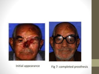
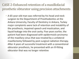
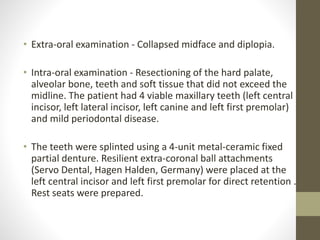
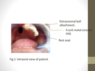
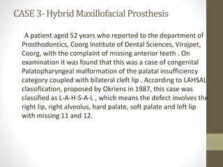
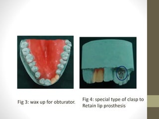
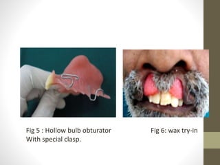
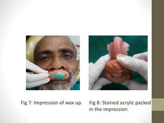
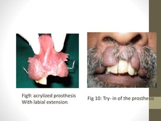
CASE 1-Magnet-Retained Facial Prosthesis 
Combined with Maxillary Obturator 
• A 65 year old diabetic patient, with chief complain of 
poor facial appearance and past history of diabetes 
mellitus.His facial tissues were affected by a fungal 
infection of rhinocerebral mucormycosis followed by 
diabetic ketoacidosis. For debridement and removal of 
necrotic tissues, ablative surgery was performed leaving 
behind a maxillary defect and communication between 
the oral,nasal and orbital cavity.
Fig: Midfacial defect 
after surgery. 
Fig 2: Intraoral view of the 
Maxillary defect
• After precise evaluation of the case, the proposed 
treatment plan was to construct a complete denture with 
obturator, as well as a facial prosthesis which would be 
attached to the obturator with cobalt samarium magnets 
(Jobmasters, Randallstown, USA).
Fig: Master cast 
In dental stone. 
Fig 4: Wax try-in with ocular 
Prosthesis.
Fig 5: Hollow acrylic 
Substructure . 
MDX4-4210-base silicone 
with laminar intrinsic stain. 
MAGNETS 
Fig 6: attached intra and 
extraoral prosthesis
Initial appearance Fig 7: completed prosthesis
CASE 2-Enhanced retention of a maxillofacial 
prosthetic obturator using precision attachments 
• A 65-year-old man was referred by his head and neck 
surgeon to the Department of Prosthodontics at the 
Ankara University, Faculty of Dentistry in Ankara, Turkey 
major complaints were lack of retention and instability of 
the prosthesis, impaired speech and mastication, and 
liquid leakage into the oral cavity. Five years earlier, the 
patient had been diagnosed with epidermoid carcinoma 
of the maxillary sinus that was treated by a unilateral 
maxillectomy followed by post-surgical radiation therapy. 
After 4 years of successful treatment with a conventional 
obturator prosthesis, he presented with an ill-fitting 
obturator that was no longer retentive
• Extra-oral examination - Collapsed midface and diplopia. 
• Intra-oral examination - Resectioning of the hard palate, 
alveolar bone, teeth and soft tissue that did not exceed the 
midline. The patient had 4 viable maxillary teeth (left central 
incisor, left lateral incisor, left canine and left first premolar) 
and mild periodontal disease. 
• The teeth were splinted using a 4-unit metal-ceramic fixed 
partial denture. Resilient extra-coronal ball attachments 
(Servo Dental, Hagen Halden, Germany) were placed at the 
left central incisor and left first premolar for direct retention . 
Rest seats were prepared.
Fig 1: Intraoral view of patient 
Extracoronal ball 
attachment. 
4 unit metal ceramic 
FPD 
Rest seat
Fig 2: final impression with 
polyvinyl siloxane. 
Fig 3: obturator frame work on 
Master model.
Fig 3: Processed prosthesis Fig 4: final fitting in mouth.
CASE 3-Hybrid Maxillofacial Prosthesis 
A patient aged 52 years who reported to the department of 
Prosthodontics, Coorg Institute of Dental Sciences, Virajpet, 
Coorg, with the complaint of missing anterior teeth . On 
examination it was found that this was a case of congenital 
Palatopharyngeal malformation of the palatal insufficiency 
category coupled with bilateral cleft lip . According to LAHSAL 
classification, proposed by Okriens in 1987, this case was 
classified as L-A-H-S-A-L , which means the defect involves the 
right lip, right alveolus, hard palate, soft palate and left lip 
with missing 11 and 12.
Fig 1: Extraoral appearance. Fig 2: Intraoral appearance
Fig 3: wax up for obturator. Fig 4: special type of clasp to 
Retain lip prosthesis
Fig 5 : Hollow bulb obturator 
With special clasp. 
Fig 6: wax try-in
Fig 7: Impression of wax up. Fig 8: Stained acrylic packed 
in the impression.
Fig9: acrylized prosthesis 
With labial extension. 
Fig 10: Try- in of the prosthesis
Fig 11: Harvested hair filled 
With self cure acrylic. Fig 12: final prosthesis in place.
Initial appearance Fig 13: final extraoral appearance

Presentation2

  • 1.
    CASE 1-Magnet-Retained FacialProsthesis Combined with Maxillary Obturator • A 65 year old diabetic patient, with chief complain of poor facial appearance and past history of diabetes mellitus.His facial tissues were affected by a fungal infection of rhinocerebral mucormycosis followed by diabetic ketoacidosis. For debridement and removal of necrotic tissues, ablative surgery was performed leaving behind a maxillary defect and communication between the oral,nasal and orbital cavity.
  • 2.
    Fig: Midfacial defect after surgery. Fig 2: Intraoral view of the Maxillary defect
  • 3.
    • After preciseevaluation of the case, the proposed treatment plan was to construct a complete denture with obturator, as well as a facial prosthesis which would be attached to the obturator with cobalt samarium magnets (Jobmasters, Randallstown, USA).
  • 4.
    Fig: Master cast In dental stone. Fig 4: Wax try-in with ocular Prosthesis.
  • 5.
    Fig 5: Hollowacrylic Substructure . MDX4-4210-base silicone with laminar intrinsic stain. MAGNETS Fig 6: attached intra and extraoral prosthesis
  • 6.
    Initial appearance Fig7: completed prosthesis
  • 7.
    CASE 2-Enhanced retentionof a maxillofacial prosthetic obturator using precision attachments • A 65-year-old man was referred by his head and neck surgeon to the Department of Prosthodontics at the Ankara University, Faculty of Dentistry in Ankara, Turkey major complaints were lack of retention and instability of the prosthesis, impaired speech and mastication, and liquid leakage into the oral cavity. Five years earlier, the patient had been diagnosed with epidermoid carcinoma of the maxillary sinus that was treated by a unilateral maxillectomy followed by post-surgical radiation therapy. After 4 years of successful treatment with a conventional obturator prosthesis, he presented with an ill-fitting obturator that was no longer retentive
  • 8.
    • Extra-oral examination- Collapsed midface and diplopia. • Intra-oral examination - Resectioning of the hard palate, alveolar bone, teeth and soft tissue that did not exceed the midline. The patient had 4 viable maxillary teeth (left central incisor, left lateral incisor, left canine and left first premolar) and mild periodontal disease. • The teeth were splinted using a 4-unit metal-ceramic fixed partial denture. Resilient extra-coronal ball attachments (Servo Dental, Hagen Halden, Germany) were placed at the left central incisor and left first premolar for direct retention . Rest seats were prepared.
  • 9.
    Fig 1: Intraoralview of patient Extracoronal ball attachment. 4 unit metal ceramic FPD Rest seat
  • 10.
    Fig 2: finalimpression with polyvinyl siloxane. Fig 3: obturator frame work on Master model.
  • 11.
    Fig 3: Processedprosthesis Fig 4: final fitting in mouth.
  • 12.
    CASE 3-Hybrid MaxillofacialProsthesis A patient aged 52 years who reported to the department of Prosthodontics, Coorg Institute of Dental Sciences, Virajpet, Coorg, with the complaint of missing anterior teeth . On examination it was found that this was a case of congenital Palatopharyngeal malformation of the palatal insufficiency category coupled with bilateral cleft lip . According to LAHSAL classification, proposed by Okriens in 1987, this case was classified as L-A-H-S-A-L , which means the defect involves the right lip, right alveolus, hard palate, soft palate and left lip with missing 11 and 12.
  • 13.
    Fig 1: Extraoralappearance. Fig 2: Intraoral appearance
  • 14.
    Fig 3: waxup for obturator. Fig 4: special type of clasp to Retain lip prosthesis
  • 15.
    Fig 5 :Hollow bulb obturator With special clasp. Fig 6: wax try-in
  • 16.
    Fig 7: Impressionof wax up. Fig 8: Stained acrylic packed in the impression.
  • 17.
    Fig9: acrylized prosthesis With labial extension. Fig 10: Try- in of the prosthesis
  • 18.
    Fig 11: Harvestedhair filled With self cure acrylic. Fig 12: final prosthesis in place.
  • 19.
    Initial appearance Fig13: final extraoral appearance