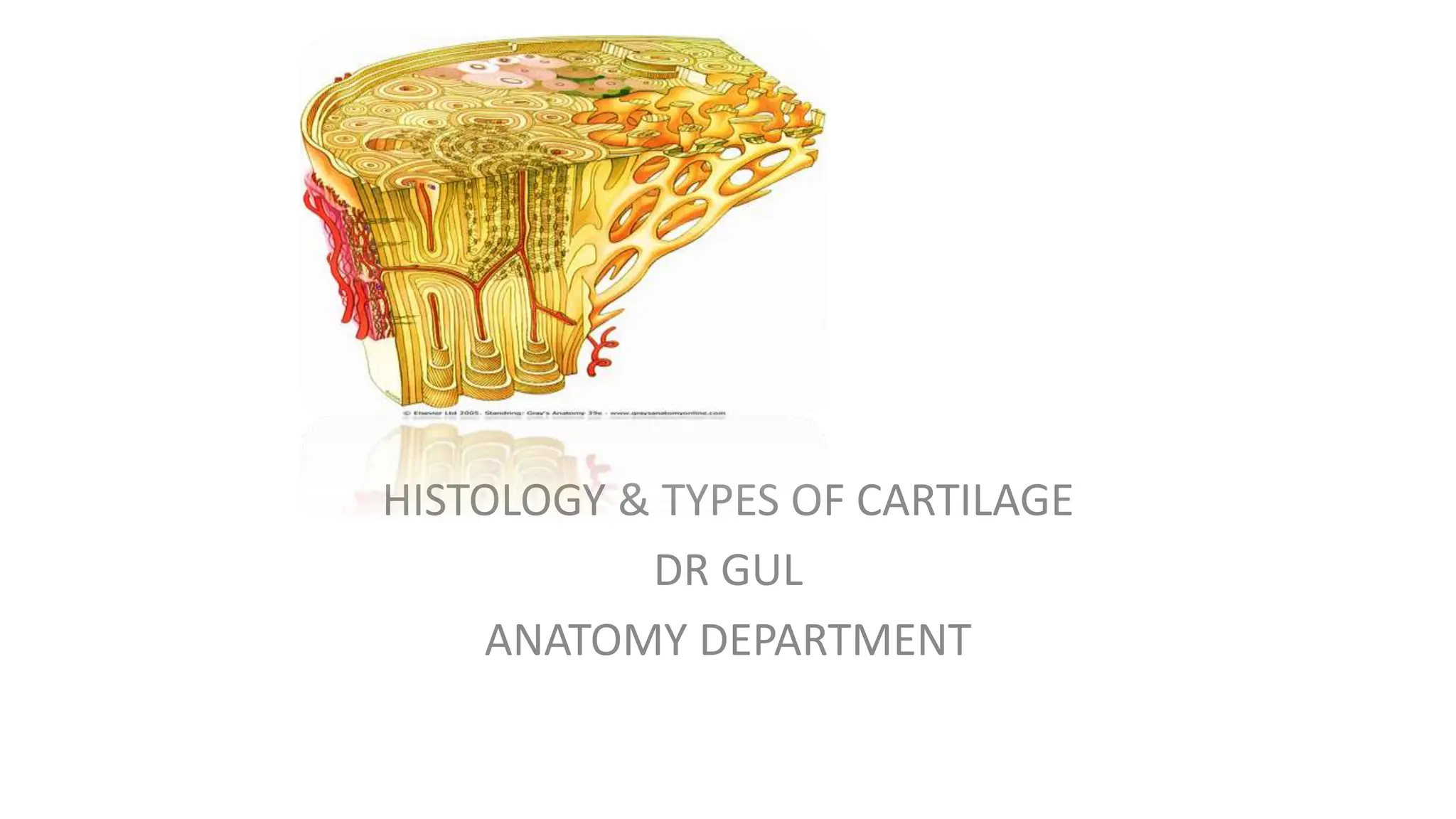
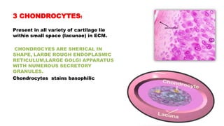
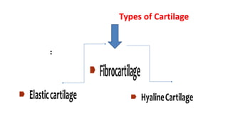
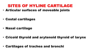
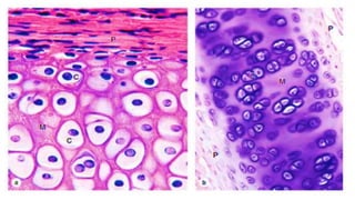
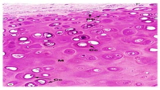
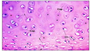
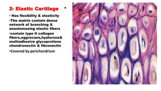
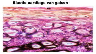
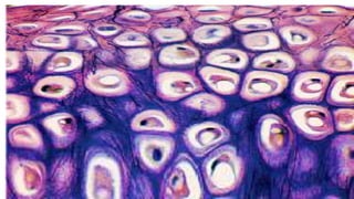
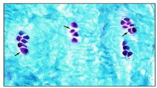
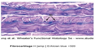
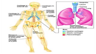
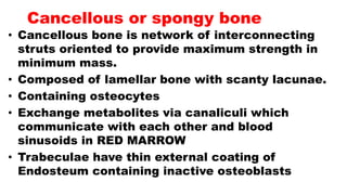
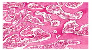
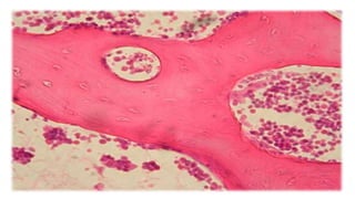
HISTOLOGY & TYPES OF CARTILAGE
DR GUL
ANATOMY DEPARTMENT
Learning objectives
• To understand cartilage
• To understand different types of cartilage
• To understand histological features of cartilage
• To discuss different cells of cartilage
CARTILAGE
• Cartilage is special type of CT
chracterized by its reslient & pliant nature
Cartilage form the skeleton and support the
body.
Cartilage form the fetal skeleton.
Cartilage is non-vascular structure and not
supplied with nervous or lymphatic tissues
Shock absorber in various places of body.
Covers articular surfaces of bones of synovial joints
PERICHONDRIUM
Perichondrium covers cartilage is dense
irregular CT.
Contains Blood vessels for blood supply to CT.
Plays an important role in the growth & repair
of cartilage.
MICROSCOPICALLY PERICHONDRIUM has two
layers …
Fibrous and Cellular
CONT;
• THE OUTER FIBROUS LAYER: consist of TYPE 1 COLLAGENE
FIBERS & FIBROBLAST.
• THE INNER CELLULAR LAYER ALSO CALLED CHONDROGENIC
LAYER which have the progenitor cells which deferentiate into
CHONDROBLAST
Composition
Chondroblasts
Chondrocytes
Chondrogenic
cells
Collagen
fibers
Elastic fibers
Proteoglycans
Glycosaminoglycans
1. Stem cells (chondrogenic cells):
• Mesenchymal
cells.
• Spindle shaped
cells with ovoid
nucleus
• small Golgi,
Few mitochondria,
and rER.
2 Chondroblasts :
• Derived from mesenchymal cells
• Ovoid, basophilic cells,
• rich with rER,
• well developed Golgi,
• numerous mitochondria,
• more secretory vesicles.
• Synthesize the fibers and the matrix .
3 CHONDROCYTES:
Present in all variety of cartilage lie
within small space (lacunae) in ECM.
CHONDROCYES ARE SHERICAL IN
SHAPE, LARDE ROUGH ENDOPLASMIC
RETICULUM,LARGE GOLGI APPARATUS
WITH NUMEROUS SECRETORY
GRANULES.
Chondrocytes stains basophilic
Types of Cartilage
:
1- Hyaline Cartilage
• Identical form resemble to frosted glass.
Covered by Perichondrium except articular surfaces.
• Matrix formed by the mainly collagen type 2 fibrils ,
multiadhesive glycoprotien,GAGs and Chondrocyte
ON THE BASIS OF STAINING DIFFERENCE, THE
EXTRACELLULAR MATRIX IS SUBDIVIDED INTO
VARIOUS TYPE TYPES:-
PERICELLULAR MATRIX: also k/as capsular matrix
outside the cell membrane of chondrocytes.
Stain deeply basophilic because of devoid of type II
collagen .
Consists chiefly sulfated protoglycans
Territorial and Interterritorial matrix
Territorial matrix contain few type II collagen & large
amount of sulfated proteoglycans
• surrounding pericellular matrix .
• Stain deeply basophilic but less intensely than pericellular
matrix.
• Interterritorial matrix . Remaining matrix is Interterritorial
matrix
• Abundant type II & type X1 collagen
• Small amount of sulphated protoglycan.
• Light basophilic stain
SITES OF HYLINE CARTILAGE
• Articular surfaces of moveable joints
• Costal cartilages
• Nasal cartilage
• Cricoid thyroid and arytenoid thyroid of larynx
• Cartilages of trachea and bronchi
2- Elastic Cartilage •
• Has flexibility & elasticity
•The matrix contain dense
network of branching &
anastomosing elastic fibers
•contain type II collagen
fibers,aggrecans,hyalurona&
multiadhesive glycoprotiens
chondronectin & fibronectin
•Covered by perichondrium
SITES OF ELASTIC CARTILAGE
• AURICLE PINNA OF EXTERNAL EAR
• EPIGLOTIS OF THE LARYNX
• EXTERNAL AUDITORY MEATUS AND AUDITORY
TUBES
Elastic cartilage van gaison
3- Fibro-cartilage
• The fibro cartilage is tough & strong resilient tissue
• Different from Hyaline or Elastic combination of hyaline
and dense CT. associated with dense CT.
• Not surrounded by periosteum
• Type 1 collagen and chondrocytes arranged in rows
• The chondrocytes are differentiated from fibrocytes.
• Located: where ever strength with flexibility is required.
SITES OF FIBROCARTILAGE
• INTERVERTIBERAL DISICS
• PUBIC SYMPHYSIS
• INTRA-ARTICULAR DISCSOF STERNOCLAVICULAR AND
TEMPOROMENDIBULAR JOINTS
• MENISCI OF KNEE JOINT
• LABERUMGLENOID (SHOULDER) AND LABRUM
ACETABULAR (HIP) JOINTS
• LIGAMENTUM TERES OF HIP JOINT
Cartilage growth
• Cartilage grows by two different
processes
• Interstitial growth
• Appositional growth
a. Interstitial growth
• involves the division of existing
chondrocytes.
• It is important in the formation of the fetal
skeleton
continues in the epiphyseal plates articular
cartilages
b. Appositional growth
• involves the differentiation of
chondroblasts and stem cells on the
inner surface of the perichondrium into
chondrocytes.
• It is responsible for increasing the girth
of the cartilage masses.
Repair of cartilage
• Repair and regeneration is poor in adults.
• In case of injury small defect may be repaired by
chondrogenic cells of perichondrium.
• Perichondrial cells cannot repair it by producing
cartilage instead they produce scar of dense CT
objectives
• To understand histological features of bones
• To discuss different types of cells present in
bones
BONE
• Bone tissue is rich in blood supply
special type of connective tissue
with calcified matrix.
• Functions:
▫ Supports and protects
▫ Bone marrow.
▫ Important storage site for calcium and other
essential minerals.
Surfaces
The outer dense shell compact bone
• Central Medullary cavity or cancellous zone of thin
interconnecting trabeculle
• The internal surfaces of bones are covered by a
thin, condensed reticular connective tissue
(endosteum) that contain bone and blood cell
precursors.
• The endosteum lines the marrow cavity and sends
extensions into the Haversian canals.
Compact bone
• Dense areas without cavities
• Parallel bony columns lines the stress exerted
area on bone
Each column is made up of concentric bony layers
or LAMELLAE
LAMELLAE contains neurovascular bundles
These neurovascular channels are called CANNALS
OF HAVERS OR HAVERSION CANALS
HAVERSION SYSTEM I…. Haversian Canals and
Lamellae is called Haversian system
Cancellous or spongy bone
• Cancellous bone is network of interconnecting
struts oriented to provide maximum strength in
minimum mass.
• Composed of lamellar bone with scanty lacunae.
• Containing osteocytes
• Exchange metabolites via canaliculi which
communicate with each other and blood
sinusoids in RED MARROW
• Trabeculae have thin external coating of
Endosteum containing inactive osteoblasts
Osteogenic cells,
Osteoblasts,
Osteocytes,
Osteoclasts
organic and
non-organic
collagen type I
fibers
1- Osteogenic (Osteoprogenitor)
cells
• Mesenchymal stem cells
• Small spindle-shaped cells with pale cytoplasm and
ovoid nuclei.
• Two types
• Osteoblast precursors have sparse rER and Golgi
complexes.
• Osteoclast precursors have abundant free ribosomes
and mitochondria.
2- Osteoblasts:
Found in in the periosteum and endosteum
• Large rounded or cuboidal cells, with deep
basophilic cytoplasm,
• well-developed RER and Golgi; and eccentric
nucleus.
• Synthesize and secrete organic components
• Osteoblasts mature called osteocytes.
3- Osteocytes
Cells are branched, smaller than osteoblasts, and not divide.
• found in cavities in the bone matrix called lacunae
Processes (branches) extend into canaliculi in the calcified matrix.
• Isolated cells Contact one another through gap junctions.
• Located near bone surfaces Maintain bone matrix.
• The death of osteocytes results in bone resorption.
4- Osteoclasts
• lie on bony surfaces in lacunae.
• • Large multinucleated cells• Acidophilic cytoplasm
containing abundant lysosomes and mitochondria and
well developed Golgi complex; and brush border of
plasma membrane facing the bone marrow.
• • Cells release acid, collagenase, and other lytic
enzymes into the compartments; these break down bone
matrix and release minerals, a process called bone
resorption.
Ossification (Bone formation)
• Bone is formed from mesenchymal
embryonic tissue by two ways:
• 1. Intra-membranous ossification
• 2. Endochondral ossification
1- Intra-membranous ossification:
• Ossification center in the form of increased the
vascularity, condensation of mesenchymal cells•
Mesenchymal cells > osteogenic cells.
• Osteogenic cells differentiate into osteoblasts; which
synthesize the organic component of bone matrix
(collagen, glycoprotein).
• osteoblasts secrete enzyme which stimulate calcium
salts to calcified
matrix.
• • The bone matrix grows in the membrane,
vascular tissues differentiate into hematopoietic
tissue (bone marrow).
• • A layer of vascular mesenchyme forming
periosteum.
• • The osteogenic stem cells in the periosteum
differentiate to osteoblasts, which start to lay
down or arrange bone lamellae (compact bone)
2- Endochondral Ossification
• Cartilage model :
• embryonic mesenchymal cells –
condensed……differentiated to
chondroblasts, chondrocytes, and
cartilage matrix.
• ▫ Primary centers of ossification:
some changes have been developed
Primary centers of ossification
• Chondrocytes at the middle of cartilage model
enlarged, and calcium salts become to deposit
around the lacunae.
• Chondrocytes die due to prevention of nutrient
diffusion through the calcified matrix, leaving empty
spaces.
• The perichondrium becomes highly vascular and
active and changed to periosteum, where the inner
vascular osteogenic cells differentiate to
osteoblasts.
• The osteoblasts start to lay down a collar of
compact bone around the shaft called sub-
periosteal collar of bone.
• The periosteum forms a periosteal bud,
which is consists of ; ca
• The vascular bud invades the sub periosteal
bone by osteoclasts.
The thin wall of the empty lacunae is broken down
forming the primary bone marrow cavity.
• As the periosteal bud invades bone and cartilage
the osteoblasts arrange themselves along the
marrow spaces and start to lay down bone matrix.
• Marrow spaces joint together forming a central
regular cavity in the middle of diaphysis
• • The microscopic structure of the developing
endochondral bones is
• characterized by 5 overlapping zones:
• 1. The zone of resting cartilage is composed of typical
hyaline cartilage and is farthest from the primary marrow
cavity.
• 2. The zone of proliferation contains columns of flattened
chondrocytes.
• 3. In the zone of hypertrophy, the chondrocytes in the
columns are enlarged and rounded.
• 4. The zone of calcification, is characterized by a more
basophilic matrix. There is often a significant overlap
between zones 3 and 4,
• which are sometimes referred to as a single zone of
hypertrophy and calcification.
• 5. The zone of ossification borders directly on the primary.
marrow cavity
• It is characterized by intensely acidophilic osteoid,
osteocytes within the bone matrix, and a monolayer of
basophilic osteoblasts
Clinical conditions
• Osteomalacia
• due to failure of normal mineralization of
osteoid
• common cause is calcium deficiency
• Osteoarthritis
• degenerative disorder with inflammatory
breakdown of articular cartilages in synovial
joints

PPT ON CARTILAGES & BONES......... .pptx

  • 1.
    HISTOLOGY & TYPESOF CARTILAGE DR GUL ANATOMY DEPARTMENT
  • 2.
    Learning objectives • Tounderstand cartilage • To understand different types of cartilage • To understand histological features of cartilage • To discuss different cells of cartilage
  • 3.
    CARTILAGE • Cartilage isspecial type of CT chracterized by its reslient & pliant nature Cartilage form the skeleton and support the body. Cartilage form the fetal skeleton. Cartilage is non-vascular structure and not supplied with nervous or lymphatic tissues Shock absorber in various places of body. Covers articular surfaces of bones of synovial joints
  • 4.
    PERICHONDRIUM Perichondrium covers cartilageis dense irregular CT. Contains Blood vessels for blood supply to CT. Plays an important role in the growth & repair of cartilage. MICROSCOPICALLY PERICHONDRIUM has two layers … Fibrous and Cellular
  • 5.
    CONT; • THE OUTERFIBROUS LAYER: consist of TYPE 1 COLLAGENE FIBERS & FIBROBLAST. • THE INNER CELLULAR LAYER ALSO CALLED CHONDROGENIC LAYER which have the progenitor cells which deferentiate into CHONDROBLAST
  • 6.
  • 7.
    1. Stem cells(chondrogenic cells): • Mesenchymal cells. • Spindle shaped cells with ovoid nucleus • small Golgi, Few mitochondria, and rER.
  • 8.
    2 Chondroblasts : •Derived from mesenchymal cells • Ovoid, basophilic cells, • rich with rER, • well developed Golgi, • numerous mitochondria, • more secretory vesicles. • Synthesize the fibers and the matrix .
  • 9.
    3 CHONDROCYTES: Present inall variety of cartilage lie within small space (lacunae) in ECM. CHONDROCYES ARE SHERICAL IN SHAPE, LARDE ROUGH ENDOPLASMIC RETICULUM,LARGE GOLGI APPARATUS WITH NUMEROUS SECRETORY GRANULES. Chondrocytes stains basophilic
  • 10.
  • 11.
    1- Hyaline Cartilage •Identical form resemble to frosted glass. Covered by Perichondrium except articular surfaces. • Matrix formed by the mainly collagen type 2 fibrils , multiadhesive glycoprotien,GAGs and Chondrocyte ON THE BASIS OF STAINING DIFFERENCE, THE EXTRACELLULAR MATRIX IS SUBDIVIDED INTO VARIOUS TYPE TYPES:- PERICELLULAR MATRIX: also k/as capsular matrix outside the cell membrane of chondrocytes. Stain deeply basophilic because of devoid of type II collagen . Consists chiefly sulfated protoglycans
  • 12.
    Territorial and Interterritorialmatrix Territorial matrix contain few type II collagen & large amount of sulfated proteoglycans • surrounding pericellular matrix . • Stain deeply basophilic but less intensely than pericellular matrix. • Interterritorial matrix . Remaining matrix is Interterritorial matrix • Abundant type II & type X1 collagen • Small amount of sulphated protoglycan. • Light basophilic stain
  • 13.
    SITES OF HYLINECARTILAGE • Articular surfaces of moveable joints • Costal cartilages • Nasal cartilage • Cricoid thyroid and arytenoid thyroid of larynx • Cartilages of trachea and bronchi
  • 17.
    2- Elastic Cartilage• • Has flexibility & elasticity •The matrix contain dense network of branching & anastomosing elastic fibers •contain type II collagen fibers,aggrecans,hyalurona& multiadhesive glycoprotiens chondronectin & fibronectin •Covered by perichondrium
  • 18.
    SITES OF ELASTICCARTILAGE • AURICLE PINNA OF EXTERNAL EAR • EPIGLOTIS OF THE LARYNX • EXTERNAL AUDITORY MEATUS AND AUDITORY TUBES
  • 19.
  • 21.
    3- Fibro-cartilage • Thefibro cartilage is tough & strong resilient tissue • Different from Hyaline or Elastic combination of hyaline and dense CT. associated with dense CT. • Not surrounded by periosteum • Type 1 collagen and chondrocytes arranged in rows • The chondrocytes are differentiated from fibrocytes. • Located: where ever strength with flexibility is required.
  • 22.
    SITES OF FIBROCARTILAGE •INTERVERTIBERAL DISICS • PUBIC SYMPHYSIS • INTRA-ARTICULAR DISCSOF STERNOCLAVICULAR AND TEMPOROMENDIBULAR JOINTS • MENISCI OF KNEE JOINT • LABERUMGLENOID (SHOULDER) AND LABRUM ACETABULAR (HIP) JOINTS • LIGAMENTUM TERES OF HIP JOINT
  • 26.
    Cartilage growth • Cartilagegrows by two different processes • Interstitial growth • Appositional growth
  • 27.
    a. Interstitial growth •involves the division of existing chondrocytes. • It is important in the formation of the fetal skeleton continues in the epiphyseal plates articular cartilages
  • 29.
    b. Appositional growth •involves the differentiation of chondroblasts and stem cells on the inner surface of the perichondrium into chondrocytes. • It is responsible for increasing the girth of the cartilage masses.
  • 31.
    Repair of cartilage •Repair and regeneration is poor in adults. • In case of injury small defect may be repaired by chondrogenic cells of perichondrium. • Perichondrial cells cannot repair it by producing cartilage instead they produce scar of dense CT
  • 32.
    objectives • To understandhistological features of bones • To discuss different types of cells present in bones
  • 33.
    BONE • Bone tissueis rich in blood supply special type of connective tissue with calcified matrix. • Functions: ▫ Supports and protects ▫ Bone marrow. ▫ Important storage site for calcium and other essential minerals.
  • 34.
    Surfaces The outer denseshell compact bone • Central Medullary cavity or cancellous zone of thin interconnecting trabeculle • The internal surfaces of bones are covered by a thin, condensed reticular connective tissue (endosteum) that contain bone and blood cell precursors. • The endosteum lines the marrow cavity and sends extensions into the Haversian canals.
  • 36.
    Compact bone • Denseareas without cavities • Parallel bony columns lines the stress exerted area on bone Each column is made up of concentric bony layers or LAMELLAE LAMELLAE contains neurovascular bundles These neurovascular channels are called CANNALS OF HAVERS OR HAVERSION CANALS HAVERSION SYSTEM I…. Haversian Canals and Lamellae is called Haversian system
  • 38.
    Cancellous or spongybone • Cancellous bone is network of interconnecting struts oriented to provide maximum strength in minimum mass. • Composed of lamellar bone with scanty lacunae. • Containing osteocytes • Exchange metabolites via canaliculi which communicate with each other and blood sinusoids in RED MARROW • Trabeculae have thin external coating of Endosteum containing inactive osteoblasts
  • 42.
  • 43.
    1- Osteogenic (Osteoprogenitor) cells •Mesenchymal stem cells • Small spindle-shaped cells with pale cytoplasm and ovoid nuclei. • Two types • Osteoblast precursors have sparse rER and Golgi complexes. • Osteoclast precursors have abundant free ribosomes and mitochondria.
  • 44.
    2- Osteoblasts: Found inin the periosteum and endosteum • Large rounded or cuboidal cells, with deep basophilic cytoplasm, • well-developed RER and Golgi; and eccentric nucleus. • Synthesize and secrete organic components • Osteoblasts mature called osteocytes.
  • 45.
    3- Osteocytes Cells arebranched, smaller than osteoblasts, and not divide. • found in cavities in the bone matrix called lacunae Processes (branches) extend into canaliculi in the calcified matrix. • Isolated cells Contact one another through gap junctions. • Located near bone surfaces Maintain bone matrix. • The death of osteocytes results in bone resorption.
  • 46.
    4- Osteoclasts • lieon bony surfaces in lacunae. • • Large multinucleated cells• Acidophilic cytoplasm containing abundant lysosomes and mitochondria and well developed Golgi complex; and brush border of plasma membrane facing the bone marrow. • • Cells release acid, collagenase, and other lytic enzymes into the compartments; these break down bone matrix and release minerals, a process called bone resorption.
  • 49.
    Ossification (Bone formation) •Bone is formed from mesenchymal embryonic tissue by two ways: • 1. Intra-membranous ossification • 2. Endochondral ossification
  • 50.
    1- Intra-membranous ossification: •Ossification center in the form of increased the vascularity, condensation of mesenchymal cells• Mesenchymal cells > osteogenic cells. • Osteogenic cells differentiate into osteoblasts; which synthesize the organic component of bone matrix (collagen, glycoprotein). • osteoblasts secrete enzyme which stimulate calcium salts to calcified matrix.
  • 51.
    • • Thebone matrix grows in the membrane, vascular tissues differentiate into hematopoietic tissue (bone marrow). • • A layer of vascular mesenchyme forming periosteum. • • The osteogenic stem cells in the periosteum differentiate to osteoblasts, which start to lay down or arrange bone lamellae (compact bone)
  • 52.
    2- Endochondral Ossification •Cartilage model : • embryonic mesenchymal cells – condensed……differentiated to chondroblasts, chondrocytes, and cartilage matrix. • ▫ Primary centers of ossification: some changes have been developed
  • 53.
    Primary centers ofossification • Chondrocytes at the middle of cartilage model enlarged, and calcium salts become to deposit around the lacunae. • Chondrocytes die due to prevention of nutrient diffusion through the calcified matrix, leaving empty spaces. • The perichondrium becomes highly vascular and active and changed to periosteum, where the inner vascular osteogenic cells differentiate to osteoblasts.
  • 54.
    • The osteoblastsstart to lay down a collar of compact bone around the shaft called sub- periosteal collar of bone. • The periosteum forms a periosteal bud, which is consists of ; ca • The vascular bud invades the sub periosteal bone by osteoclasts.
  • 55.
    The thin wallof the empty lacunae is broken down forming the primary bone marrow cavity. • As the periosteal bud invades bone and cartilage the osteoblasts arrange themselves along the marrow spaces and start to lay down bone matrix. • Marrow spaces joint together forming a central regular cavity in the middle of diaphysis
  • 57.
    • • Themicroscopic structure of the developing endochondral bones is • characterized by 5 overlapping zones: • 1. The zone of resting cartilage is composed of typical hyaline cartilage and is farthest from the primary marrow cavity. • 2. The zone of proliferation contains columns of flattened chondrocytes. • 3. In the zone of hypertrophy, the chondrocytes in the columns are enlarged and rounded.
  • 58.
    • 4. Thezone of calcification, is characterized by a more basophilic matrix. There is often a significant overlap between zones 3 and 4, • which are sometimes referred to as a single zone of hypertrophy and calcification. • 5. The zone of ossification borders directly on the primary. marrow cavity • It is characterized by intensely acidophilic osteoid, osteocytes within the bone matrix, and a monolayer of basophilic osteoblasts
  • 59.
    Clinical conditions • Osteomalacia •due to failure of normal mineralization of osteoid • common cause is calcium deficiency • Osteoarthritis • degenerative disorder with inflammatory breakdown of articular cartilages in synovial joints